
















![【ベーシック】体外で3Dの心臓をつくる![心臓オルガノイド]](https://i.ytimg.com/vi/f3Z6Y0Fs548/maxresdefault.jpg)



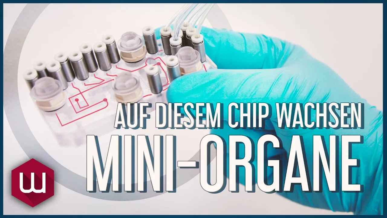












The Dynamic Organ System is an open organ-on-chip platform consisting of a pump, biochips with dual chambers separated by porous membranes, reservoirs, tubing, and connectors. Each biochip contains two interconnected chambers (left and right) with top and bottom channels separated by a porous membrane, enabling the creation of complex organ models like vasculature and epithelium. The setup process involves attaching plugs to close static sites, connecting reservoirs to the biochip via ports, and linking tubing to both the biochip and reservoir lids. The complete workflow includes biochip stabilization with ethanol, membrane coating, cell seeding at appropriate concentrations, incubation until confluence, and finally connecting the perfusion equipment to the pump for medium perfusion. This system allows researchers to build human in vitro disease and infection models using standard laboratory equipment without capital spending.
DynamicOrgan System: Open Organ-on-Chip Platform Demo
Added:hello and welcome to the demonstration of our Dynamic organ system which consists of the pump and the developer kit within the kit you can find the tubing which is attached to the chip to peruse medium the connectors or adapters to connect the tubing to the chip the small reservoirs which are also attached to the chip and can hold medium or any treat you would like to have and the plugs to close the static site lastly we have here the biochip which you also can find within the kit now I would like to explain you the chip in more detail each chip consists of two Chambers a left and a right one which can be used for duplicates seever treatment conditions or even different organ moders which could be interconnected each chamber consists of two channels a top channel and a bottom Channel separate by a porous membrane which you can also see here in this model in this example we have a vascul in the top channel and an eperium in the bottom Channel each Channel have then different individual parts to access the top and the bottom Channel now I would like to walk you through the samply of the perfusion equipment to do so I've laid out all the different components over here let's start with the plugs to close the bottom channel in this example the top channel will be used for the perfusion so next the reservoirs can be attached to the biochip via the ports lastly the tubing can be attached to the biochip by attaching the connectors into the ports and the other ending of the tubing into the lid of the reservoirs with this the perfusion equipment is ready and the chip can now be attached to the pump to do so at first we have to disconnect the CET from the pump and attach the tubing to the CET then the CET can be put back onto to the pump and the same procedure then for the second tubing make sure that everything fits nicely and with this the perfusion setup is ready and perfusion can be started in the following videos we would like to walk you through each step required to enable cell seating in the biochips growth of a Confluence cell layer and ultimately to start perfusion of an organ model the first step is the stabilization of the biochip with ethanol to prepare for coating and cell seeding after two washing steps with Ultra Pure Water the biochip can be stored in a sterile petri dish the second step is the coding of the biochip membrane to prepare for cell seeding coding solution will be applied to all channels used for cell seeding and incubate as usual the coating of the biochip is directly followed by the seaing of cells before loading of cells the biochips will be washed and prepared for cell medium cells are harvested and repaired as usual and then loaded at appropriate concentrations into the biochip after loading of cells biochips are placed in an incubator more cell types could be added as soon as respective layers become confluent lastly once the cells are grown to Confluence the model is ready to be perfused the perfusion equipment is set up as shown before the reservoirs and tubing are connected to the biochip and the hood setup is connected to the peristaltic pump inside the incubator and perfusion is started after this stage supernatant could be sampled for further analysis or treatment treat ments apply depending on the aim of your research thank you for watching should there be any questions we're always happy to help
Up Next
Microfluidic Automation: Pressure Control and Flow Systems
@Fluigent
117 views•2025-11-24

Triumph of Orthodoxy Icon: Byzantine Art & History Explained
@BenCallan
2.1K views•2024-08-06

FastAPI vs Flask vs Django: Choosing the Right Python Web Framework
@TechWithTim
302.5K views•2024-05-26

Game of Thrones Opening Credits: A Cinematic Analysis
@gameofthrones
46.3M views•2011-04-18
Related Study Plans & Knowledge Roadmaps
Structured learning paths in General & Interdisciplinary Studies