Respiratory distress involves compensatory mechanisms like increased breathing rate and work of breathing (retractions, accessory muscle use, nasal flaring, grunting) to maintain oxygenation and ventilation; respiratory failure occurs when these compensatory mechanisms fail, characterized by inadequate oxygenation and ventilation despite efforts, often presenting with desaturation, cyanosis, paradoxical respirations, stridor, and critically, a change in mental status from agitation to lethargy as CO2 levels rise.
Recognizing Respiratory Distress vs Failure | Pediatric Critical Care
Added:Deep Dive
Basic pediatric respiratory anatomy and physiology, including how children's airways and chest wall compliance differ from adults.

Pediatric vs Adult Anatomical & Physiological Differences | Anesthesia PG Class
5.8K
Pediatric airway anatomy differs significantly from adults: newborns have large heads with prominent occiputs requiring shoulder roll for alignment, large tongues relative to oral cavities, floppy omega-shaped epiglottises, and larynx positioned at C3-C4 levels (vs C5-C6 in adults). The narrowest airway portion is the cricoid cartilage. Trachea is narrower (4mm vs 8mm in adults) with 75% cross-sectional area reduction from minimal edema. Respiratory system has immature characteristics including compliant chest walls, immature intercostal muscles, and decreased lung compliance. Pulmonary surfactant production begins at 20 weeks but peaks at 36 weeks, explaining why preterm infants are prone to respiratory distress syndrome.
Click to watch →

THỰC HÀNH LÂM SÀNG BỆNH HÔ HẤP NHI KHOA - PGS.TS.BS. Nguyễn Tiến Dũng
194
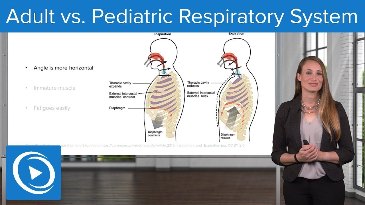
Children's respiratory system differs significantly from adults: nasal passages are highly vascularized and narrow, prone to obstruction; sinuses develop gradually (maxillary sinuses appear as spots at birth, develop by age 2-6); Waldeyer's ring is prominent in infants, with adenoids regressing by age 4; trachea is narrower than adults, making croup an emergency; bronchioles lack cartilage and have minimal smooth muscle, causing rapid airway obstruction in bronchiolitis; chest wall compliance is reduced in infants due to horizontal rib positioning, leading to rapid respiratory failure.
Click to watch →
Adult vs. Pediatric Respiratory System – Pediatric Nursing | Lecturio Nursing
36.2K
The pediatric respiratory system differs significantly from adults in several key ways: (1) Chest shape: Infants have horizontally attached ribs and a rounder chest that matures to a flatter adult shape, with the trachea bifurcating at T3 in children versus T6 in adults; (2) Respiratory muscles: The diaphragm is the primary inspiratory muscle in infants due to immature intercostal muscles, and young babies cannot compensate for respiratory distress because their abdominal muscles don't mature until 3-4 months; (3) Upper airway: Children have proportionally larger heads, smaller mouths, larger tongues, and higher larynx positions, making them obligate nose breathers with narrower airways; (4) Airway diameter: An infant's airway is approximately 4mm versus 8mm in adults, meaning even minor inflammation can cause 50% reduction in lumen size; (5) Bronchial walls: Pediatric bronchi have less muscle tissue and immature beta-adrenergic receptors, making them more prone to collapse and less responsive to bronchodilators; (6) Surfactant: Produced by type II pneumocytes starting at 23 weeks gestation, premature infants lack sufficient surfactant leading to increased surface tension and respiratory distress; (7) Alveoli: At birth, very few alveoli are functional, with most development occurring within the first 2 years, making them more susceptible to collapse and atelectasis.
Click to watch →
Pediatric Respiratory Distress
225.9K
Pediatric airways differ significantly from adults in both anatomy and physiology. Anatomically, children have smaller airways with proportionally larger structures including the tongue, tonsils, and adenoids relative to their oral cavity. The epiglottis is long and floppy, making it vulnerable to swelling (as seen in croup), and the larynx is positioned more superiorly and anteriorly. Tracheal cartilage is underdeveloped and floppy, causing easy collapse when the neck is flexed. Physiologically, children have increased metabolic rate, higher oxygen consumption, and smaller lung volumes, creating potential for rapid decompensation. Unlike adults where cardiac events typically precede arrest, respiratory events are the primary cause of pediatric cardiopulmonary arrest.
Click to watch →

Sistem Respirasi Pada Anak : Pnemonia, Bronkitis, Bronkiolitis & Covid 19
113
Children's respiratory anatomy differs significantly from adults: airways are narrower and shorter, cartilage is softer and more flexible (prone to collapse), and breathing rates are faster (infants ~60 breaths/min vs. adults ~15 breaths/min). Ventilation relies heavily on diaphragm movement. The epiglottis is longer and more flexible, increasing airway collapse risk. Newborns have only 10% of adult alveoli, limiting gas exchange capacity. Even 1mm airway obstruction can reduce cross-sectional area by 75%, causing rapid breathing increase. These anatomical differences make children more vulnerable to respiratory distress and require different clinical assessment approaches.
Click to watch →
Deep Dive
Normal pediatric vital signs, specifically age-appropriate respiratory rates, heart rates, and oxygen saturation levels.

Normal Pediatric Vital Signs | EMT Anatomy & Physiology | Medic Materials
13.8K
Pediatric vital signs change significantly with age, with neonates having the highest respiratory rates (30-53 bpm) and heart rates (100-205 bpm awake, 90-160 bpm asleep), while blood pressure progressively increases and respiratory/heart rates decrease as children mature; by adolescence, vital signs approach adult normal ranges (respiratory rate 12-20 bpm, heart rate 60-100 bpm awake, blood pressure 110-130/64-83 mmHg).
Click to watch →

Pediatric Vital Signs
6.6K
Pediatric vital signs change significantly with age: respiratory rate decreases from 35-55 breaths per minute in newborns to 12-18 breaths per minute in children over 12 years; heart rate decreases from 100-150 beats per minute in newborns to 55-85 beats per minute in older children; blood pressure starts lower than adults (65-85/45-55 mmHg) and rises to adult levels (110-135/65-85 mmHg) by age 12; newborns have higher heart rates (100-160 bpm) and respiratory rates (35-60 breaths per minute) compared to adults, with oxygen saturation needing to be 95% or greater.
Click to watch →

Signos vitales en niños
3.9K
Pediatric vital signs include heart rate (125-150 bpm in newborns, decreasing to 60-100 bpm in adolescents), respiratory rate (30 breaths/min in infants, 12-20 in adolescents), blood pressure (60/40 mmHg in infants, 100-110/60 mmHg in adolescents), temperature (36.5-37.5°C), and oxygen saturation (95%+ normal, <90% indicates respiratory distress); measurement techniques differ from adults, with heart rate measured at the apex (5th intercostal space, midclavicular line) for infants rather than radial pulse, and respiratory rate measured for a full minute rather than 15-second intervals.
Click to watch →

Reta Final TJDFT Técnico Judiciário - Enfermagem Pós-Edital: Saúde Pública - Prof. Thaysa Vianna
4.7K
Pediatric vital signs vary by age: For infants 0-2 months: respiratory rate 40-60 breaths/min, heart rate 70-170 bpm (average 120 bpm). For infants 2-11 months: respiratory rate up to 50 breaths/min. For children 12-59 months: respiratory rate up to 40 breaths/min. For children 6-8 years: respiratory rate up to 30 breaths/min. For children over 8 years: respiratory rate up to 20 breaths/min. Heart rate: 4 years 80-120 bpm (average 100 bpm), 6 years 75-115 bpm (average 100 bpm), 8 years 70-110 bpm (average 90 bpm), 10 years 70-110 bpm (average 90 bpm). These parameters are essential for distinguishing normal from abnormal pediatric vital signs.
Click to watch →

การวัดสัญญานชีพและการเช็ดตัวลดไข้ในเด็ก
349
Normal vital signs vary by age: Body temperature 36.5-37.4°C, Oxygen saturation 95-100% for all ages. Heart rate: newborns 100-180 bpm, children 1-5 years 80-100 bpm, children over 10 years 60-90 bpm. Respiratory rate: newborns 40-60 bpm, children 1-5 years 22-35 bpm, children over 10 years 14-22 bpm. Blood pressure: infants ≤90/60 mmHg, children ≤105/70 mmHg, older children ≤120/80 mmHg, adolescents ≤135/90 mmHg.
Click to watch →
Deep Dive
The components of the Pediatric Assessment Triangle (PAT), with an emphasis on evaluating 'work of breathing'.

UPDATE PALS 2026
354
The Pediatric Assessment Triangle (PAT) is a rapid assessment tool used to evaluate a child's condition within seconds by observing general appearance, work of breathing, and circulation to skin. It consists of three components: (1) Appearance - assessing consciousness level, interaction with environment, muscle tone, and signs of respiratory distress; (2) Work of Breathing - evaluating breathing rate, use of accessory muscles, retractions, nasal flaring, and abnormal breath sounds; (3) Circulation to Skin - examining skin color, capillary refill time, and perfusion status. The assessment should be completed in less than 30 seconds and can be repeated to detect changes in patient condition.
Click to watch →

EMR Chapter 17 Pediatric Emergencies
309
The Pediatric Assessment Triangle (PAT) has three components: (1) Appearance - general appearance indicating how well the heart and lungs are working and how well the central nervous system is functioning, (2) Work of Breathing - abnormal breath sounds, abnormal positioning, retractions of the neck or chest, and flaring of the nostrils, (3) Circulation to the Skin - paleness (inadequate blood flow), mottling (patchy skin discoloration), and cyanosis (bluish discoloration from low oxygen). Reassess the child's appearance regularly as it can change quickly.
Click to watch →

SURVEI PRIMER & SEKUNDER PADA ANAK || dr. Stephen Diah Iskandar, Sp.A
292
The Pediatric Assessment Triangle (PAT) consists of three components: (1) Appearance: assesses tone, interaction, comfort, eye contact, and cry quality; (2) Work of Breathing: evaluates respiratory effort including nasal flaring, retractions, abnormal body position, and additional breath sounds; (3) Circulation: checks for cyanosis, pallor, mottling, and capillary refill time. Clinical interpretation: if all three components are normal, the patient can be observed; if circulation is abnormal, the patient has shock requiring immediate intervention; if only work of breathing is abnormal, it indicates respiratory distress; if appearance and work of breathing are abnormal, it indicates respiratory failure requiring airway management. The PAT helps quickly identify which organ system is failing and guides immediate management priorities.
Click to watch →

Pediatric Advanced Life Support (PALS) Systematic Approach
217.8K
The Pediatric Assessment Triangle (PAT) is used for the initial assessment during the first quick observation from the doorway of a seriously ill or injured child. It focuses on three key areas: Appearance (level of consciousness and ability to interact), Breathing (work of breathing, position, and audible breath sounds like stridor, grunting, and wheezes), and Circulation/Color (overall circulatory status, skin color including palor, mottling, cyanosis, flushing, petechiae, or purpura). This non-touch assessment helps identify general types of respiratory, circulatory, or neurological issues requiring urgent treatment within seconds of encountering the child.
Click to watch →

Kleine Kinder, große Krämpfe - Selim Aksoy
210
The Pediatric Assessment Triangle (PAT) is a rapid assessment tool consisting of three components: (1) Appearance - assessing consciousness level, responsiveness, and interaction with the environment; (2) Work of Breathing - evaluating respiratory effort, presence of stridor, and breathing pattern; (3) Circulation to Skin - examining skin color, capillary refill time, and perfusion. This tool helps quickly determine if a child is stable or requires immediate intervention.
Click to watch →
Deep Dive
Fundamental concepts of pulmonary gas exchange, including the physiological differences between oxygenation and ventilation.

Oxygenation Exam 2VOPPT
101
Oxygenation is the process of getting oxygen into the blood, occurring in the alveoli where oxygen moves from inhaled air into the bloodstream while carbon dioxide moves out. Ventilation is the act of moving air in and out of the lungs. A critical distinction exists: one can have good ventilation (air reaching the lungs) but poor oxygenation (oxygen not entering the blood). For example, in pneumonia, air can reach the lungs but fluid blocks oxygen from entering the blood. Understanding this difference is essential for proper patient care and treatment planning.
Click to watch →

Top NBME Concepts - Respiratory (USMLE Step 1)
103.5K
Ventilation (gas exchange of CO2) and oxygenation (gas exchange of O2) are separate physiological processes that should be considered independently. This separation allows for better understanding of different pathophysiological mechanisms. For example, high altitude affects oxygenation (low PaO2) but not ventilation (normal PaCO2), while COPD affects both.
Click to watch →
Diffusion vs Perfusion limited Gases
5.4K
This section introduces the fundamental concepts of pulmonary gas exchange. The blood-gas barrier consists of multiple layers including surfactant, alveolar cells, basement membranes, interstitial space, and endothelial cells. Both oxygen and carbon dioxide transport via simple diffusion, moving from high to low concentration gradients. Oxygen moves from alveoli to capillaries while CO2 moves in the opposite direction. Understanding these basic mechanisms is essential for comprehending more complex aspects of respiratory physiology.
Click to watch →

Oxigenação vs. Ventilação: Você sabe a Real Diferença?
189
Ventilation is the mechanical process of moving air into and out of the lungs, primarily responsible for CO2 elimination rather than oxygenation. Atmospheric air contains approximately 78% nitrogen and 21% oxygen. While ventilation is necessary for oxygenation (since oxygen must enter the lungs), increasing ventilation beyond normal levels does not proportionally increase oxygen uptake because the oxygen fraction remains constant at 21%. Instead, increased ventilation primarily enhances CO2 elimination. To improve oxygenation, oxygen concentration must be enriched beyond atmospheric levels using devices like oxygen masks, which can increase oxygen fraction to 70% or higher. This distinction is critical for understanding respiratory physiology and clinical management.
Click to watch →

Insuficiencia Respiratoria aguda By ARUMED
75.3K
This section covers the foundational principles of pulmonary gas exchange. Gas exchange occurs through three mechanisms: ventilation (air movement), perfusion (blood flow), and diffusion (gas transfer across the alveolar-capillary membrane). Oxygen diffuses passively from high to low partial pressure areas, while CO2 moves in the opposite direction. The alveolar-capillary membrane must remain thin and intact for efficient exchange. The oxygen-hemoglobin dissociation curve describes how hemoglobin binds and releases oxygen. Rightward shifts (acidosis, hypercapnia, fever) increase oxygen release to tissues; leftward shifts (alkalosis, hypothermia) decrease release. Understanding these principles is essential for diagnosing and managing respiratory failure.
Click to watch →
Prerequisite Knowledge
- Concept 01Basic pediatric respiratory anatomy and physiology, including how children's airways and chest wall compliance differ from adults.
- Concept 02Normal pediatric vital signs, specifically age-appropriate respiratory rates, heart rates, and oxygen saturation levels.
- Concept 03The components of the Pediatric Assessment Triangle (PAT), with an emphasis on evaluating 'work of breathing'.
- Concept 04Fundamental concepts of pulmonary gas exchange, including the physiological differences between oxygenation and ventilation.
Subsequent Learning
- Step 01Pediatric Advanced Life Support (PALS) algorithms and protocols for managing respiratory distress, failure, and arrest.
- Step 02Selection and clinical application of pediatric oxygen delivery devices, non-invasive ventilation (CPAP/BiPAP), and bag-valve-mask ventilation.
- Step 03Pathophysiology, clinical presentation, and targeted management of specific pediatric respiratory illnesses such as croup, bronchiolitis, asthma, and epiglottitis.
- Step 04Advanced monitoring techniques in pediatric critical care, including arterial blood gas (ABG) interpretation and end-tidal CO2 (EtCO2) capnography.
Distress Signs
Failure Signs
Distress Signs
0:06Playing Section
- 1
Respiratory distress uses compensatory mechanisms, increasing work of breathing.
- 2
Key signs include retractions, nasal flaring, grunting, and anxiety.
- 3
Tachycardia and upright posture often accompany distress.
The Continuum Model and Limitations of Binary Categorization
While distinguishing between respiratory distress and respiratory failure is a foundational concept in pediatric training, some medical educators and clinicians argue that this binary classification is oversimplified and can lead to cognitive errors. In practice, respiratory compromise exists on a rapid, dynamic continuum. Relying strictly on these two categories can cause clinicians to delay intervention, waiting for classic signs of 'failure' (such as bradypnea or altered mental status) to appear before escalating care. Furthermore, physical signs of distress and failure have high inter-observer variability. Critics advocate for a continuum-based approach that emphasizes early, objective monitoring (such as point-of-care ultrasound and capnography) and preemptive non-invasive respiratory support, rather than focusing on a distinct transition point between distress and failure.
Deep Dive
Pediatric Advanced Life Support (PALS) algorithms and protocols for managing respiratory distress, failure, and arrest.

How to perform PALS | Pediatric Advanced Life Support |
142
PALS is an advanced life support course for healthcare providers who will serve as lead providers in pediatric cardiac arrest situations, building upon BLS and ACLS knowledge. The core algorithm focuses on identifying and treating reversible causes (H's and T's: Hypovolemia, Hypoxia, Hydrogen ion excess, Hypothermia, Hypoglycemia, Tension pneumothorax, Toxins, Thrombosis, Tamponade). For pediatric cardiac arrest, oxygenation and ventilation are the top priorities, with defibrillation at 2 Joules per kilogram for VFib/pulseless VT, epinephrine at 0.1 mg/kg every 3-5 minutes, and amiodarone at 5 mg/kg every 3-5 minutes. For symptomatic bradycardia in infants under 12 months with heart rate below 60 bpm, CPR should be initiated as if in cardiac arrest. For SVT, adenosine at 0.1 mg/kg (max 6 mg) is the first-line medication, followed by amiodarone or synchronized cardioversion if refractory.
Click to watch →

Pediatric Advanced Life Support
18.6K
The American Heart Association's Pediatric Advanced Life Support (PALS) guidelines provide standardized algorithms for managing pediatric cardiac emergencies, emphasizing that time is critical—every minute of delayed treatment increases neurologic compromise and mortality risk. For cardiac arrest, providers must initiate high-quality CPR within 10 seconds using a 15:2 compression-to-ventilation ratio at 100-120 compressions per minute, with depth of one-third anteroposterior chest diameter. Non-shockable rhythms (asystole, pulseless electrical activity) require immediate epinephrine administration at 0.1 mL/kg IV/IO every 3-5 minutes, while shockable rhythms (ventricular fibrillation, pulseless ventricular tachycardia) demand immediate defibrillation with escalating energy doses (2 J/kg first shock, 4 J/kg second, ≥4 J/kg max 10 J/kg). For symptomatic bradycardia, treatment depends on whether cardiopulmonary compromise exists; if present, CPR and epinephrine are indicated, while atropine may be used for AV block. The 5 Hs and 5 Ts framework (hypovolemia, hypoxia, hydrogen ion, hypo/hyperkalemia, hypothermia, tension pneumothorax, cardiac tamponade, toxins, pulmonary thrombosis, coronary thrombosis) guides identification of reversible causes.
Click to watch →

Chapter 2 PALS and Pediatric Assessment - American Health Care Academy
333
Pediatric Advanced Life Support (PALS) provides a systematic approach for healthcare providers to assess and treat children with respiratory distress, respiratory failure, or shock using the ABCDE approach (Airway, Breathing, Circulation, Disability, Exposure) combined with the SAMPLE evaluation (Signs and Symptoms, Allergies, Medications, Past Medical History, Last Meal, Events) and tertiary assessments including laboratory tests (ABG, VBG, hemoglobin) and non-laboratory studies (pulse oximetry, capnography, chest X-rays) to prevent cardiopulmonary failure and cardiac arrest.
Click to watch →
Pediatric Advanced Life Support (PALS) Systematic Approach
217.8K
The PALS systematic approach is a structured method for assessing and managing seriously ill or injured children, following the evaluate-identify-intervene sequence. It consists of four components: initial assessment using the Pediatric Assessment Triangle (PAT) to quickly identify life-threatening conditions through appearance, breathing, and circulation/color; primary assessment using A-B-C-D-E (Airway, Breathing, Circulation, Disability, Exposure) with real-time treatment of life-threatening abnormalities; secondary assessment using the SAMPLE mnemonic (Signs and symptoms, Allergies, Medications, Past medical history, Last meal, Events); and diagnostic tests. Key concepts include recognizing respiratory cases (distress vs. failure) and shock cases (hypovolemic, distributive, obstructive, cardiogenic), understanding that shock does not always present with hypotension, and knowing age-specific hypotension thresholds (neonate: 60mmHg, infant: 70mmHg, child 1-10: age×2+70, child >10: 90mmHg).
Click to watch →

PALS 01: Introduction
74.4K
Pediatric Advanced Life Support (PALS) follows the ABCs: Airway (mouth, nose, trachea, bronchi, lungs) gets oxygen from outside to lungs; Breathing moves oxygen from lungs into blood; Circulation pumps oxygenated blood to end organs. In children, respiratory failure (airway/breathing problems) most commonly leads to cardiac arrest, unlike adults where cardiac arrest typically starts first. Warning signs of respiratory failure include abnormal respiratory rate, increased effort, and cyanosis. Shock is inadequate oxygen delivery to tissues, progressing from compensated (tachycardia, cool extremities, decreased pulses) to uncompensated (altered mental status, decreased urine output, metabolic acidosis, weak pulses, tachypnea).
Click to watch →
Deep Dive
Selection and clinical application of pediatric oxygen delivery devices, non-invasive ventilation (CPAP/BiPAP), and bag-valve-mask ventilation.
Clase: VMNI en paciente crítico pediátrico.
8.5K
NIV encompasses two primary modalities: CPAP (Continuous Positive Airway Pressure) and BiPAP (Bilevel Positive Airway Pressure). CPAP delivers continuous positive pressure throughout the respiratory cycle, primarily recruiting alveoli and reducing atelectasis without inspiratory pressure support. BiPAP provides two distinct pressure levels—higher IPAP during inspiration and lower EPAP during expiration—with ramp features adjustable for pathology type (longer ramps for restrictive conditions, shorter for obstructive). Sensitivity settings determine ventilator responsiveness to patient inspiratory effort. CPAP is indicated when respiratory drive maintains adequate gas exchange; BiPAP supports altered respiratory drive with guaranteed backup respiratory rate. Both modalities require careful parameter selection based on patient condition and response.
Click to watch →

Non-Rebreather Mask and Manual Ventilation Bag by R. Pascucci | OPENPediatrics
59.2K
This video explains two manual ventilation systems for pediatric respiratory support: the non-rebreather mask, which delivers high-concentration oxygen through a reservoir bag that prevents room air entrainment and CO2 rebreathing via one-way valves, and the self-inflating bag, which operates without a fresh gas source and includes a Popoff valve that releases pressure at 40-45 cm H2O for safety; the Mapleson/McGill circuit (anesthesia bag) requires a fresh gas source but allows seamless transitions between spontaneous breathing, CPAP, and full mechanical ventilation by relying on adequate fresh gas flow (2.5-3 times minute volume) to eliminate CO2.
Click to watch →

25 MOST IMPORTANT ANAESTHESIA CLINICAL QUESTIONS FOR 26TH JULY FMG EXAM
16.9K
Non-invasive ventilation uses mask interface instead of endotracheal intubation. Key criteria: patient must be conscious and able to cooperate, and must have good mask seal. Indications include acute exacerbation of COPD (BPAP first-line), acute cardiogenic pulmonary edema, and obstructive sleep apnea. Modes include CPAP (continuous positive airway pressure) and BiPAP (Bilevel positive airway pressure). Contraindications include apnea, severe facial trauma, claustrophobia, and inability to protect airway. Oxygen delivery devices are categorized as low-dependency (variable FiO2 dependent on patient breathing pattern) or high-dependency (fixed FiO2 independent of breathing). Low-dependency devices: nasal cannula delivers 25-45% FiO2 at 2-6 L/min; simple face mask delivers 30-60% FiO2 at 6-10 L/min; face mask with reservoir bag delivers 70-90% FiO2 at 10-15 L/min. High-dependency devices: Venturi mask delivers fixed FiO2 (25-60%) independent of breathing; High Flow Nasal Cannula delivers 21-100% FiO2.
Click to watch →

Como Interpretar Gráficos da Ventilação Mecânica Neonatal e Pediátrica @ft.claudioalbuquerque
318
CPAP (Continuous Positive Airway Pressure) maintains continuous positive pressure in the airways, transmitted to intrapulmonary regions to keep upper airways open, maintain small airways patent, and stabilize alveoli. Initial neonatal CPAP is approximately 6 cm H2O, while pediatric patients may require 8-10 cm H2O. This continuous pressure facilitates airflow, increases gas exchange surface area, and improves oxygenation. In neonatal respiratory distress syndrome, surfactant deficiency leads to increased alveolar surface tension and collapse tendency; CPAP counteracts this by providing continuous pressure. BiPAP (Bilevel Positive Airway Pressure) provides two pressure levels: IPAP (Inspiratory Positive Airway Pressure) for inspiration and EPAP (Expiratory Positive Airway Pressure) for expiration. The delta P (IPAP minus EPAP) provides pressure support that increases tidal volume and reduces work of breathing. BiPAP is particularly beneficial for patients with difficulty generating adequate tidal volume, such as those with asthma, bronchiolitis, or neuromuscular diseases.
Click to watch →

FUNDAMENTAL OF NURSING For NORCET 8 | Most Important MCQs By Naresh Sir | Z to F Nursing
22.3K
This comprehensive section covers oxygen delivery devices used in clinical practice. Venturi mask delivers passive pressure (not positive pressure) and provides precise oxygen concentration control, making it ideal for COPD patients requiring 2 liters/minute. Nasal cannula delivers 1-6 liters/minute with corresponding FIO2 values (24-44%). CPAP maintains continuous positive pressure during both inhalation and exhalation, while BiPAP provides high pressure during inhalation and low pressure during exhalation. The section emphasizes that without humidification bottles, oxygen delivery should not exceed 4 liters/minute.
Click to watch →
Deep Dive
Pathophysiology, clinical presentation, and targeted management of specific pediatric respiratory illnesses such as croup, bronchiolitis, asthma, and epiglottitis.
Laryngotracheobronchitis (LTB) & croup: Nursing Process (ADPIE)
16.7K
Croup (laryngotracheobronchitis) is a viral upper airway inflammation affecting children aged 3 months to 6 years, typically caused by parainfluenza, adenovirus, influenza, or RSV. The infection spreads through airborne droplets and causes inflammation, edema, and mucus secretion in the subglottic area—the narrowest part of the pediatric airway with non-expandable cartilage. This leads to inspiratory stridor, barking cough, and increased work of breathing. Diagnosis relies on clinical assessment, with chest X-ray potentially showing a steeple sign. Treatment includes racemic epinephrine and albuterol for airway opening, dexamethasone for inflammation, and acetaminophen for fever, along with humidified oxygen and possible intubation for severe cases.
Click to watch →

Epiglottitis, Croup, Bronchiolitis - Pediatric Nursing - Respiratory Disorders | @LevelUpRN
248.6K
This video explains three pediatric respiratory disorders: Epiglottitis is a life-threatening inflammation of the epiglottis (a cartilage flap at the top of the larynx) caused by bacterial or viral infection, more common in unvaccinated children aged 2-5, presenting with high fever, sore throat, hoarse voice, drooling, and respiratory distress; treatment requires immediate airway management through intubation, antibiotics, and corticosteroids, with critical nursing care emphasizing keeping the patient calm and avoiding supine positioning. Croup (acute laryngotracheobronchitis) is a viral infection (often parainfluenza) causing inflammation of the larynx, trachea, and bronchioles, characterized by a barking cough, inspiratory stridor, and respiratory distress; treatment includes nebulized epinephrine, corticosteroids, and family education on preventing agitation and using cool-mist humidifiers. Bronchiolitis is a common viral lung infection in children caused by RSV, resulting in bronchiolar inflammation and mucus production, presenting with cough, sneezing, fever, wheezing, and prolonged expiration; treatment is primarily supportive with oxygen therapy and suctioning of secretions.
Click to watch →

Respiratory Paediatrics 1 | Acute Respiratory infections, Croup, Epiglottitis
7.1K
Croup (laryngotracheobronchitis) is a viral infection causing inflammation and swelling of the larynx and trachea, primarily affecting children under 5 years. The hallmark features include barking cough, stridor, and subcostal/intercostal retractions. Etiology is predominantly parainfluenza viruses, with influenza B as another cause. Clinical diagnosis is based on characteristic findings; lateral neck X-ray may show the 'steeple sign.' Management involves nebulized epinephrine for rapid airway dilation, systemic corticosteroids (dexamethasone) to reduce inflammation, oxygen therapy for hypoxia, and intubation for severe cases. Hospitalization is indicated for progressive stridor, toxic appearance, or impaired consciousness.
Click to watch →

50 High Yield Pulmonology Questions | Mnemonics And Proven Ways To Memorize For Your Exam!
143.4K
This section addresses acute pediatric respiratory emergencies including croup, epiglottitis, and bronchiolitis. Croup (laryngotracheitis) is most commonly caused by parainfluenza virus type 1, presenting with barking cough and stridor, treated with nebulized epinephrine and dexamethasone. Acute epiglottitis presents with the three Ds (drooling, dysphagia, distress) and requires immediate airway management without provocative procedures. The steeple sign shows subglottic narrowing. Bronchiolitis is primarily caused by RSV. Pertussis progresses through catarrhal, paroxysmal, and convalescent stages, with macrolides as first-line antibiotics.
Click to watch →

360 Degree Series 🔥| 360 Degree Most Imp MCQs #1569 | NORCET & All Nursing Exam| Siddharth Sandu Sir
14.1K
Croup is a group of respiratory disorders, with laryngotracheobronchitis being the most common (viral infection) and acute epiglottitis being the most dangerous. Acute epiglottitis presents with drooling, tripod position, and stridor due to airway obstruction. Management includes humidified oxygen, oxygen saturation monitoring, and keeping the child calm. Tongue depressor examination is contraindicated as it can rupture the epiglottis. The characteristic 'steeple sign' or 'thumb sign' is seen on chest X-ray. Other types include spasmodic laryngitis (allergic) and tracheitis (bacterial).
Click to watch →
Deep Dive
Advanced monitoring techniques in pediatric critical care, including arterial blood gas (ABG) interpretation and end-tidal CO2 (EtCO2) capnography.

Monitoring in ICU and ABG interpretation (Based on case studies) (PACT in the context of COVID-19)
1.8K
This lecture provides a comprehensive overview of monitoring strategies and arterial blood gas interpretation for critically ill pediatric patients. The content covers hemodynamic monitoring including Pediatric Early Warning Score (PEWS), clinical assessment parameters (heart rate, CRT, urine output, sensorium), invasive and non-invasive blood pressure monitoring, lactate and ScvO2 for microcirculatory assessment, and fluid responsiveness evaluation. For respiratory monitoring, the lecture addresses clinical assessment of respiratory distress versus respiratory failure, pulse oximetry principles, end-tidal CO2 monitoring, and lung ultrasound techniques. The arterial blood gas interpretation section presents a systematic stepwise approach including clinical context assessment, pH analysis, primary disorder identification, compensation evaluation using Boston rules and Winter's formula, anion gap calculation with albumin correction, and delta gap ratio for detecting mixed acid-base disorders. The presentation emphasizes that while advanced monitoring technologies are valuable, serial clinical trend monitoring remains essential for early recognition of deterioration in resource-limited settings.
Click to watch →

MD/DNB Pediatrics | Nelson Based Teaching | ABG Interpretation | by Dr. Abinaya on the RAAONLINE App
24.5K
This lecture teaches systematic ABG interpretation for pediatric critical care, covering normal values (pH 7.35-7.45, PCO2 35-45 mmHg, bicarbonate 22-26 mEq/L), buffer systems (bicarbonate-carbonic acid, proteins, phosphate, bone), and compensation patterns: respiratory compensation occurs rapidly (within 24 hours) while metabolic compensation takes 2-4 days; for metabolic acidosis, expect 1.2× bicarbonate change in PCO2; for metabolic alkalosis, expect 6× bicarbonate change in PCO2; for acute respiratory acidosis, expect 1× PCO2 change in bicarbonate; for chronic respiratory acidosis, expect 3× PCO2 change in bicarbonate; use anion gap (Na - Cl - HCO3) and delta ratio to identify mixed disorders.
Click to watch →

Capnography by C. Smallwood | OPENPediatrics
24.2K
Capnography is a critical monitoring tool in pediatric critical care with multiple clinical applications including endotracheal tube placement verification, ventilation monitoring for hypo/hyperventilation detection, early identification of life-threatening events like apnea and airway obstruction, disease severity assessment, and CPR quality monitoring. Capnography graphically displays CO2 partial pressure or concentration against time, while capnometry shows only numeric values. End-tidal CO2 represents the CO2 concentration at exhalation end, typically 0-5 mmHg below arterial CO2 across all ages. Exhaled tidal volume comprises dead space volume (airway, mechanical, and alveolar dead space with no CO2) and alveolar volume (gas exchange portion containing CO2). Understanding these fundamentals enables clinicians to interpret capnograms effectively for patient management.
Click to watch →

Como monitorizar uma criança? (Respiratório e Hemodinâmico)
230
This segment explores advanced monitoring technologies in pediatric critical care. Volumetric capnography provides graphical CO2 analysis for ARDS management, guiding ventilator strategy adjustments. Electrical Impedance Tomography (EIT) uses chest sensors to visualize lung ventilation in real-time without radiation, detecting atelectasis and consolidation. Lung ultrasound is becoming essential, with A-lines as normal findings and B-lines indicating pulmonary edema or atelectasis. Studies show lung ultrasound predicts bronchiolitis severity. Basic hemodynamic monitoring includes heart rate, non-invasive BP, peripheral perfusion, and urine output (normal 3-5 ml/kg/hour). Serial lactate assesses tissue perfusion. Point-of-care echocardiography provides ejection fraction (normal 50-75%), chamber dimensions, and pulmonary artery pressure (normal 14-18 mmHg).
Click to watch →

Monitoring Gas Exchange (Mechanical Ventilation - Lecture 5)
67.4K
This lecture explains three essential methods for monitoring gas exchange in mechanically ventilated patients: Arterial Blood Gas (ABG) analysis provides direct measurements of pH, PaO2, and PaCO2 to assess acid-base balance and oxygenation; Pulse Oximetry offers continuous real-time monitoring of oxygen saturation using light absorption principles at 660nm and 940nm wavelengths, though it has limitations including interference from nail polish, methemoglobin, and motion artifacts; Capnography continuously measures CO2 in expired air through infrared spectroscopy, producing a characteristic waveform with four phases where End-Tidal CO2 serves as a surrogate for arterial CO2 and abnormalities in waveform shape (such as the 'shark fin' pattern indicating high airway resistance) or the PaCO2-ETCO2 gradient (>5mmHg suggesting low cardiac output or V/Q mismatch) provide valuable diagnostic information about cardiopulmonary function.
Click to watch →
Distress Signs
Failure Signs
Distress Signs
0:06- 1
Respiratory distress uses compensatory mechanisms, increasing work of breathing.
- 2
Key signs include retractions, nasal flaring, grunting, and anxiety.
- 3
Tachycardia and upright posture often accompany distress.
The Continuum Model and Limitations of Binary Categorization
While distinguishing between respiratory distress and respiratory failure is a foundational concept in pediatric training, some medical educators and clinicians argue that this binary classification is oversimplified and can lead to cognitive errors. In practice, respiratory compromise exists on a rapid, dynamic continuum. Relying strictly on these two categories can cause clinicians to delay intervention, waiting for classic signs of 'failure' (such as bradypnea or altered mental status) to appear before escalating care. Furthermore, physical signs of distress and failure have high inter-observer variability. Critics advocate for a continuum-based approach that emphasizes early, objective monitoring (such as point-of-care ultrasound and capnography) and preemptive non-invasive respiratory support, rather than focusing on a distinct transition point between distress and failure.
Basic pediatric respiratory anatomy and physiology, including how children's airways and chest wall compliance differ from adults.
Normal pediatric vital signs, specifically age-appropriate respiratory rates, heart rates, and oxygen saturation levels.
The components of the Pediatric Assessment Triangle (PAT), with an emphasis on evaluating 'work of breathing'.
Fundamental concepts of pulmonary gas exchange, including the physiological differences between oxygenation and ventilation.
Pediatric Advanced Life Support (PALS) algorithms and protocols for managing respiratory distress, failure, and arrest.
Selection and clinical application of pediatric oxygen delivery devices, non-invasive ventilation (CPAP/BiPAP), and bag-valve-mask ventilation.
Pathophysiology, clinical presentation, and targeted management of specific pediatric respiratory illnesses such as croup, bronchiolitis, asthma, and epiglottitis.
Advanced monitoring techniques in pediatric critical care, including arterial blood gas (ABG) interpretation and end-tidal CO2 (EtCO2) capnography.
68.5K views482likes4:16@OPENPediatricsOriginal Release: 2017-06-30
Recognizing Respiratory Distress and Failure by Dr. Monica Kleinman.
When we say respiratory distress, we're really referring to the use of compensatory mechanisms to maintain adequate oxygenation and ventilation when something, one of many things, is causing respiratory compromise.
Typically, the compensation consists of an increase in the rate and work of breathing.
That increase will increase minute ventilation, allowing the child to exhale more carbon dioxide when gas exchange is impaired.
Signs of respiratory distress include retractions, sternal retractions, for instance, because of the flexible, compliant breastbone, as well as intercostal retractions.
And this is an example of increased effort resulting in muscles exaggerating the spaces between the ribs or in the sternum.
Use of accessory muscles, such as the strap muscles in the neck, nasal flaring, and grunting, an expository noise that's designed to apply some PEEP.
[GRUNTING SOUND] Children in respiratory distress typically are anxious.
It's an uncomfortable feeling, and with that stress, they oftentimes have tachycardia.
These are some examples, as well, of signs of respiratory distress.
The child who wants to sit upright and is unable to lie flat comfortably.
The child on the left looks anxious, and the child on the right is demonstrating increased work of breathing.
Respiratory failure, on the other hand, is at the other end of the spectrum and is defined by inadequate oxygenation and ventilation.
Essentially occurs when compensatory mechanisms fail to maintain gas exchange.
Interestingly, respiratory failure can occur without preceding respiratory distress, meaning it can happen when there is depression of respiratory effort, typically by something like a poisoning, which depresses mental status, or seizures, or severe weakness leading to inadequate respiratory effort.
Signs of respiratory failure, when the compensatory mechanisms are no longer able to sustain gas exchange, include desaturation or cyanosis despite supplemental oxygen.
The appearance of increased effort leading to poor air entry, in other words, lots of work for very little gain.
This may be characterized by a sound of gasping when the child inspires.
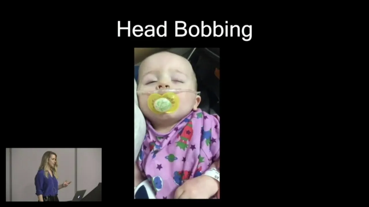
[GASPING SOUND] Head bobbing, which is an exaggerated form of accessory muscle use in which the head literally will bob as the muscles of the neck and shoulders are trying to participate in gas exchange.
Seesaw or paradoxical respirations, where the abdomen and chest move in the opposite ways than they usually do.
And sounds of severe obstruction, such as stridor.
[STRIDOR SOUND] The hallmark of respiratory failure, to distinguish it from respiratory distress, is a change in mental status.
When oxygen levels fall and carbon dioxide rises, this is a very stressful feeling and results in sympathetic stimulation and agitation.
As carbon dioxide continues to rise, it has a sedating effect on the central nervous system, leading to a child who is lethargic, sleepy, or obtunded.
The child who appears to have increased work of breathing, desaturation, and sleepiness or obtundation is one whom you need to be very concerned has transitioned from respiratory distress to respiratory failure and may progress to respiratory arrest and cardiac arrest.
Up Next

13:24
Croup in Kids: Symptoms, Home Care, and When to Seek Help
@pedsdoctalk
567.7K views•2021-10-22

48:47
Graphic Medicine: Comics for Collaborative Healthcare Communication
@nationalpatientadvocate
189 views•2023-12-04

10:28
Neuroanatomy: Central and Peripheral Nervous System Divisions Explained
@AKLECTURES
136.2K views•2014-09-20

3:05
Stages of Labor and Vaginal Birth | Childbirth Animation
@nucleusmedicalmedia
52.1M views•2017-08-18
Related Study Plans & Knowledge Roadmaps
Structured learning paths in Medicine
medicine
Abnormal LFTs: Diagnostic Workup & Pathology
01Liver Anatomy and Basic Physiology
02Introduction to Liver Function Tests (LFTs)
03Hepatocellular vs. Cholestatic Patterns
+ 3 more learning modules
12h6 modules
Explore Roadmap
medicine
Analyzing Blood Smears: RBC, WBC & Platelets
01Introduction to Blood Components & Smear Preparation
02Normal Morphology of Blood Cells
03Erythrocyte Pathologies & Morphological Variants
+ 2 more learning modules
12h5 modules
Explore Roadmap
medicine
Mechanical Ventilation: Waveforms & Alarms
01Anatomy and Physiology of Breathing
02Introduction to Mechanical Ventilation & Core Modes
03Ventilator Settings and Waveform Interpretation
+ 2 more learning modules
12h5 modules
Explore Roadmap
medicine
Neuro Exam: Localization & Stroke Syndromes
01Foundations of Neuroanatomy and Vascular Supply
02The Neurological Examination: Step-by-Step
03Principles of Neurological Localization
+ 3 more learning modules
18h6 modules
Explore Roadmap