
























The EFAST exam is a systematic point-of-care ultrasound examination used to evaluate hypotensive and critical trauma patients by assessing three body compartments: the thoracic cavity (for pneumothorax via lung sliding assessment), the pericardial sac (for pericardial effusion), and the abdominal cavity (for free fluid detection in Morrison's pouch, paracolic gutters, and dependent areas). The exam uses binary questions to guide assessment and employs a curvilinear probe for efficient scanning, with the right upper quadrant being most sensitive for free fluid detection in supine patients, though the pelvis becomes the gravity-dependent area in upright patients.
EFAST Exam: Point-of-Care Ultrasound for Trauma & Critical Care
Added:Basic physics of medical ultrasound, including concepts of echogenicity (hyperechoic, hypoechoic, anechoic) and acoustic shadowing.
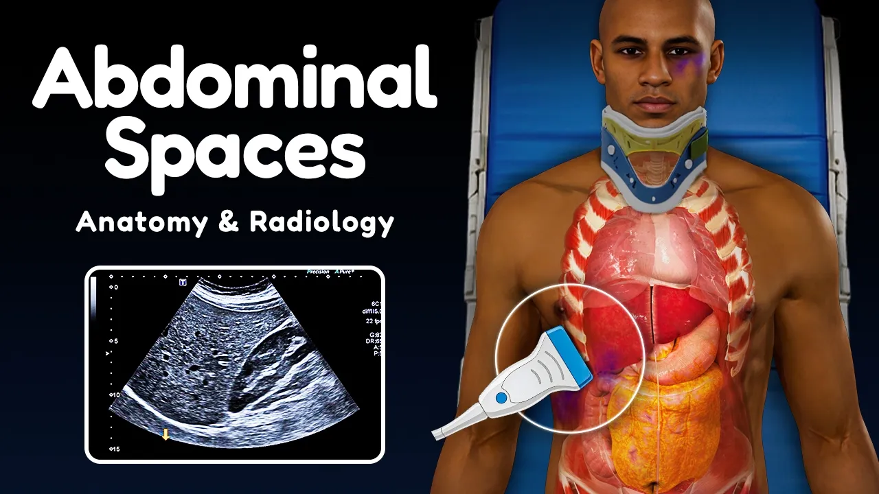
Gross anatomy of the thorax, abdomen, and pelvis, specifically focusing on the pleural space, peritoneal cavity, pericardium, and dependent recesses like Morison's pouch.
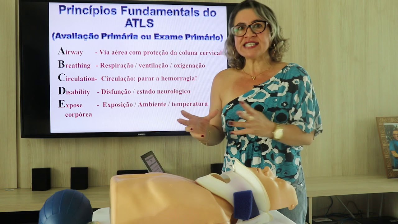
Fundamental understanding of trauma resuscitation protocols, such as Advanced Trauma Life Support (ATLS) and the primary survey.
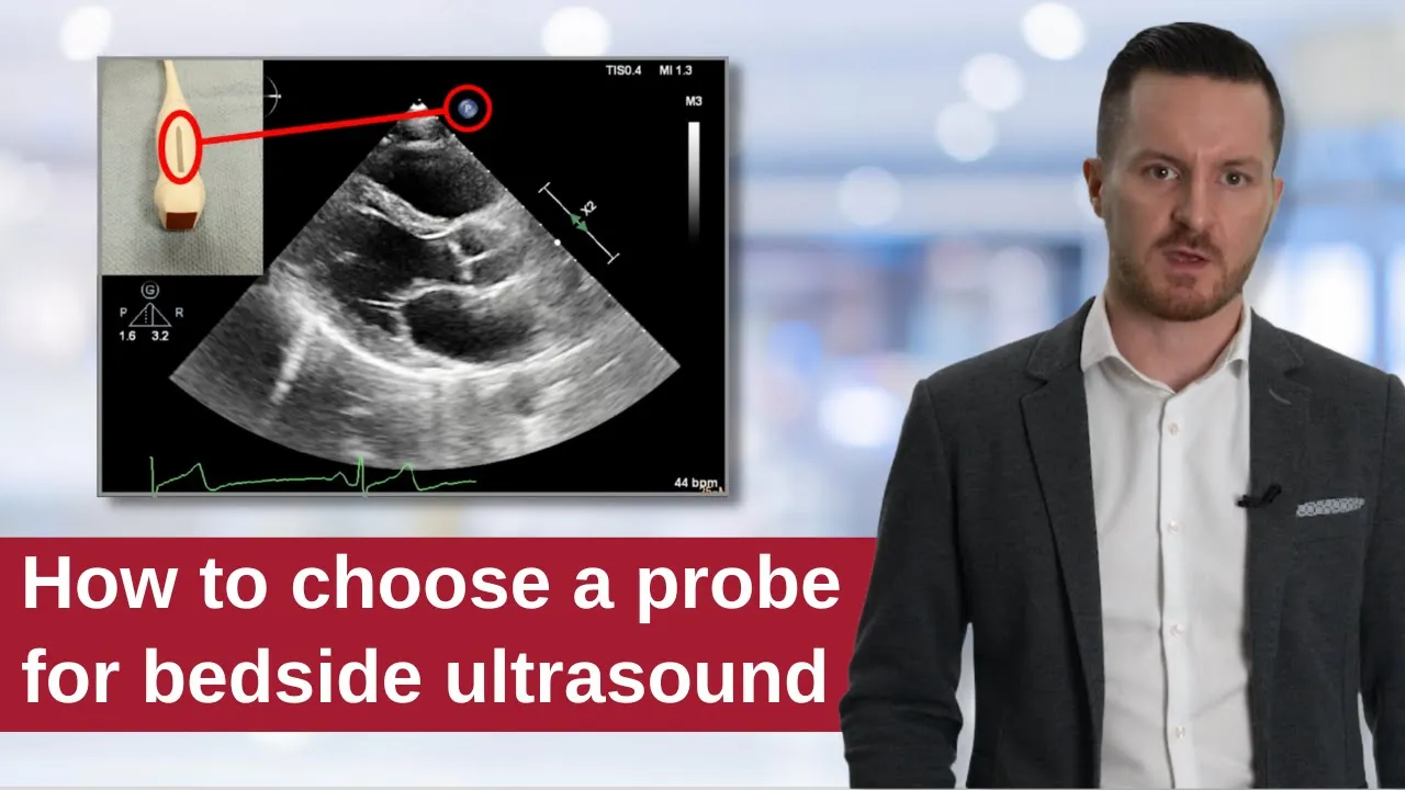
Basic ultrasound 'knobology' and probe selection, specifically the differences between curvilinear, phased array, and linear transducers.
The RUSH (Rapid Ultrasound for Shock and Hypotension) exam to evaluate undifferentiated shock states.
Ultrasound-guided emergency procedures, such as pericardiocentesis, thoracentesis, and chest tube placement.
Advanced evaluation of cardiac function using point-of-care ultrasound, including qualitative assessment of left ventricular ejection fraction and inferior vena cava (IVC) collapsibility for fluid responsiveness.
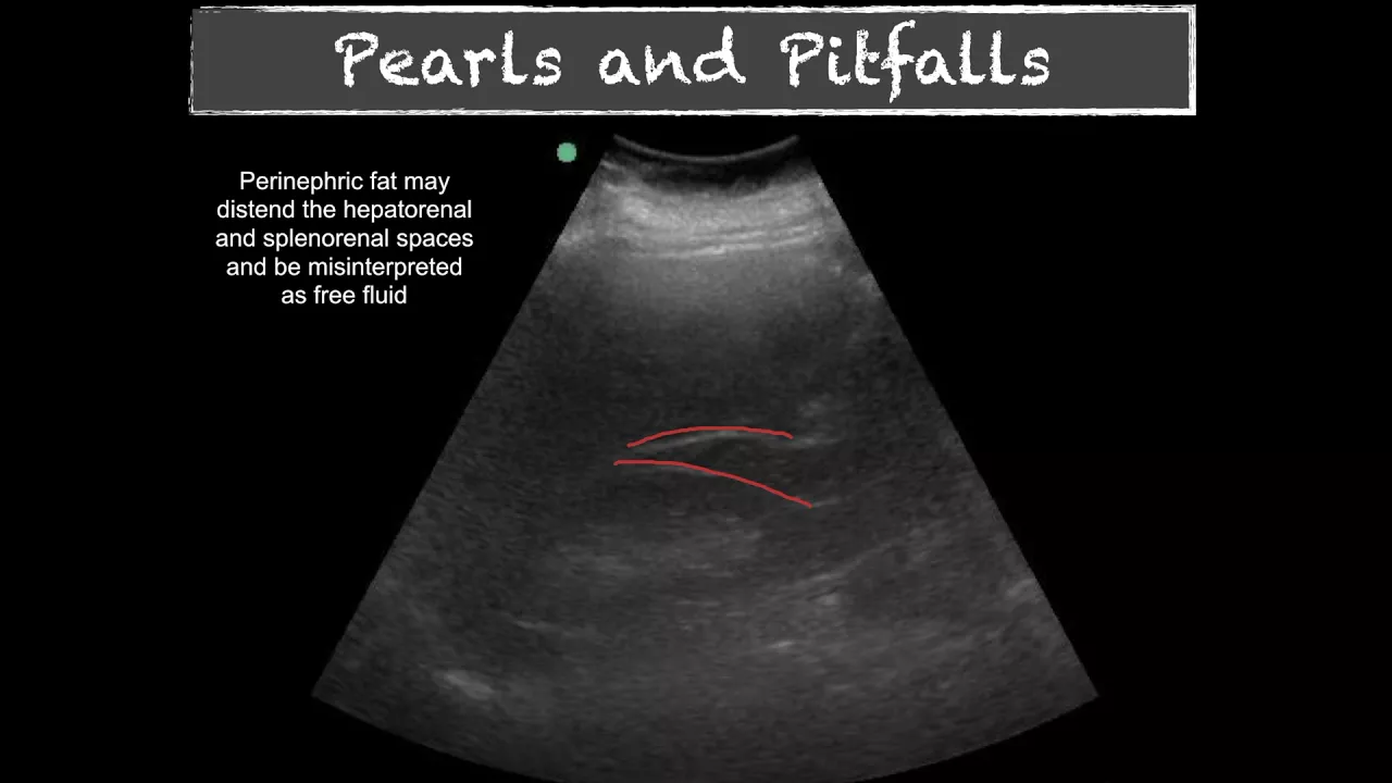
How to recognize and troubleshoot EFAST pitfalls, such as distinguishing pre-existing ascites from acute hemoperitoneum, or identifying subcutaneous emphysema.
Integration of EFAST findings into definitive clinical decision-making pathways, such as determining the immediate need for emergency laparotomy versus CT imaging.
78.2K views1.6Klikes17:25Original Release: 2021-06-26
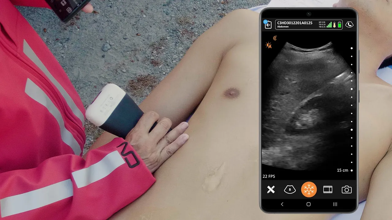
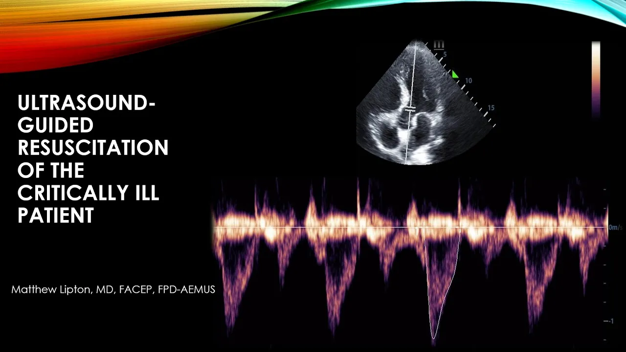
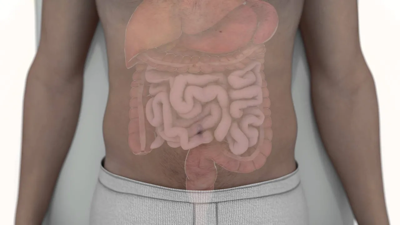
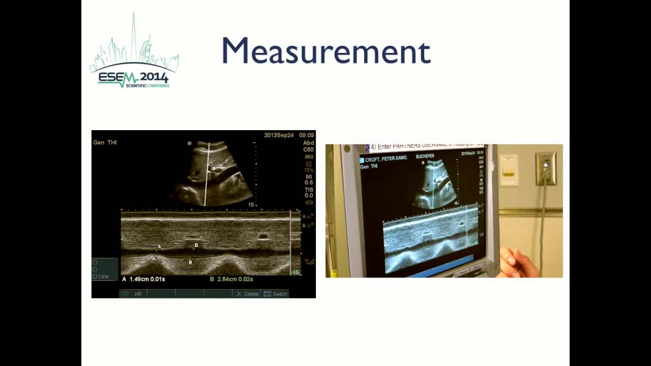
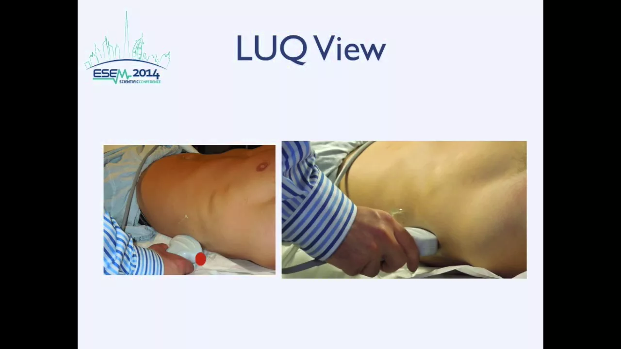
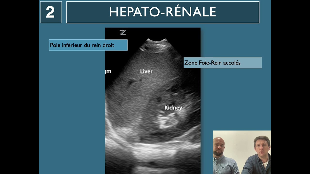
welcome to pocus geek i'm jared marks and in this video we're going to talk about the efast exam the efast exam is crucial to the evaluation of trauma patients that are hypotensive and critical within your emergency department and it's a great way to evaluate for hemoperitoneum or causes of shock within the patient understanding how to get the right images to increase your sensitivity and your positive predictive value are important and in this video we're going to discuss how to better perform this exam the extended focused assessment with sonography and trauma is one of the oldest applications used within emergency ultrasound to evaluate critical care patients it's one of the core applications that's led to the development of other other critical care protocols to evaluate patients and even though it's a study that was first started in trauma patients these other protocols are focused on medical patients and the efas can be quite helpful in evaluating these patients also when you're performing this exam i would recommend that you use a curvilinear probe you want a low frequency probe that allows you to evaluate all the areas you're going to without having to switch probes and slow you down during your exam you'll have much of your medical team waiting on you to perform this exam and give them a diagnosis and being able to move through it quickly and efficiently is going to be in your best interest and the patient's best interest the indications for performing any faster trauma obviously when we talk about that portion but you can use this in medical patients and any patient that is critical especially hypotensive tachycardic or respiratory distress you can use components of this exam to evaluate them and help expedite care it does have a role in penetrating trauma but that is typically around the lungs and heart and when we're looking to see if there is either a pneumothorax or a large pericardial effusion that's causing them to decompensate when we're doing this exam uh keep in mind especially in blunt trauma what we're going to be doing is looking at these three areas of the body and the pericardial sac in the heart the lungs and the thoracic cavity and then the abdominal cavity and so when we talk about doing clinical ultrasound we have binary questions we're going to introduce three binary questions during this lecture but the first one is is there free fluid and we're going to look in all these three areas to evaluate for free fluid when i go throughout this exam i like to go in a clockwise manner around the abdomen this allows us to work from the most sensitive to the least sensitive areas on the abdomen for evaluating for free fluid and when i start in the right upper quadrant you can go from anywhere along the costal margin on the anterior abdomen over to the mid to posterior axillary line i personally like to do the posterior or the mid axillary line and we're going to essentially evaluate the pleural space and diaphragm down through the inferior tip of the liver or kidney which is whatever most inferior and by doing so methodically through this area we will increase our sensitivity and be able to find that free fluid now what we have here and we can see is that we're going to try to evaluate that diaphragm area and the lung we're going to really focus on finding that spine line and watching that insert into the diaphragm and when we look at this here we see that right through here is that spine line and then this is the diaphragm right here and by looking right here we can see that there's no free fluid even though this is anechoic because the spine does not continue above it that means that there's no free fluid here and so when we go back to our list we're going to evaluate the pleural space for free fluid we see that and there's no free fluid then we're going to start working down through morrison's pouch in this area and evaluating for fluid and we may not always get that in one view so we may need repeat views and we can see that area through here we want to take a little caution where there's this rib shadow but hopefully we can see in our next image that we get a little bit more of that and we come down around the inferior tip of the liver and in this case we're also seeing the inferior tip of the kidney now what this means to us is that this is called the paracolic gutter and this is the first area that will see free fluid in the abdomen and we'll see free fluid collect right in this area and after it's collected here it will start to track up through morrison's pouch after we've finished evaluating the right upper quadrant then we're going to go to the sub xiphoid area and whether you follow the protocol that i am teaching you here or you follow your own i would suggest to you that you make the right upper quadrant and sub-zyphoid be your your first views because these will most often explain why a patient's hypotensive for you now in addition to evaluating the pericardial sac for free fluid we may be able to evaluate the heart to see if that functions normal now this may not always be possible with the curvilinear probe and maybe at the end we'll have to come back with the phased array and do more of a full cardiac exam now when we're looking at the cardiac evaluation we want to see the entire pericardium to evaluate for free fluid and then we really need to see the in the interior myocardial walls and that will help us to assess for function and with our pro marker pointed to the patient's right we're going to look up towards the patient's left mid clavicular line to left shoulder and we'll get a view like this and what we're going to see is that we can see all four chambers of the heart and that right ventricle is going to be abutting right up against the liver wall and right through this area is where we're going to look and evaluate for free fluid now it doesn't mean that you won't get free fluid back here because you can that would typically be more of a isolated finding but we're going to evaluate right here for free fluid and along the right side of the heart because if there is an acutely an acute pericardial effusion or blood within the pericardial sac that can compromise their function and the filling of the right ventricle and lead to hypotension and making our patient a critical patient now in this case we can see the inner walls of that left ventricle we can see it close down it closes down well this is a normal appearing ejection fraction if you want to learn more about cardiac ultrasound you can watch the other lecture which i'll include a link up here in the corner which can further help you assess the function of the left ventricle now at times when we're doing this we may not be able to see the subxiphoid view maybe it's not a view that's obtainable in the patient and that's okay we do have the option of doing a parasternal view if you're going to do that i would recommend switching to a phased array probe shooting between the ribs that will just make it easier i would typically save that till after you finish evaluating the rest of the abdomen changing back and forth on probes even in the best machines is going to take 30 seconds at a time essentially and so you want to really get through the exam and then come back to this at end at the end and you can focus on it and even if you get a good subsidied view you can come back and look at the paris journal to further evaluate the ejection fraction if you needed or the cardiac activity after finishing the sub xiphoid view we're going to continue to move clockwise around the abdomen and we're going to go to the left upper quadrant the left upper quadrant's a little bit more difficult than the right upper quadrant you can't do the right um the anterior abdomen like you can on the right because the spleen is so much smaller you really need to get really posterior on these patients and what i like to do is go all the way to the posterior posterior axillary line and then i will fan a little bit anterior and if i don't see the spleen there then i will drag the probe just a little anterior between the posterior axillary line and the mid axillary line and then fan posterior and that will most often help you see the spleen and get the view you need now again much like the right upper quadrant we're going to evaluate from the pleural space all the way to the inferior pole of the kidney and the the spleen which is ever is most inferior and this will give us a good thorough assessment of the area so with our probe in this area we're going to see that we can see the spleen right here in kind of that rose color and we're going to look right above that and see our lung and so when we look here this is our diaphragm and again we have this spine line coming through here and it disappears into the diaphragm now this does not look like free fluid here this is not quite a continuation of the spine line this is a normal appearing exam and the difference between looking at this in a still image and dynamic as we can see whether that diaphragm is erasing the vertebral bodies which it will be now the difference in this exam is that fluid will typically collect here around the pole of the spleen or here before we see it through the splenoreno recess and once it does collect here it is going to go suprasplenic or subdiaphragmatic through this area before it will ever fill the splenorenal recess here the spleen will essentially have to be covered in fluid uh before you'll see it in this area so keep that in mind that this area here this pole and this pole are your most critical and then if you can you want to try to see along this diaphragm now that can be quite a bit more difficult with the ribs being in the way now after you get this view we want to be complete so we're going to come down here's the tip of the spleen right here where we're going to look for free fluid and then we're going to come down and look at this inferior pole of the kidney and this is again our paracolic gutter which is going to be a little bit more sensitive for us after completing the left upper quadrant we're going to continue to move through the abdomen in a clockwise motion and we're going to go down and visualize the pelvis now when we visualize the pelvis we want to identify the bladder we're going to identify any other organs we can in males the prostate and females the uterus and then we want to look cranial to these [Music] to these objects because we need to look into the intraperitoneal cavity and both of these lie within the pelvis now when we start in the in a male patient the pro marker will be directed towards the umbilicus or towards the head drag down right above the pubic bone and when you once you hit that pubic bone start to evaluate for the anatomy and what we see here is our bladder and inferior on the body we see the prostate uh and seminal vesicles and it's important to remember that those are not part of the intraperitoneal cavity and so we're not looking for fluid around them instead we're going to look up into the abdomen into this area within the intraperitoneal cavity for free fluid and so again when we look at this image we see prostate hear seminal vesicles but remember that's the cut off of where the pelvis is is about right here and then up here in the abdomen is where we want to look for that free fluid so make sure you start in a long access it helps to identify those structures and then you can look for free fluid along that wall now at times i will do a short access especially in male patients sometimes i'll do it in female in a if it's a retroverted uterus but what i'll do is i'll use the prostate in males or the cervix and use that as a landmark to then start fanning superior or moving dragging superior towards the umbilicus and help me to evaluate for free fluid once i hit the intraperitoneal cavity in a female patient you know instead we're going to look for the uterus to help us identify what we're doing and we'll get a image like this if they have an anterior verted uterus or anterior flexed uterus as in this case and what we're going to be doing is looking for free fluid along this posterior wall of the uterus now in females especially of reproductive age they can have free fluid and we actually see just a tiny bit right there in this patient and that free fluid can be normal um if they're still ovulating uh how we key into that is we cut the uterus into thirds and so if we look at this this would be a third and this would be a third and anything in the lower third could be physiologic fluid and normal if it gets into the middle to upper third you need to consider that pathological until proven otherwise now we will have patients uh female patients that do not have an anterior flexed uterus and instead we'll see this view and how where we want to look for fluid in them is along this posterior bladder wall and right in this area and by doing that this is where our fluid is going to collect is right here and it's going to be sitting just anterior this is the most dependent area that we'll see in the pelvis in a patient earlier i stated that the right upper quadrant is the most sensitive view for evaluating for free fluid in the abdomen and although that is true if your patient's been sitting upright ambulated into your emergency department then that may not be true and what you have to do is evaluate the pelvis in these patients early and i would maybe do that as my first view in those select cases because they've been standing upright or sitting upright and that has caused the pelvis to be the gravity dependent uh area for free fluid and so i would start and evaluate there first after we've finished evaluating the abdomen we're going to jump up and continue to evaluate the thorax now we've already evaluated for free fluid in the thorax but now we also have a third binary question and is that is is there lung sliding present if there's lung sliding present then that rules out a pneumothorax and so this is important especially in our supine patients because an ultrasound is much more sensitive than a chest x-ray in detecting if there is a pneumothorax present and so what we want to do is we're going to identify the pleural line we're going to look for lung sliding along that line and if there's no lung sliding then we can go evaluate for what's called a lung point now because this is such a large area what we want to do is scan mid-clavicular line bilaterally and we're going to evaluate for lung sliding at each intercostal space and we're going to get a view similar to what we're going to see here in each one of those views but we want to stop at each rib space or if we're using a curvilinear probe sometimes we can see more than one and evaluate for free fluid and what we're going to do is identify that pleural line we need to recognize the ribs and then we'll be able to evaluate along the pleural line for lung sliding so as we can see here along this pleural line right here we're going to just watch for that to shimmer back and forth i've had people say it looks like ants marching on a log others just say it looks like it's shimmering to them or sparkling whatever it looks like to you it's just important to recognize that now be careful and make sure your hands nice and stable otherwise you won't be able to tell if that movement's present one trick you can do if you can't tell if it's present is to hit your zoom function and then zoom in on this area and hopefully that helps you see that it's moving back and forth just a little bit and it's not going to move in large portions but we can see right in here just a little flicker we can see it over in here and that's the lung sliding occurring at that pleural line if you still have difficulty telling you can switch to a linear probe just like i said in the cardiac you could switch to the cardiac probe and you can evaluate forward lung sliding this will be a little more sensitive and i'm less hesitant to switch to this probe at this point because i've typically finished the rest of the exam during that time in summary we ask binary questions when we do pocus and on a knee fast exam we're going to evaluate for free fluid in the thorax pericardial space and in the abdomen in addition we can assess whether the cardiac function is normal and also if there's presence of long sliding just remember if lung sliding is not present that doesn't mean there's a pneumothorax instead you need to go evaluate for a lung point which will be covered in another video and have a link within the description below i hope you found that video helpful in understanding how you can improve your quality for an e-fast exam if you have any questions about this or other point-of-care ultrasound related questions please feel free to reach out to me at pocusgeek gmail.com or you can comment below also check out one of our other videos listed here
Up Next

27:12
EFAST Ultrasound Exam: Step-by-Step POCUS Tutorial
@TakeoKun
128K views•2016-08-29

48:47
Graphic Medicine: Comics for Collaborative Healthcare Communication
@nationalpatientadvocate
189 views•2023-12-04

10:28
Neuroanatomy: Central and Peripheral Nervous System Divisions Explained
@AKLECTURES
136.2K views•2014-09-20

3:05
Stages of Labor and Vaginal Birth | Childbirth Animation
@nucleusmedicalmedia
52.1M views•2017-08-18
Related Study Plans & Knowledge Roadmaps
Structured learning paths in Medicine
medicine
Abnormal LFTs: Diagnostic Workup & Pathology
01Liver Anatomy and Basic Physiology
02Introduction to Liver Function Tests (LFTs)
03Hepatocellular vs. Cholestatic Patterns
+ 3 more learning modules
12h6 modules
Explore Roadmap
medicine
Analyzing Blood Smears: RBC, WBC & Platelets
01Introduction to Blood Components & Smear Preparation
02Normal Morphology of Blood Cells
03Erythrocyte Pathologies & Morphological Variants
+ 2 more learning modules
12h5 modules
Explore Roadmap
medicine
Mechanical Ventilation: Waveforms & Alarms
01Anatomy and Physiology of Breathing
02Introduction to Mechanical Ventilation & Core Modes
03Ventilator Settings and Waveform Interpretation
+ 2 more learning modules
12h5 modules
Explore Roadmap
medicine
Neuro Exam: Localization & Stroke Syndromes
01Foundations of Neuroanatomy and Vascular Supply
02The Neurological Examination: Step-by-Step
03Principles of Neurological Localization
+ 3 more learning modules
18h6 modules
Explore Roadmap