Forensic anthropologists estimate age at death by examining six key skeletal indicators: skull fusion patterns (helpful for infants to young adults), tooth development and eruption stages (baby teeth replaced by permanent teeth around age 12, wisdom teeth appearing around 18), the pubic symphysis which thins and smooths over time until about age 50, long bone growth plates that close by approximately age 25, rib morphology changes from smooth to pockmarked with age, and overall bone density reduction due to calcium loss; these methods allow estimation within specific windows: ±2 years for those under 30, ±5 years for ages 30-70, and less precision for those over 70.
Estimating Age at Death from Skeletal Remains: 6 Methods
Added:Deep Dive
Basic human osteology, including the identification and anatomical terminology of major bones such as the skull, pelvis, ribs, and long bones.
DrSkeleton | Introduction à l'Osteologie humaine
6.1K
The human skeleton consists of 206 bones, classified into four types: long bones (longer than width, found in limbs), short bones (cube-shaped, in wrists and ankles), flat bones (protective, like skull and ribs), and irregular bones (complex shapes, like vertebrae). The skeletal system serves multiple functions including support, protection, movement, blood cell production, and mineral storage. Anatomical terminology includes: linea (long line on bone), sulcus (groove), facet (joint surface), tubercle (small projection), and fossa (depression).
Click to watch →
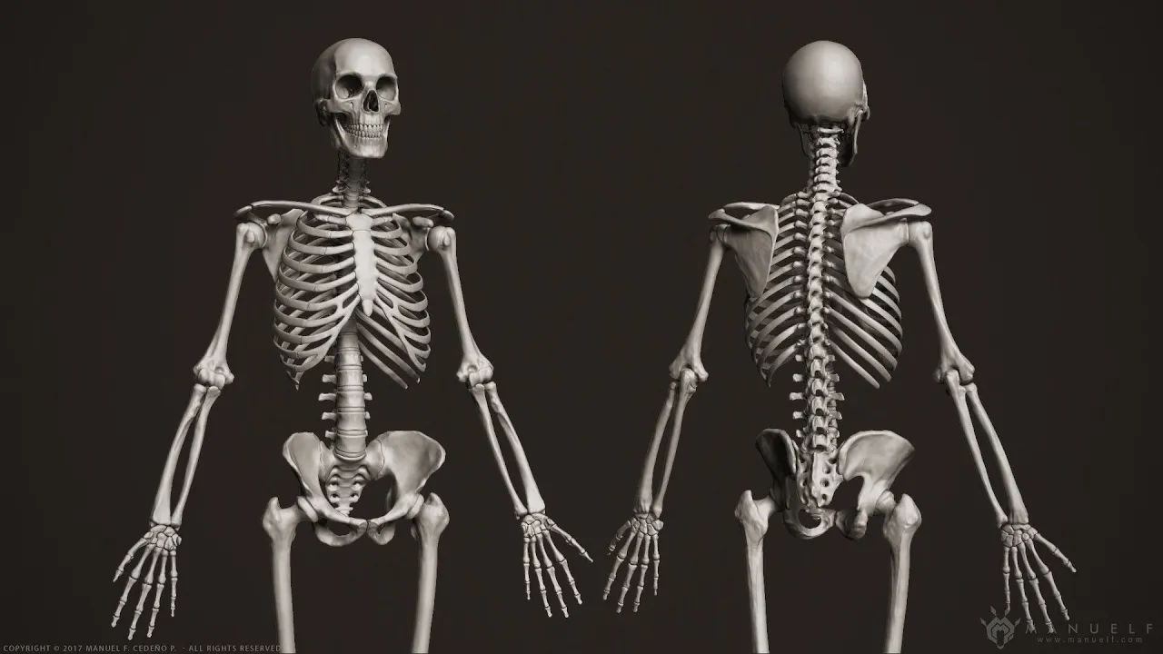
Human Osteology: Axial & Appendicular Skeleton Explained
63K
Human osteology is the study of bones essential to biological anthropology, enabling comparison of skeletal remains across species and understanding evolutionary adaptations. The human skeleton is divided into two primary parts: the axial skeleton (skull, ribcage, and vertebral column) and the appendicular skeleton (limbs and pelvis). Key anatomical terminology includes directional terms like proximal/distal (relative to trunk), medial/lateral (relative to sagittal midline), anterior/posterior (relative to coronal plane), and superior/inferior (above/below). The skull contains eight cranial bones held by fibrous sutures (coronal, sagittal, and lambdoidal) that don't fully fuse until age two, with the foramen magnum's position indicating locomotion type. The vertebral column consists of cervical (7), thoracic (12), lumbar (5), sacral (5 fused), and coccygeal (3-5 fused) vertebrae, protecting the spinal cord. The appendicular skeleton includes the pectoral girdle (clavicles and scapulae), upper limb bones (humerus, radius, ulna, carpals, metacarpals, phalanges), and lower limb bones (femur, tibia, fibula, tarsals, metatarsals, phalanges), all arranged symmetrically on both sides of the body.
Click to watch →

Class 6 Basic Science | Morning Booster - Annual Exam 2025 | Xylem Class 6
37.2K
The human skeleton contains several major bones that can be identified. The skull (A) protects the brain and consists of multiple bones joined together with limited movement, except for the lower jaw. The ribs (B) are 12 pairs of curved bones that form a protective cage around the chest cavity. The femur (C) is the thigh bone, which is the strongest and longest bone in the human body. These bones serve different structural and protective functions in the body.
Click to watch →

Введение в остеологию | Позвонки | Нормальная анатомия | Медвуза
22.7K
The instructor introduces the osteology course covering upper limb, lower limb, and skull. The session lasts 1-1.5 hours with recordings on YouTube and VK. Students should prepare a working notebook. The instructor systematically teaches fundamental anatomical directional terms: anterior, posterior, dorsal, ventral, caudal, cranial, distal, proximal, superior, inferior, medial, lateral, major, minor, basal, and apical. The instructor provides mnemonic devices for memorization and encourages students to share their own associations.
Click to watch →

Clavicle complete Lecture | Anatomy | Clinical features and determination | Batch 1.O Chalk Talk
3K
Osteology is the study of bones, with 'osteo' meaning bones and 'logy' meaning study. The human body contains 206 bones divided into the axial skeleton (80 bones) and appendicular skeleton (126 bones). The axial skeleton lies around the body's central axis and includes the skull (28 bones), 12 pairs of ribs (24 bones), sternum (1 bone), and vertebral column (26 bones). The ribs are classified as true ribs (7 pairs), false ribs (3 pairs), and floating ribs (2 pairs). The appendicular skeleton consists of four limbs (60 bones each), pectoral girdle (4 bones: 2 clavicles and 2 scapulae), and pelvic girdle (2 hip bones). Girdles connect the appendicular skeleton to the axial skeleton.
Click to watch →
Deep Dive
The biology of bone growth and development, specifically the concepts of endochondral ossification and epiphyseal fusion.

Anatomie générale 1 - Cours d'ostéologie générale
345.1K
Endochondral ossification is the primary mode of bone development in most ossification centers, particularly in the limbs. It involves the formation of a cartilaginous model first, composed of cartilage tissue with chondrocytes. Ossification centers then appear within this cartilaginous model. In long bones, a diaphyseal nucleus appears in the middle, while proximal epiphyseal nuclei appear at the extremities. The residual cartilage between these centers forms metaphyses, which contain growth plates (epiphyseal cartilage) responsible for longitudinal bone growth. Growth hormone drives this developmental process.
Click to watch →
Endochondral Ossification
560.7K
Endochondral ossification is the primary method of bone formation in the body, occurring in all bones below the skull except the clavicle. The process begins with vascularization of the perichondrium, followed by mesenchymal cell differentiation into osteoblasts that form a bone collar at the primary ossification center. The remaining cartilage calcifies and dies, creating a central cavity that is invaded by a periosteal bud containing osteogenic cells. Osteoclasts degrade the central cartilage while osteoblasts deposit spongy bone, eventually forming the medullary cavity. Epiphyseal plates develop at the ends, with secondary ossification centers appearing after birth. The final mature bone contains articular cartilage on joint surfaces and red marrow within the spongy bone of the epiphyses.
Click to watch →

Ossification | Bone Formation | Histogenesis of Bone | Bone Histology | Embryology of the Skeleton
435K
Bones form through two primary ossification processes: intramembranous ossification, where bone develops directly from mesenchyme without a cartilage template (forming skull and facial bones), and endochondral ossification, where cartilage serves as a template that gets replaced by bone (forming long bones like femur and humerus). In endochondral ossification, mesenchyme forms hyaline cartilage, which undergoes hypertrophy, calcification, and eventual replacement by bone through primary and secondary ossification centers separated by the epiphyseal growth plate, enabling longitudinal bone growth until fusion occurs.
Click to watch →
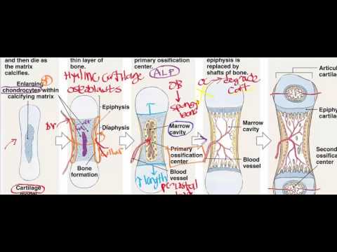
Endochondral Ossification
146.6K
Endochondral ossification is the primary method of bone formation for all bones beneath the skull (except the clavicle) that occurs in utero and involves the transformation of a hyaline cartilage model into bone through a series of stages: mesenchymal cells differentiate into osteoblasts to form a bone collar, chondrocytes enlarge and secrete alkaline phosphatase to calcify the cartilage matrix, blood vessels penetrate to cause chondrocyte death creating a central clearing, and periosteal buds containing osteoblasts and osteoclasts replace the dead cartilage with spongy bone, eventually forming the marrow cavity; additionally, epiphyseal cartilage continues to produce cartilage at the growth plates until approximately age 20, enabling longitudinal bone growth, while the articular cartilage at the epiphyses prevents bone-on-bone friction but can deteriorate to cause osteoarthritis.
Click to watch →
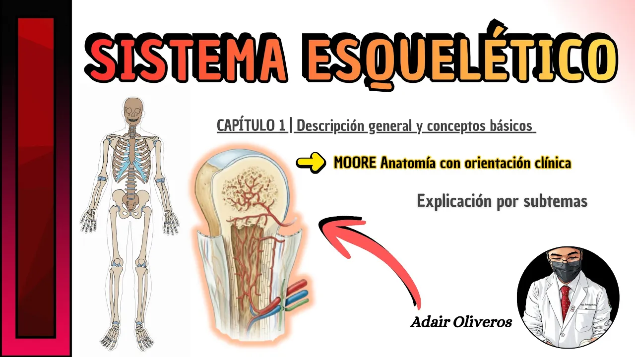
SISTEMA ESQUELÉTICO | MOORE Anatomía con orientación clínica #anatomia #mooreanatomía #medstudent
148
Most bones require many years to grow and mature. The humerus begins ossifying at approximately 8 weeks (end of embryonic period) and continues until about 20 years of age. All bones derive from mesenchyme through two processes: intramembranous ossification (directly from mesenchyme) and endochondral ossification (from cartilage derived from mesenchyme). In endochondral ossification, mesenchymal cells condense and differentiate into chondroblasts forming an initial cartilage mold that grows and calcifies. Periosteal capillaries grow inward, forming the periosteal bud with osteogenic cells that initiate the primary ossification center. Bone tissue replaces most cartilage in the diaphysis. Secondary ossification centers appear after birth in the epiphyses. Epiphyseal arteries grow toward developing cavities. The widened diaphysis adjacent to the epiphysis is the metaphysis. When growth ceases, the epiphyseal plate is replaced by bone in both sides (synostosis), leaving only the epiphyseal line. Longitudinal growth occurs at both sides of the epiphyseal plates, with fusion progressing from puberty to maturity.
Click to watch →
Deep Dive
Fundamentals of dental anatomy, including the human dental formula, tooth eruption sequences, and the difference between deciduous and permanent dentition.

TISHLARNING UMUMIY ANATOMIYASI
247
Deciduous teeth formula is 2-1-2 (2 incisors, 1 canine, 2 molars per quadrant). Permanent teeth formula is 2-1-2-3 (2 incisors, 1 canine, 2 premolars, 3 molars per quadrant). Humans have 20 deciduous teeth and 32 permanent teeth. Eruption sequence: first molars at age 6, premolars at 10-12, canines at 11-12, and third molars (wisdom teeth) at 17-21 (upper) or 12-26 (lower). The third molars often fail to erupt and may be extracted.
Click to watch →
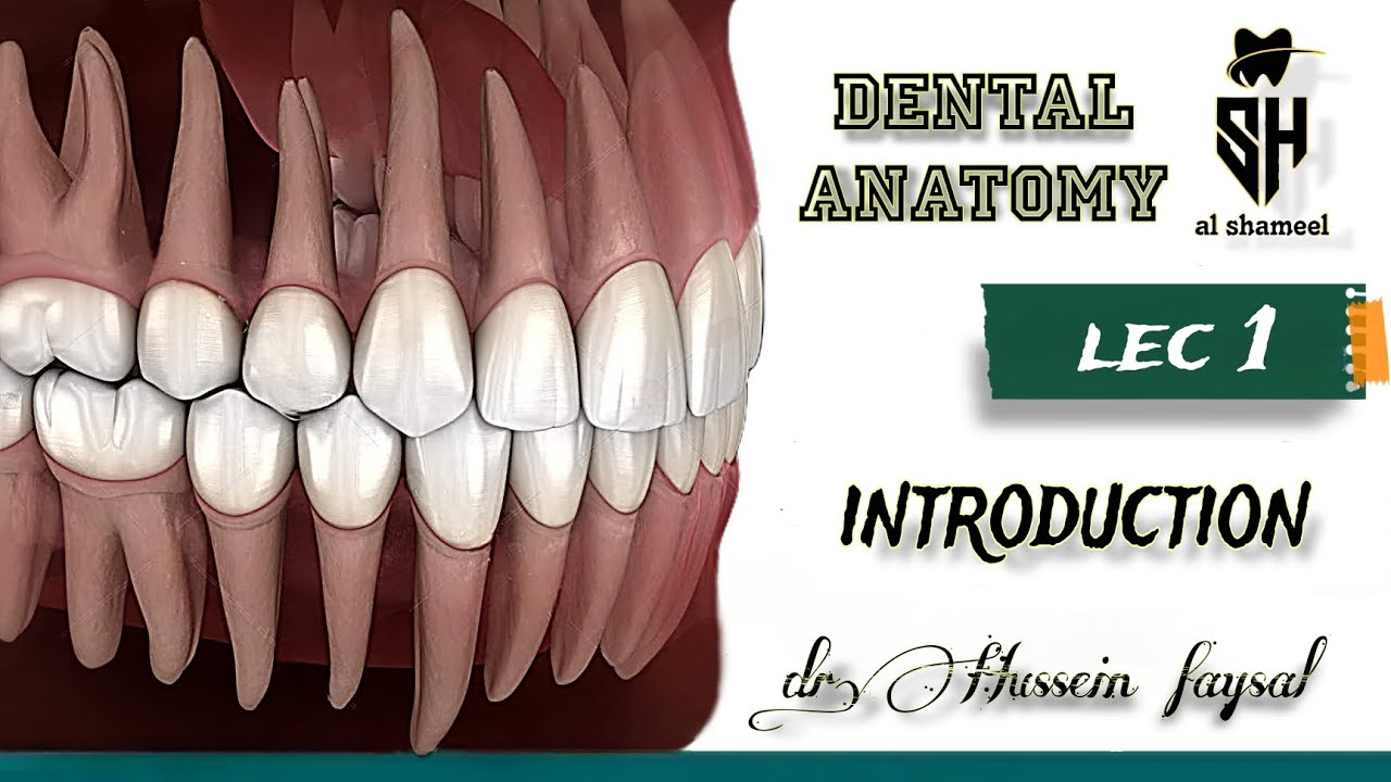
dental anatomy Introduction lec.1 l تشريح أسنان المرحله الاولى . المحاضره الاولى المقدمة طب الأسنان
29.8K
This lecture introduces fundamental dental anatomy concepts including tooth classification (incisors, canines, premolars, and molars), dental terminology (dentin, dental, heterodont, diastema, diphodont), and the two dentition systems: deciduous (20 teeth, formula 2/2, 1/1, 2/2, 0/0 per quadrant, erupting from 6 months to 2.5 years) and permanent (32 teeth, formula 2/2, 1/1, 2/2, 3/3 per quadrant, erupting from 6 years to 20 years). Key concepts include the transition period at age 6, the eruption sequence of permanent teeth, and the distinction between maxillary (fixed) and mandibular (movable) jaws.
Click to watch →
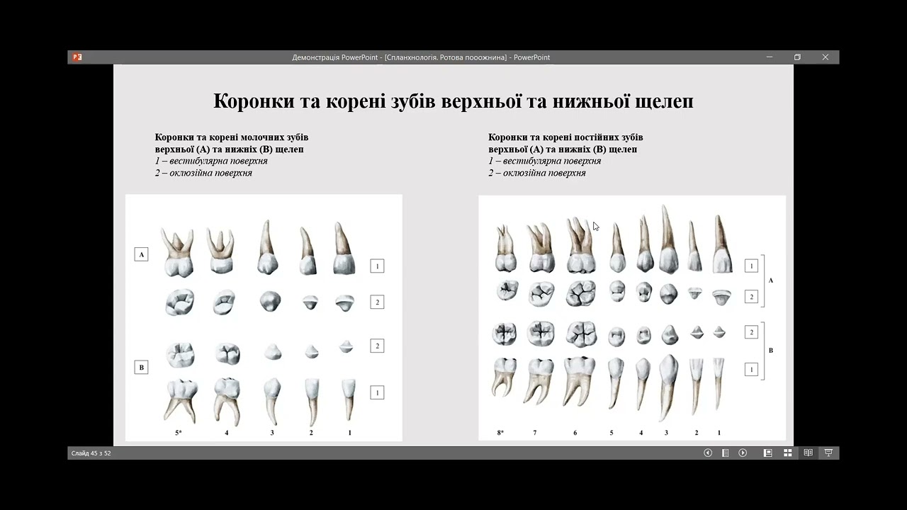
Анатомія зубів. Зубощелепна система людини.
8.6K
Permanent teeth include: (1) Incisors (4 per arch) - chisel-shaped with sharp cutting edges, conical roots; (2) Canines (1 per side) - longest, most massive, conical crown; (3) Premolars (2 per side) - two cusps, may have bifurcated roots; (4) Molars (3 per side) - largest occlusal surface, multiple cusps (4-5), multiple roots (2-3). Third molars (wisdom teeth) are smallest, may fuse, and absent in 10% of population. The dental formula (2 1 2 3) represents tooth count per arch. Tooth numbering systems include sequential numbering from midline and universal two-digit system. Deciduous eruption begins around 6 months, completing by age 3. Permanent eruption begins around age 6: first lower molars, upper incisors, canines, premolars, second molars, and wisdom teeth (17-25 years).
Click to watch →

Introduction to Dental Anatomy (Part 1)
8.4K
Dental anatomy studies tooth development, morphology, function, and identity in the human dentition. Primary teeth (20 total) erupt from 6 months to 2.5 years, while permanent teeth (32 total) erupt from 6 years to 12 years (except third molars at 18-22 years). Teeth are classified into three types: incisors (small, single roots, present in both dentitions), premolars (larger than incisors, multiple roots, only in permanent dentition), and molars (largest, multiple roots, present in both dentitions). Maxillary teeth are in the upper jaw, mandibular in the lower jaw. Tooth formulas summarize dentition: primary (2/2 incisors, 1/1 canines, 2/2 molars), permanent (2/2 incisors, 1/1 canines, 2/2 premolars, 3/3 molars).
Click to watch →
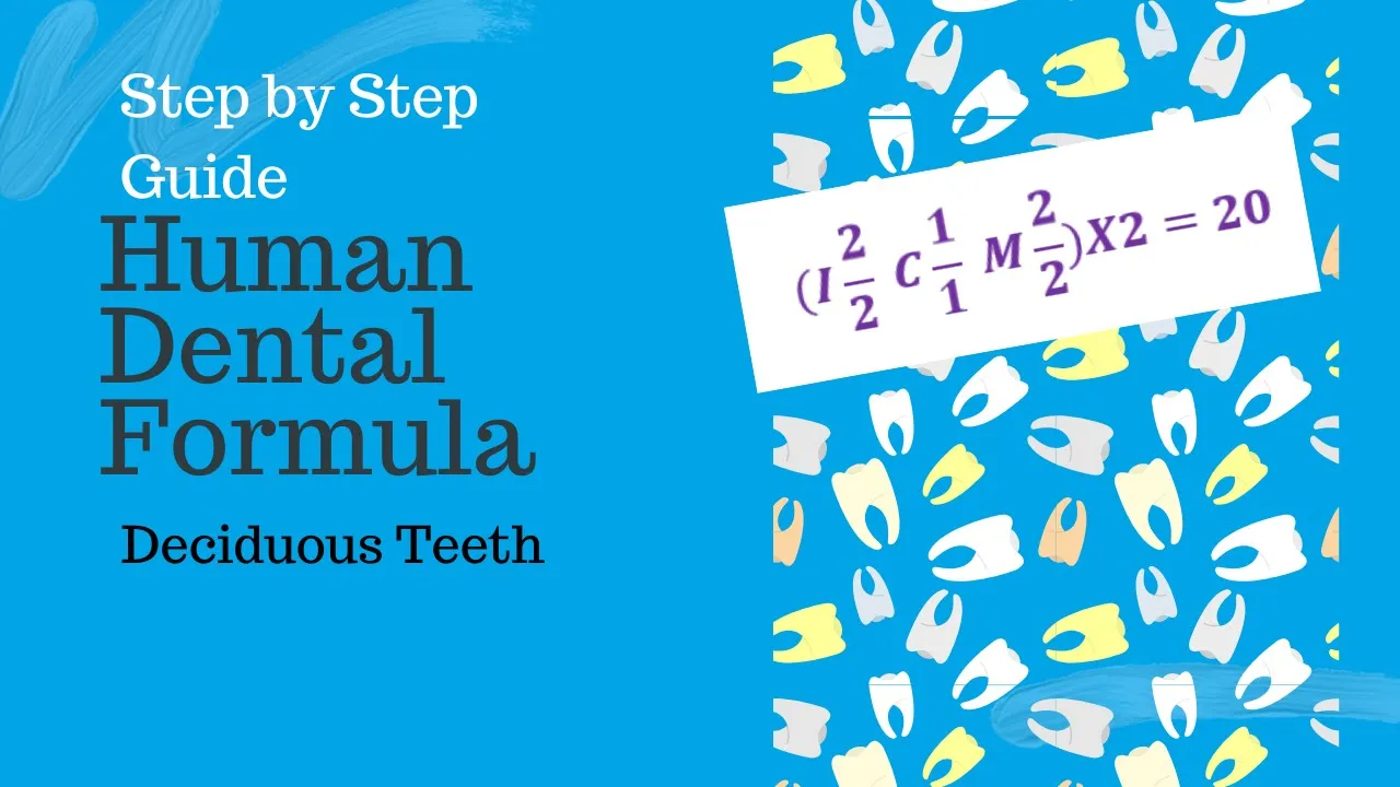
Human Dental Formula Deciduous Dentition | Dental Formula
10.7K
The human deciduous dental formula is written as 2I, 1C, 2M for each quadrant, meaning each quadrant contains two incisors, one canine, and two molars, resulting in a total of 20 deciduous teeth (10 per side).
Click to watch →
Deep Dive
The concept of the 'biological profile' in forensic anthropology and the general distinction between chronological age and biological age.

Jakie tajemnice skrywają ludzkie kości? | 7 metrów pod ziemią
398.3K
Age estimation in forensic anthropology provides biological age rather than chronological age. For children, dental development is key: X-rays reveal which teeth are present and whether wisdom teeth have developed. The mandibular symphysis changes shape with age, becoming more irregular in younger individuals. For adults, the pubic symphysis becomes smoother with age, and degenerative changes may indicate older age. Cranial suture closure was historically used but is less reliable due to individual variation. Forensic anthropologists always provide age ranges rather than specific ages.
Click to watch →

老化生物学者だけど「加齢について」質問ある? | Tech Support | WIRED Japan
95.1K
Chronological age is simply the number of years since birth, as shown on identification documents. Biological age, however, measures the actual physiological state of an organism. These two concepts are distinct: a person chronologically 50 years old might have a biological age more similar to someone who is 60 years old. This distinction is important because it means we can potentially slow the rate at which biological changes accumulate or even reverse them, potentially adding years of life without the painful aspects of aging.
Click to watch →

Rejuvenation Roundup Podcast - July 2022
2.3K
Chronological age is simply the amount of time elapsed since birth, while biological age represents what age your body appears to be when analyzed physiologically. If scientists had no idea when you were born but studied your biology, they would determine your biological age, which may differ from your chronological age—you could be identified as younger or older than you actually are. With advances in healthcare and longevity science, humans may slow or reverse aging, creating growing gaps between chronological and biological age. In the future, someone's chronological age could be completely disconnected from their biological age.
Click to watch →

Diferença de Idade Cronológica e Idade Biológica
857
Chronological age is the number of years a person has lived, while biological age reflects the actual physiological condition of the body, which can be influenced by lifestyle factors such as nutrition, exercise, and supplementation; a person's biological age may differ significantly from their chronological age, with some 50-year-olds appearing and functioning like 40-year-olds while others may have accelerated biological aging.
Click to watch →

Explorations Chapter 15 Forensic Anthropology
2.2K
Forensic anthropology determines biological profiles (sex, age, stature, ancestry) from skeletal remains by analyzing specific bone features: sex is estimated from the pelvis and skull (males have thicker, more angular jaws and narrower subpubic angles, while females have wider pelvic brims for childbirth); age is determined through epiphyseal unions and dental eruption in children, and pubic symphysis patterns in adults; stature is calculated using long bone lengths with ethnicity-specific formulas; ancestry is inferred from skull features like nasal aperture width, dental characteristics, and zygomatic arch shape. Trauma analysis distinguishes antemortem injuries (with healing evidence) from postmortem damage, while pathology reveals diseases affecting bone development such as osteosarcoma, rickets, and achondroplasia.
Click to watch →
Prerequisite Knowledge
- Concept 01Basic human osteology, including the identification and anatomical terminology of major bones such as the skull, pelvis, ribs, and long bones.
- Concept 02The biology of bone growth and development, specifically the concepts of endochondral ossification and epiphyseal fusion.
- Concept 03Fundamentals of dental anatomy, including the human dental formula, tooth eruption sequences, and the difference between deciduous and permanent dentition.
- Concept 04The concept of the 'biological profile' in forensic anthropology and the general distinction between chronological age and biological age.
Subsequent Learning
- Step 01Methods for estimating other key components of the biological profile, such as biological sex, ancestry, and stature from skeletal remains.
- Step 02Skeletal pathology and trauma analysis, focusing on how diseases, occupational stress, and trauma affect bone remodeling and age indicators.
- Step 03Taphonomy and forensic chemistry, exploring how environmental factors influence bone preservation and the use of radiocarbon dating or stable isotope analysis.
- Step 04Quantitative and statistical methods in forensic anthropology, including the use of software like FORDISC to calculate transition analysis and probability intervals for age.
- Step 05Application of these methods in real-world contexts, such as disaster victim identification (DVI), historical bioarchaeology, and human rights investigations.
Skeletal Clues
Age Markers
Rib & Bone
Skeletal Clues
0:00Playing Section
- 1
Forensic experts use bone traits to estimate age in decomposed remains.
- 2
Six key indicators include skull, teeth, pelvis, long bones, ribs, and density.
Statistical Bias and Population-Specificity in Morphological Age Estimation
While macroscopic methods (like analyzing the pelvis, ribs, and cranial sutures) are standard in forensic anthropology, they face significant criticism regarding accuracy, subjectivity, and statistical reliability. A major critique is the 'reference population mimicry' bias, where traditional techniques inadvertently categorize unknown skeletons into the age distribution of the historic reference collection used to develop the method. Furthermore, morphological bone changes vary widely based on genetics, health, and lifestyle, leading to massive margins of error—especially in adults over 50. To counter these limitations, modern forensic specialists advocate for 'Transition Analysis,' which applies Bayesian statistics to eliminate reference-sample bias and provide more accurate probabilistic age ranges. Additionally, critics increasingly favor objective biochemical methods, such as aspartic acid racemization or DNA methylation, to bypass the inherent subjectivity and population-specific limitations of visual skeletal inspections.
Deep Dive
Methods for estimating other key components of the biological profile, such as biological sex, ancestry, and stature from skeletal remains.

Full Brian Laundrie Autopsy
27.3K
This section covers skeletal sex, ancestry, and stature estimation. Male traits observed on the cranium included large mastoid processes, blunt supraorbital margin, pronounced globular well-developed nuchal region with inion hook, and robust mental eminence. The ossa coccyx presented with narrow sub-pubic angle, greater sciatic notch, tall and narrow helix, absent ventral arc, and narrow pelvic inlet. Statistical analysis estimated the skeletal remains were male. For ancestry, the cranium presented with a long nasal spine, patent nasal sill, narrow nasal aperture, angled nasals, S-shaped maxillary sutures, Carabelli's cusp, anterior bulging transverse palatine sutures, and simple vault sutures. Statistical analysis estimated the decedent was white. For stature, measurements of the radius (264 millimeters) and ulna (277 millimeters) were used with the Forensic Discriminant Function 3.1, calculating forensic stature for white male ancestry groups as 66.2 to 74.9 inches (approximately 5 feet 6 inches to 6 feet 2 inches).
Click to watch →
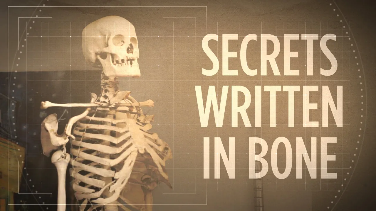
Whose Bones Are These? Investigating A Classroom Skeleton
282.5K
Forensic anthropology enables reconstruction of an individual's complete biological profile through systematic skeletal analysis. This process involves four key components: sex determination through pelvic structure, mastoid process size, and cranial features; age estimation by examining bone fusion patterns (humans start with ~300 bones, reducing to ~206 by age 40); height calculation using long bone measurements; and ancestry assessment through cranial measurement comparison to global databases. Combined with geochemical analysis revealing dietary habits and radiocarbon dating establishing time periods, these methods transform anonymous bones into detailed biological profiles, allowing researchers to reconstruct aspects of an individual's life history including geographic origins and lifestyle patterns.
Click to watch →

Revisão de Véspera Polícia Federal - Perito Criminal - Área 21: Antropologia Forense
802
Biological profile estimation includes sex, age, ancestry, and stature. Sex estimation uses pelvic and long bone characteristics with antroposcopic (1-5 scale) and metric (DSP2 software) methods. Age estimation for young adults uses epiphyseal fusion (iliac crest 16-22 years, third molar eruption, sacroiliac fusion ~25 years, clavicle medial face). For adults, methods include USB for pubic symphysis, Rouart's method, and Hertner's method. Dental age estimation uses Lamedã method for individuals over 30 years. Population affinity uses cranial and dental methods including Fordisc, Ancestr, and Azudas. Stature estimation uses long bone measurements with population-specific validation. Post-mortem interval estimation uses bone weathering stages, aquatic/terrestrial environment effects, and accumulated degree days for entomological analysis.
Click to watch →
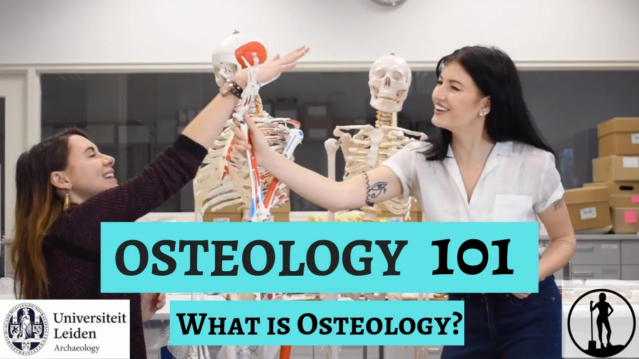
OSTEOLOGY 101: What is Osteology? Part 1 of 5 | Dig it With Raven X Leiden University Archaeology
8.1K
Osteologists determine three key aspects of biological profile from skeletons: age, sex, and stature. Age estimation provides a range rather than exact numbers, with distinct groupings for adults (young adult, late young adult, middle adult, older adult). Accuracy decreases significantly after age 50. Sex determination examines morphological differences in the face and pelvis, categorizing findings along a spectrum from male to female with intermediate categories. Stature estimation uses mathematical formulas applied to long bones (femur, tibia, humerus), with multiple methods averaged for reliability. These techniques allow researchers to create detailed biological profiles of individuals from archaeological contexts, revealing patterns about past populations.
Click to watch →
Public Lecture - Forensic anthropology - Professor Dame Sue Black
112.3K
Forensic anthropologists determine age, sex, and ancestry from skeletal remains. For example, a male individual aged 20-25 years with Caucasian ancestry (pertaining to the Caucasus Mountains region) can be identified. Height estimation ranges from 5'5" to 5'8". These biological profiles are entered into police missing persons databases, which may yield hundreds of potential matches requiring further analysis.
Click to watch →
Deep Dive
Skeletal pathology and trauma analysis, focusing on how diseases, occupational stress, and trauma affect bone remodeling and age indicators.
Explorations Chapter 15 Forensic Anthropology
2.2K
Trauma analysis distinguishes antemortem (healing evidence, muscle stress patterns) from postmortem injuries (no healing). Types include sharp force trauma, blunt force trauma, projectile wounds, and thermal damage. Bone pathology examines diseases affecting bone development: congenital conditions, traumatic injuries, degenerative diseases (osteoporosis, rickets), infectious diseases, metabolic disorders, and tumors. Examples include osteosarcoma, osteogenesis imperfecta, and achondroplasia.
Click to watch →
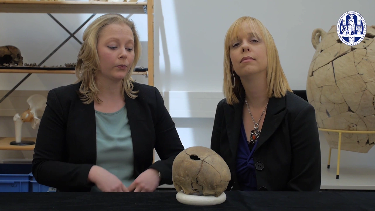
2.3 Paleopathology Trauma
2.4K
Skeletal trauma analysis is a subfield of paleopathology focusing on bone injuries. Beyond fractures, it includes dislocations, atrophy from disrupted blood supply, and artificial modifications like cranial deformation. Fracture patterns reveal past activities—different occupations (farmers vs. city workers, hunters vs. agriculturalists, sailors vs. land-dwellers) produce distinct fracture risks. Bone fractures heal through four stages: initial hematoma formation, soft callus development (1-2 weeks), hard callus mineralization (3 weeks to 3 months), and final remodeling (up to 7 years). Healing time varies by fracture type, bone area, age, infection, immobilization, and nutrition.
Click to watch →

우리가 잠든 사이, 발밑에선... 신도시 아파트 지하에서 쏟아진 5,000구의 유골|취미는 과학|알고e즘
379.3K
Repetitive occupational activities create characteristic skeletal changes called stress markers or Harris lines. These appear as dense bands within bone tissue resulting from repeated mechanical stress that temporarily interrupts normal bone remodeling. Different occupations produce different patterns: kneeling and squatting create specific stress markers in the tibia and femur. These markers serve as permanent records of an individual's lifetime physical activities and occupational history.
Click to watch →
Morfofisiologia Comparada: Sistema de Sustentação, parte 2 #09
234
Bone mass remains stable until age 25-35, then gradually decreases. Women experience accelerated bone loss after menopause due to estrogen deficiency, leading to earlier and greater fracture risk. Rickets results from vitamin D and calcium deficiency, causing impaired bone mineralization and deformities in children. Osteoporosis involves excessive bone resorption relative to formation, leading to decreased density and fracture risk. Osteopetrosis represents the opposite extreme with excessive bone formation. Trauma includes contusions (soft tissue injury), luxations (joint displacement), and fractures (bone breaks). Fractures stimulate healing through osteoblast recruitment. Bones adapt to mechanical stress through remodeling, becoming stronger at stress points (Wolff's law).
Click to watch →
Implications of Palaeopathology on Human Skeletal Analysis | Prof Veena Mushrif-Tripathy | ASC IITGN
331
Joint diseases (osteoarthritis, spondylitis, temporomandibular joint issues) result from breakdown of articular cartilage and reactive bone formation, commonly affecting older populations. Metabolic disorders manifest through specific skeletal indicators: rickets (vitamin D deficiency) shows porous skull texture; scurvy (vitamin C deficiency) causes extreme jaw porosity; Harris lines appear as growth-arrested lines in developing bones indicating childhood illness; enamel hypoplasia appears as white defects on teeth showing when children were sick during tooth development. Occupational stress markers reveal habitual activities through skeletal modifications: maxillary sinusitis from inhaling impurities during biofuel use; squatting facets on tibiae indicating prolonged squatting positions; clavicular changes from repetitive load-carrying activities showing increased robustness.
Click to watch →
Deep Dive
Taphonomy and forensic chemistry, exploring how environmental factors influence bone preservation and the use of radiocarbon dating or stable isotope analysis.
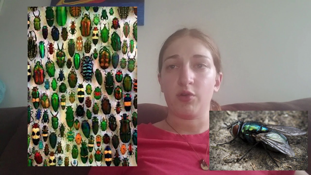
The (Disturbing) Basics of Taphonomy
183
The skeletal stage (3+ weeks post-mortem) consists primarily of bones and dried skin with odor nearly vanished. Most arthropods have departed, though a few may remain. Bone, collagen, hair, and nails degrade significantly slower than flesh and organs, preserving skeletal remains. Environmental factors dramatically affect decomposition rates—warm environments accelerate decomposition within weeks, while cold environments preserve bodies longer due to reduced arthropod activity. Adipocere forms in warm, moist, low-oxygen environments approximately one month post-mortem, creating a waxy preservative material that can protect body parts for decades. Mummification occurs in warm/dry or cool/dry climates or high-salt soils, preserving skin for extended periods. Despite being gruesome, taphonomy is essential for forensics, requiring individuals with powerful stomachs to study decomposition processes and their environmental interactions.
Click to watch →

Radioactive Fossils | HMNS BEYOND BONES PODCAST
184
Taphonomy studies conditions affecting organisms from death to discovery. Fossil preservation varies dramatically based on local groundwater chemistry, mineral availability, and burial conditions—even within the same geological formation. River systems create varied environments where tributaries deposit different minerals. Stable isotope analysis reveals ancient water sources through bone chemistry. Fossil mineralization involves complex, cyclical processes over millions of years: bones can undergo partial mineralization, then demineralization if groundwater becomes acidic, followed by remineralization. Pressure from overlying sediment causes distortion and fracturing. Surface exposure accelerates deterioration through freeze-thaw cycles causing fissures. Ice age fossils near the surface can disintegrate upon disturbance, requiring immediate stabilization with consolidants. Complete petrification requires complete mineral replacement throughout the structure.
Click to watch →

Antropología forense e isótopos estables
133
Stable isotopes (elements with different atomic masses that do not decay) accumulate in human tissues through diet and environment, allowing forensic anthropologists to determine geographic origin and dietary habits of unidentified remains by analyzing isotope ratios in bones, teeth, hair, and nails; for example, nitrogen isotopes indicate trophic level (meat consumption), oxygen isotopes reveal proximity to water bodies, and carbon isotopes indicate plant consumption patterns, with different tissues providing different time windows (bone collagen: 10-20 years, hair/nails: 30-40 days, dental enamel: childhood).
Click to watch →

El Rincón del Disidente | LA SEMILLA DEL MAL: CUANDO LOS ASESINATOS SE DISFRAZAN DE RITUAL
2.9K
Forensic anthropology begins with questions, not symbols. The body is a document, not a symbol - containing biological profile (age, sex, height), trauma evidence, and postmortem alterations. Taphonomy studies how environmental factors (animals, climate, water, sedimentation, fire) alter remains and create false evidence. Vertebrate animals can destroy bone features and create pseudolesions. Time since death estimation has known error rates. The environment can write lies over the body. This scientific approach provides temporal windows and limits, making it more reliable than beliefs that sell certainties.
Click to watch →

La Mystérieuse Femme Sans Nom | Les Eaux Troubles Du Crime | True Crime Stories
89.5K
Taphonomy studies what happens to bodies between death and discovery, including water movement, currents, and decomposition. Water temperature affects decomposition rates, with colder temperatures preserving remains longer. In this case, the body remained in the river for 1-2 years, undergoing significant transformation including skin detachment and bone damage from currents and rocks.
Click to watch →
Deep Dive
Quantitative and statistical methods in forensic anthropology, including the use of software like FORDISC to calculate transition analysis and probability intervals for age.
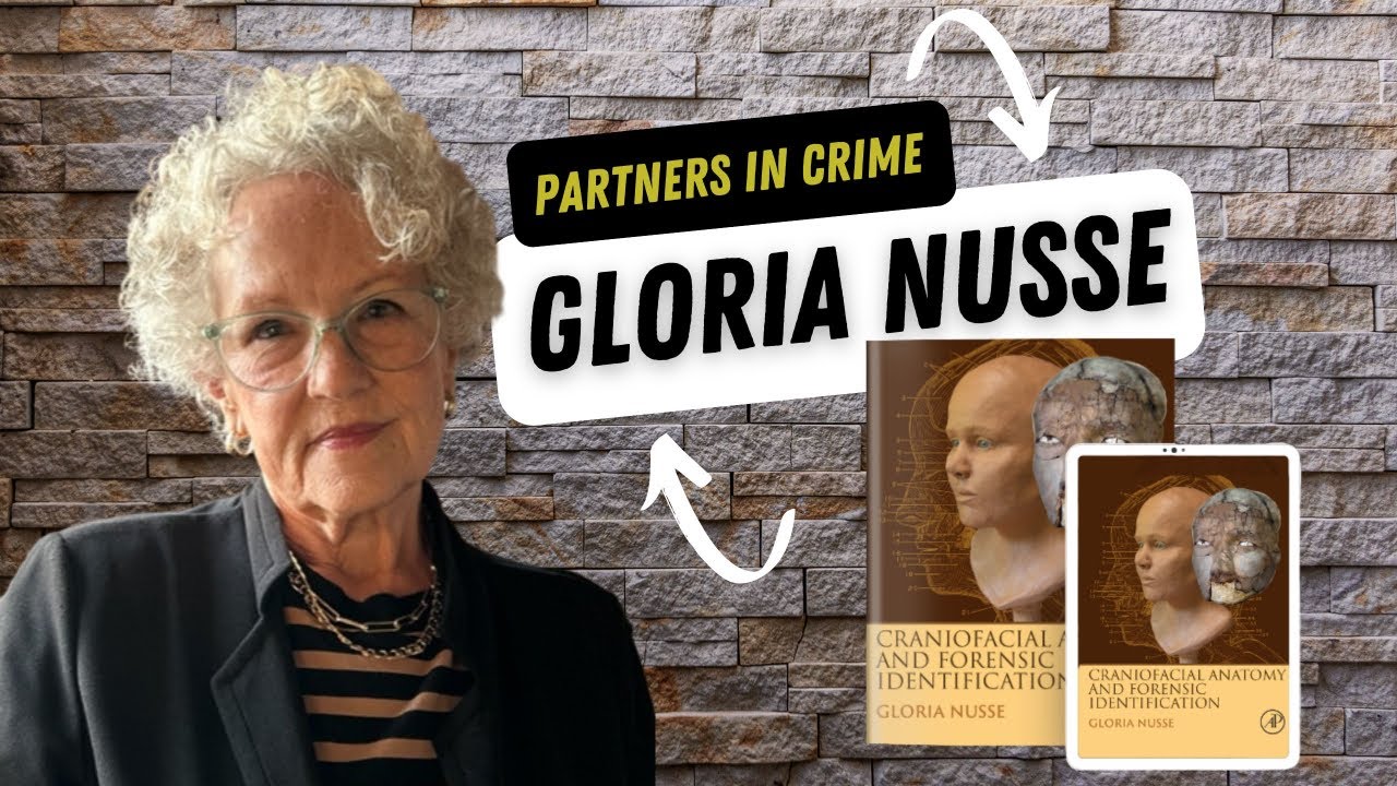
Partners in Crime Presents: Gloria Nusse, Forensic Anatomy in Action
935
Forensic anthropologists use software programs like FORDISK to analyze skeletal measurements. Data from multiple skeletons is entered into the program, which graphs and compares measurements against established databases. This computer-aided analysis helps determine ancestry and other characteristics by comparing unknown skeletons to reference populations.
Click to watch →

Bones from Pacific island likely those of Amelia Earhart
124
The researcher used a new technology called FORDISC to estimate the sex and ancestry of the remains. This technology allows researchers to analyze bone measurements with realistic assumptions about who could have been on Nikumaroro island during the relevant time period.
Click to watch →
Resolução da Prova de Perito Odonto-legal de Sergipe
1.1K
Forensic anthropology uses specialized software for population affinity analysis: Ancestry (23 cranial measurements), ForenTIC (binary population systems), ChronoAge (dental age estimation), Severe (17 cranial characteristics), DSP2 (10 pelvic measurements for sex), Ford (34 cranial measurements), and 3D Morph (3D geometric morphometry). Dental age estimation becomes challenging in adults due to closed apices. Third molars are valuable for forensic age estimation, particularly in criminal responsibility cases.
Click to watch →

The Death of Richard III: CSI Meets History
96.7K
The team did not do a mathematical calculation of the odds of finding Richard III, but when the DNA work was published, they did a statistical analysis of the probability that it was the remains of Richard III. When adding together all factors (skeleton from the right era, male, with battle wounds, scoliosis, found in the high-status burial area, and matching DNA), the probability came out at 99.9999% certain. This left room for conspiracy theories but provided strong scientific evidence. The speaker explained that ethnicity is determined by anthropologists using multiple features, particularly the shape of the skull. Different ethnicities have different prominence of features such as eyebrows and eye sockets, and different prominences of other skull features.
Click to watch →

Forensic Science
2.6K
Forensic anthropology ranges from simple visual examination (determining if remains are human and estimating age) to high-tech statistical and analytical techniques. Age estimation methods are being developed with statistical approaches for better accuracy. Geneticists can extract ancient DNA from bones for identification purposes.
Click to watch →
Deep Dive
Application of these methods in real-world contexts, such as disaster victim identification (DVI), historical bioarchaeology, and human rights investigations.

Resurrected by Flood Water | Buried Secrets | Episode 1
51.1K
Forensic anthropology employs systematic methodologies to identify human remains after catastrophic events. The process begins with calculating the minimum number of individuals (MNI) by sorting all recovered bones by skeletal element type (humerus, femur), dividing them by body side (left versus right), and seriating them by size. For sex determination, pelvic bones serve as the primary indicator. Age estimation uses long bone measurements compared against established population standards, with additional markers like the pubic symphysis and sternal end of the fourth rib providing more precise estimates. Pathological conditions such as osteoarthritis, healed fractures, and cancerous lesions offer further identification clues. In the Harden case, these methods helped identify 119 individuals from approximately 3,400 recovered bones, demonstrating how scientific analysis transforms fragmented remains into identifiable individuals.
Click to watch →

El lector de huesos: El caso Lasa y Zabala
1M
Forensic anthropology serves as a critical tool in human rights investigations, enabling the identification of victims and the documentation of torture methods through skeletal analysis. In the Lasa and Zabala case, forensic expert Paco Etxeberria identified the remains of two ETA members killed by GAL (state-sponsored terrorist group) in 1983, revealing that the perpetrators had buried the victims with cal viva (quicklime) in an attempt to destroy evidence. The forensic analysis demonstrated that cal viva actually preserves soft tissues rather than destroying them, and the victims' remains showed clear evidence of torture including ligatures around the mouth and eyes, as well as bullet wounds. This case established that state actors can be held accountable for human rights violations, though the legal process faced challenges in proving torture due to the deteriorated condition of the remains.
Click to watch →

¡HEBER no pega una, ACHE destapó la olla y ZUBÍA cayó muy bajo! | Resumen de la Semana #106 | ZIN TV
80.5K
Forensic anthropology plays a crucial role in investigating human rights violations by analyzing human remains to determine cause of death and identify victims. In this case, investigators discovered a skull and other bone fragments in a clandestine grave containing lime and a stone slab, indicating a deliberate attempt to conceal the burial. The process involves collecting evidence, conducting forensic analysis, and working to identify the remains to provide closure to families and establish accountability for perpetrators.
Click to watch →
Forensic human identification | The Search
189.7K
DVI involves forensic anthropologists working at mass fatality scenes to identify victims through systematic processes. Their roles include: identifying human remains at incident sites (distinguishing human from animal fragments), performing triage in mortuaries to categorize remains, and conducting detailed analysis to determine sex, age, ethnicity, and height. They also document pathological conditions and other identifying features. This work requires collaboration across multiple stages of the identification process.
Click to watch →

Mother Helps Killer Son Cover Up Horrific Crime | New Detectives | Real Responders
182.6K
Forensic anthropologists use skeletal remains to estimate victim characteristics. Dr. Elizabeth Murray determined the victim's age range was 25-45 years by examining the collar bone, rib cage, and pelvis. For height estimation, she used an unusual method since long bones were missing: she measured the clavicle and derived stature from it. The victim was taller than average, between 5'6" and 6'1". DNA analysis confirmed the victim was Cheryl Durkin, transforming her from a missing person to a homicide victim.
Click to watch →
Skeletal Clues
Age Markers
Rib & Bone
Skeletal Clues
0:00- 1
Forensic experts use bone traits to estimate age in decomposed remains.
- 2
Six key indicators include skull, teeth, pelvis, long bones, ribs, and density.
Statistical Bias and Population-Specificity in Morphological Age Estimation
While macroscopic methods (like analyzing the pelvis, ribs, and cranial sutures) are standard in forensic anthropology, they face significant criticism regarding accuracy, subjectivity, and statistical reliability. A major critique is the 'reference population mimicry' bias, where traditional techniques inadvertently categorize unknown skeletons into the age distribution of the historic reference collection used to develop the method. Furthermore, morphological bone changes vary widely based on genetics, health, and lifestyle, leading to massive margins of error—especially in adults over 50. To counter these limitations, modern forensic specialists advocate for 'Transition Analysis,' which applies Bayesian statistics to eliminate reference-sample bias and provide more accurate probabilistic age ranges. Additionally, critics increasingly favor objective biochemical methods, such as aspartic acid racemization or DNA methylation, to bypass the inherent subjectivity and population-specific limitations of visual skeletal inspections.
Basic human osteology, including the identification and anatomical terminology of major bones such as the skull, pelvis, ribs, and long bones.
The biology of bone growth and development, specifically the concepts of endochondral ossification and epiphyseal fusion.
Fundamentals of dental anatomy, including the human dental formula, tooth eruption sequences, and the difference between deciduous and permanent dentition.
The concept of the 'biological profile' in forensic anthropology and the general distinction between chronological age and biological age.
Methods for estimating other key components of the biological profile, such as biological sex, ancestry, and stature from skeletal remains.
Skeletal pathology and trauma analysis, focusing on how diseases, occupational stress, and trauma affect bone remodeling and age indicators.
Taphonomy and forensic chemistry, exploring how environmental factors influence bone preservation and the use of radiocarbon dating or stable isotope analysis.
Quantitative and statistical methods in forensic anthropology, including the use of software like FORDISC to calculate transition analysis and probability intervals for age.
Application of these methods in real-world contexts, such as disaster victim identification (DVI), historical bioarchaeology, and human rights investigations.
2.7K views87likes4:10@DeathScienceOriginal Release: 2021-04-22
now let's talk about estimating the age of the deceased just based on their bones and let's assume that the deceased is either extremely decayed or completely skeletonized now to the untrained eye this is incredibly difficult to estimate the age except for the forensic anthropologist this individual can determine the age sex uh ancestry just by looking at certain specific traits found on certain parts of the bone itself now before we get started be sure to dig up that like button hit subscribe and you can learn more at www.deathscience.org so there are six special traits that we will be looking for on the human remains to identify the age one the skull two the teeth three the pelvis four the long bones five the ribs and six the bone density number one let's talk about the skull skulls are usually only truly helpful with determining the age of infants to young adults see infants are born with skulls that have many different pieces and over time they fuse together it was once strongly believed that when adult sutures fuse that indicates old age but it's now debated whether or not that can be relied on for determining the age of adults and elderly since there's such a wide variety of factors involved number two let's talk about the teeth children grow up with about 20 baby teeth and develop 32 permanent teeth at around the age of 12.
wisdom teeth appear at about the age of 18 years old but for determining the age of adults based on their teeth it's a little bit more difficult primarily because we we can lose our teeth at any point in our lives number three let's talk about the pelvis right in the front there's a little piece of cartilage in between where your pelvic bones meet it's called the symphysis and it thins out over time till about the average age of 50 years old the more zigzaggy it looks and it will smooth out over time number four let's talk about the long bones bones like the lung arm and leg bones in these bones there are growth plates that allow the bones to continue growing until typically about the age of 25 years old and at which time they will close since the bones are done growing number five let's dive into the ribs usually looking at the sternal area like where the ribs meet the breastbone in the young they're usually round and smooth and throughout age they become more tampered and pockmarked some examiners can observe these parts in an individual under 30 years old and estimate the age at which the deceased died to within a two-year window which is pretty alright in my opinion and for the deceased that are between 30 to 70 years old the examiners can estimate about five years to their age at death and over 70 years old not much happens there and then number six the overall bone density as we get older our bones lose calcium and become less dense an x-ray of the remains can help see the density but we have to understand that osteoporosis and malnutrition may give a false positive and let us know in the comments have you ever seen a real human skeleton in your life you can learn more at www.deathscience.org bye
Up Next

22:32
Forensic Anthropology Lab: Sex, Age, Ancestry & Stature Estimation
@prodikedokteranfkkmkugm
425 views•2020-10-29

38:41
Astrobiology Through an Anthropological Lens: Field Insights
@georgiatechastrobiology
128 views•2021-04-14
4:45
Proxemics Explained: The Study of Personal Space
@Vox
536K views•2016-07-05

10:38
Psychology of Internet Trolling and Pranks Explained
@AzzyLand
10.7M views•2017-12-20
Related Study Plans & Knowledge Roadmaps
Structured learning paths in Anthropology
anthropology
Space Anthropology: Analogs, Culture & Habitats
01Foundations of Space Anthropology & Cosmic Imaginaries
02Extreme Isolation & Micro-Societies in Orbit
03Space Analog Habitats & Earth-Based Simulations
+ 2 more learning modules
15h5 modules
Explore Roadmap
anthropology
Legal Anthropology: Customary Law & Pluralism
01Foundations of Legal Anthropology and Social Justice
02Legal Pluralism: Coexisting Legal Systems
03Customary Law and Indigenous Dispute Resolution
+ 2 more learning modules
15h5 modules
Explore Roadmap
anthropology
Global Tourism: Gaze, Authenticity & Heritage
01Introduction to the Sociology of Tourism
02The 'Tourist Gaze' (John Urry)
03Staged Authenticity in Tourism
+ 3 more learning modules
14h6 modules
Explore Roadmap
anthropology
Modern Kinship (ART, Adoption & Chosen Family)
01Foundations of Kinship and Family
02Assisted Reproductive Technologies (ART) & Relatedness
03Transnational Adoption and Global Kinship
+ 2 more learning modules
12h5 modules
Explore Roadmap