




















![CRINA presentation Oct 19 [Advances in single-cell technology]](https://i.ytimg.com/vi/BLbyMiv1QnE/maxresdefault.jpg)

To measure fluorescence in bacterial cultures, first grow overnight cultures in LB medium, then wash cells with PBS to eliminate fluorescent background from LB, dilute cultures to bring optical density (OD) into the linear range of 0.1-0.9, and use a 96-well plate reader with appropriate excitation and emission filters to quantify fluorescence per cell by normalizing total fluorescence against OD measurements.
Measuring Fluorescence in Bacterial Cultures: A Lab Protocol Overview
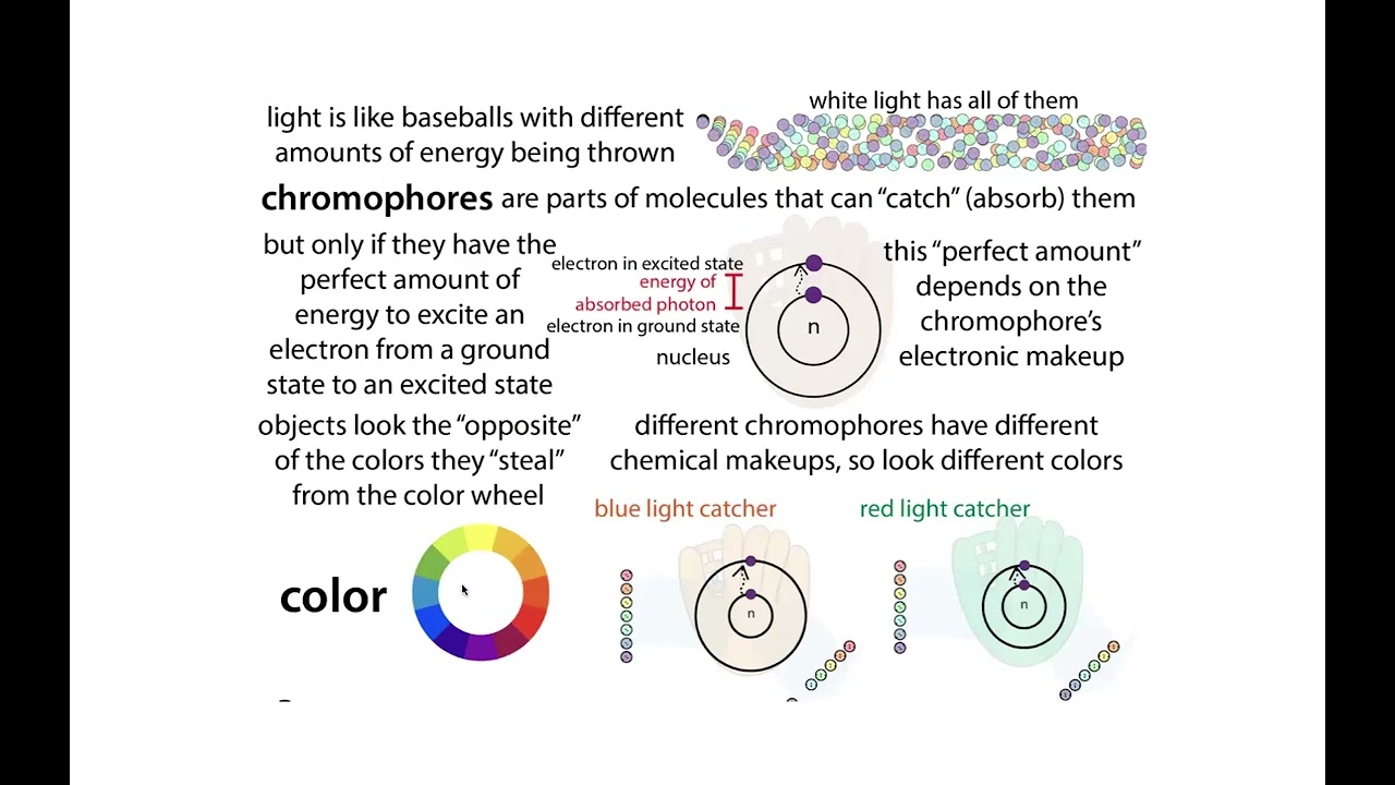
Added:Basic principles of fluorescence, including the concepts of excitation and emission wavelengths, Stokes shift, and fluorophores (e.g., GFP).
Fundamentals of bacterial cell culture, including growth phases (log vs. stationary), aseptic technique, and optical density (OD600) measurement.
The basic operation of microplate readers and how to handle and load 96-well plates.
The mathematical concept of normalization and why dividing a signal by cell density is necessary to determine per-cell expression.
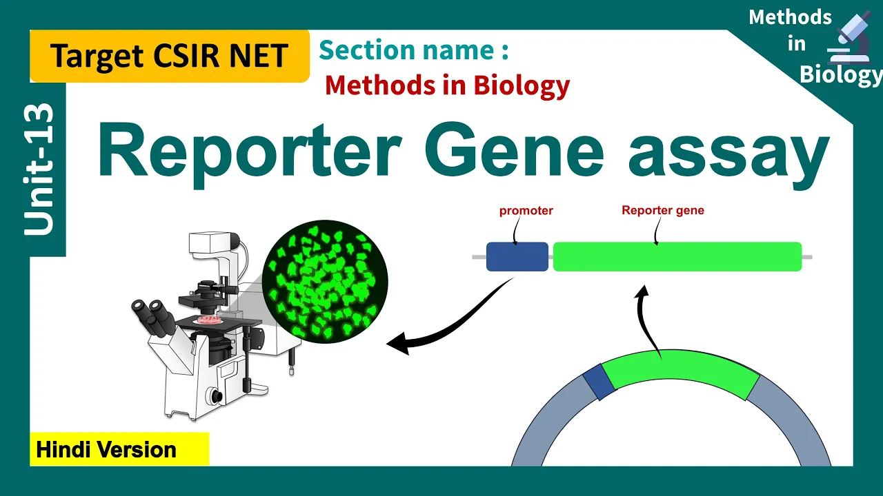
Designing and executing quantitative gene expression assays using fluorescent reporter proteins under different promoter strengths.
Troubleshooting common issues in fluorescence assays, such as background autofluorescence, photobleaching, and the inner filter effect.
Performing time-course (kinetic) fluorescence assays to track real-time promoter activity and population dynamics over several hours.
Applying high-throughput screening (HTS) methodologies to screen mutant libraries or evaluate chemical libraries in drug discovery.
Transitioning from population-level plate reader measurements to single-cell resolution analysis using flow cytometry.
3.6K views81likes14:55Original Release: 2017-09-08
hi everybody welcome back to synthetic biology 1.
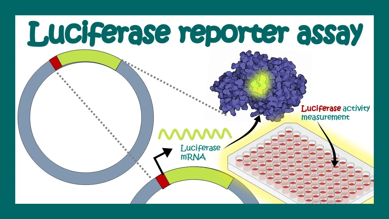
today we're going to be measuring fluorescence in bacterial cultures and to help us learn how to do that i have prepared a little game here before me you see two plates of bacteria marked s27 and s28 one of these bacteria i have transformed with a construct that gives red fluorescence rfb and the other one i have not so by measuring fluorescence today we are going to determine which of these two bacterial cultures is fluorescent uh and we're also going to try to be as quantitative as possible about measuring that fluorescence so at the end of the day we want to know how much more fluorescent is the fluorescent culture first step take these single colonies from these plates and grow up an overnight culture in lb this is something that we've seen before so i grab an inoculating loop like this one pick a single culture single colony off the plate start a culture these are 25 mil cultures put them in the incubator 37 degrees overnight and the next day they look like this right these are saturated cultures we can tell because uh they're they're very cloudy they look like saturated cultures so these are good they've been growing overnight now they're ready to use next step take a one mil aliquot of each of these cultures our fluorescent measurement technology is very sensitive does not require a large volume of cells one mil is going to be more than enough take my one ml of cells transfer it to a 1.5 ml eppendorf tube like this one and don't forget to label okay one mil of cells the next thing we need to do with these cells is wash them so we grew our cultures in lb because that's a standard media formulation but unfortunately for us lb looks like this right it's this yellow golden color in the lab it's also fluorescent right so lb contains vitamins and and other small molecules that are that are fluorescent which means that if we do our fluorescence measurements in lb we'll have a high background coming just from the lb itself so that means we need to remove the lb and in this case what we're going to do is we're going to wash the lb out and replace it with pbs phosphate buffered saline which is just a neutral solution that looks like this it's perfectly clear and has no fluorescence background so we take our one mil of cells into the centrifuge spin them down 30 seconds maximum speed pop them out pour off the spent lb and then what i'm going to do is invert these tubes on a sterile kimwipe and kind of blot them this will absorb just those last few drops of lb it's also cleaner like i don't have lb on my hands when i'm done i like it dry them out a little bit and you can see the pellet uh stays very firmly stuck to the bottom of the tube now we resuspend equal volume of pbs you'll notice that i pipette up and down to break up the pellet and then vortex just to make sure all the the cells are completely homogeneous in the pbs do next step in order to get accurate fluorescence measurements i need to dilute these cultures right so remember that we started with an overnight saturated culture of e coli and lb so that means we expect the od to be three or five or even higher right so this is a this is a very dense culture it's almost opaque there's just no way for light to penetrate a culture like that which means that we can't get accurate od readings and we can't get accurate fluorescence readings so i'm going to take these cultures and i'm going to dilute them by a factor of 10.
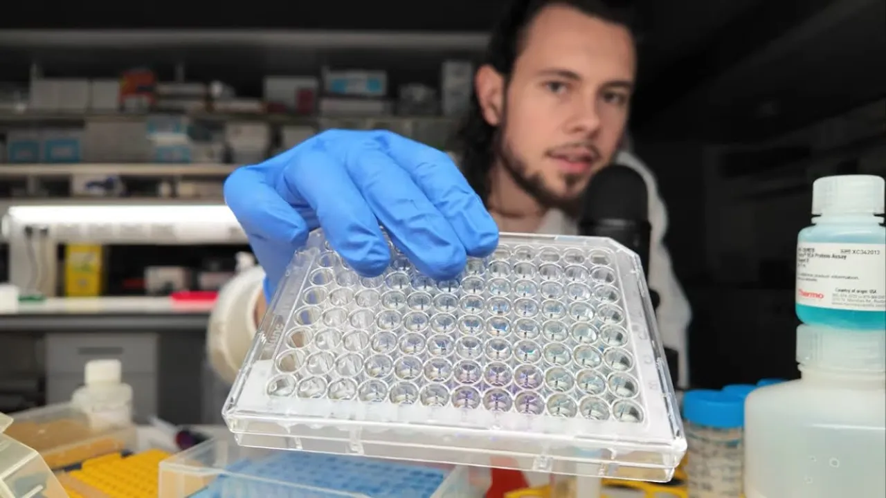
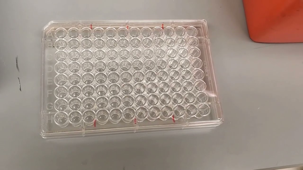
also in pbs this is going to bring them down into the the linear range of our od measurements and our fluorescence measurements right so you figure saturated culture has an od of about three if i do dilute it by one to ten it'll have an od of about 0.3 and what we want is something in the range of 0.1 up to 0.9 this is the linear range of od for most absorbance and fluorescence measurements them another vortex okay and now looking at a culture like that uh i can just sort of tell by eye that this is where we want to be for an absorbance measurement or a fluorescence measurement it's cloudy but i can still see through it uh which means it's it's going to be roughly in the linear range of our od measurements but just to be safe i'm going to take a quick measurement of the od here on my my bench top spectrophotometer a few hundred microliters of these cells load them into a cuvette and i've already prepared this spectrophotometer i've blanked it with a blank of just pure pbs so i've got an od of 0.37 for the first culture and 0.38 for the second culture and that that makes sense that's about where i expected them to be normally of course i would write them down but uh you guys will remember right now you'll notice that both of these ods that we're starting with are not necessarily exactly the same right and this is important because you naturally you would expect more cells to produce more fluorescence right and when we're taking a fluorescence measurement generally what we're interested in is not the total amount of fluorescence but the fluorescence per cell right so that's why uh when we do these fluorescence measurements we're going to measure simultaneously the od which we know is proportional to the number of cells so that we can normalize by the number of cells right so the the interesting unit in this case is not the total fluorescence but it's the fluorescence divided by the od or the fluorescence per cell if you like now to take our fluorescence measurements we are going to use a 96 well plate that looks like this so this is a 96 well plate if you haven't seen it before it's basically just 96 tiny little test tubes that we use whenever we want to do 96 experiments at once instead of just one experiment at once okay and in this case i've chosen a model that has a clear bottom this is very important because it's going to allow light to pass through so that we can take absorbance measurements and it's got black walls which means that those black walls will soak up any extra light that we uh that we uh use to take our fluorescence measurements and it will uh it will help prevent that that that that stray light from coming back into our detector and showing up as background so just as an example um here's a clear plate right this is something that you don't want to use for fluorescence measurements because you can have shell cells shining across the gap and sort of contaminating their neighbors with their fluorescence and a white plate is also not a great choice because it's going to reflect a lot more of the light potentially giving a higher background signal so we use our black plates these have a working volume of about 200 microliters so i'll load in 200 microliters of s27 200 microliters of s28 and of course 200 microliters of pure pbs with no cells in it and this will be my blank all right let's go measure some fluorescence here we are in the dark room i've set up our illuminator tool we've got two green excitation filters tuned to the wavelength of rfp excitation and one red emission filter tuned to the wavelength of rfp emission what this is going to allow us to do is to stimulate specifically the red fluorescent protein and then observe specifically the fluorescence light that is emitted by rfp fluorescence with this setup just by eye we'll be able to solve the mystery of which of our two strains is transformed with the red fluorescent protein so let's start by taking a look at the plates that we began with so here i'm loading on the petri dish struck out with single colonies of s27 doesn't look like there's anything there and now when i add the s28 plate you can see very clearly very bright red fluorescent colonies so this is s28 this definitely resolves the mystery of which strain is fluorescent the answer is s28 i also brought into the dark room a small bottle of lb and pbs so if i put these under the lights you can see for lb there is a weak but detectable fluorescent signal so this is something that we don't want to include in our readings we want to measure fluorescence only from bacteria which is the reason why we washed the lb away and replaced it with this pbs which has absolutely no detectable fluorescent signal at all so the final thing that i'll load on here are our two one mil aliquots of cells that we prepared s27 and s28 and you can clearly see a very strong fluorescent signal that is specific to the s28 strain so this is the same fluorescent signal that the plate reader is going to read although in a quantitative way and when we take this fluorescent signal and divide it by the absorbance or the od of the strain that we loaded onto the plate reader we'll get a quantitative measurement of the fluorescence per cell you
Up Next

10:50
Mycorrhizal Fungi and Vineyard Health: Nutrient Uptake Explained
@westovervineyardadvising7937
124 views•2025-07-09
14:12
Quantitative Real-Time PCR (qPCR): Principle, Method & Data Analysis
@animatedbiologywitharpan
129.2K views•2023-09-06

23:33
Microbial Degradation of Plastics: Biodegradation Pathways & Sustainability
@majeedhammad
2.9K views•2021-04-11

16:04
CRISPR and Genetic Engineering: How Gene Editing Works and Why It Matters
@kurzgesagt
30.5M views•2016-08-10
Related Study Plans & Knowledge Roadmaps
Structured learning paths in Biotechnology
biotechnology
AAV Production: Transfection, Prep & qPCR
01Introduction to Gene Therapy and AAV Biology
02Mammalian Cell Culture: Suspension HEK293 Cells
03Upstream Bioprocessing: Triple-Transfection Optimization
+ 2 more learning modules
18h5 modules
Explore Roadmap
biotechnology
Building Gut-on-a-Chip: PDMS, Cells & TEER
01Foundations of Microfluidics and Gut Anatomy
02Cleanroom Fabrication & PDMS Soft Lithography
03Epithelial Cell Culture and On-Chip Seeding
+ 2 more learning modules
26h5 modules
Explore Roadmap
biotechnology
Optimizing CFPS: E. coli Extract & Yield
01Fundamentals of Molecular Biology and Translation
02Introduction to Cell-Free Protein Synthesis (CFPS)
03Preparing E. coli Crude Lysate (The Extract)
+ 2 more learning modules
19h5 modules
Explore Roadmap
biotechnology
Haematococcus PBR: Design, Stress & Astaxanthin
01Biology and Life Cycle of Haematococcus pluvialis
02Bench-Scale Photobioreactor (PBR) Design and Engineering
03Culture Media Formulation and Inoculation
+ 2 more learning modules
15h5 modules
Explore Roadmap