When evaluating patients for extubation after a spontaneous breathing trial, clinicians must assess four key areas: respiratory status (using RSBI <105 indicating possible success, <80 suggesting ~95% success probability, along with oxygenation tolerance at FiO2 <40% and PEEP 5-8 cmH2O), cardiovascular stability, neurologic alertness and ability to follow commands, and psychological factors like anxiety reduction; the single most important criterion is whether there has been significant alleviation or reversal of the primary cause for mechanical ventilation.
Key Weaning Parameters for Mechanical Ventilation Monitoring
Added:Deep Dive
Basic respiratory anatomy and physiology, including the mechanics of breathing, gas exchange, and lung compliance.

Respiratory System- Dr. Jessica Guerrero
2.4K
Inspiration is an active process requiring diaphragm contraction and external intercostal muscle contraction, increasing thoracic volume and decreasing pressure below atmospheric to draw air in. Expiration is typically passive, with muscles relaxing and volume decreasing to increase pressure and expel air. Atelectasis is lung collapse; pneumothorax involves air/fluid in pleural cavity reducing compliance. Three factors affect ventilation: airway resistance (increased by inflammation), surfactant (reduces surface tension), and lung compliance (elasticity). Healthy lungs have high compliance; poor posture, scoliosis, or scar tissue from emphysema decrease compliance. Lung volumes include tidal volume, inspiratory reserve, expiratory reserve, and residual volume (~150mL). Capacities combine these volumes. Anatomical dead space refers to air not participating in gas exchange.
Click to watch →

Basic Anatomy & Physiology for Nurse โดยนายแพทย์ดำรงค์ สุกิจปัญญาโรจน์
30.2K
The respiratory system includes the upper respiratory tract (nose, pharynx, larynx) and lower respiratory tract (trachea, bronchi, lungs). The nasal cavity warms, humidifies, and filters inspired air. The larynx contains cartilages including the thyroid, cricoid, and epiglottis. The epiglottis prevents food from entering the trachea during swallowing. The alveoli are thin-walled sacs where gas exchange occurs. Each alveolus is surrounded by capillaries. The respiratory membrane (alveolar-capillary barrier) is extremely thin for efficient diffusion. Pleural membranes (visceral and parietal pleura) reduce friction during breathing. Lung compliance is the ease of lung expansion. Elastic recoil is provided by elastic fibers in lung tissue. Surfactant reduces surface tension and prevents alveolar collapse. Inspiration is primarily active, involving diaphragm contraction (increasing thoracic volume) and external intercostal muscle contraction. Expiration is normally passive, resulting from elastic recoil of lungs and chest wall. Pulmonary ventilation moves air in and out of the lungs. Tidal volume is the amount of air moved per breath (approximately 500 mL). Gas exchange follows physical laws: diffusion occurs from high to low concentration, and partial pressure determines gas movement. Oxygen diffuses from alveoli into blood; carbon dioxide diffuses from blood into alveoli. Hemoglobin binds oxygen in the lungs (high O2 partial pressure) and releases it in tissues (low O2 partial pressure). The dissociation curve is sigmoidal, allowing efficient loading and unloading of oxygen. Carbon dioxide is transported in blood as bicarbonate (approximately 70%), bound to hemoglobin (approximately 20%), and dissolved in plasma (approximately 10%). The chloride shift facilitates CO2 transport. Respiratory control centers are located in the brainstem: the medulla oblongata (primary rhythm generator) and pons (modulates breathing). These centers receive input from chemoreceptors (detecting O2, CO2, pH) and mechanoreceptors.
Click to watch →

Basic Respiratory Physiology (MEDEX/MCAI Preparatory Course 2023)
1.5K
This extensive section covers the complete foundation of respiratory physiology. The lungs perform critical non-respiratory functions including immune defense through mucus and macrophages, fibrinolysis, thermoregulation, surfactant synthesis, acid-base regulation, and drug metabolism. The respiratory control system involves central chemoreceptors in the medulla detecting CSF pH changes from CO2 diffusion, and peripheral chemoreceptors in carotid/aortic bodies responding to arterial oxygen tension. The CO2-ventilation relationship is linear within normal ranges but flattens at extreme hypercapnia. Oxygen stimulates ventilation only when PaO2 falls below 60 mmHg. In COPD/OSA patients, the ventilatory curve shifts right, requiring higher CO2 thresholds. Functional Residual Capacity (FRC) is the lung volume at end-expiration where lung elastic recoil equals chest wall outward pull (~30 ml/kg), preventing alveolar collapse by containing nitrogen that splints alveoli open. Closing capacity (CC) is the volume where airways begin collapsing during expiration; normally CC < FRC, but in newborns CC is very high (~90% TLC), explaining their higher PaO2 values. CC gradually decreases until age 44, then increases, exceeding FRC by age 66 even in upright position. Lung compliance is the change in volume per unit pressure change, measured as static (~200 ml/cm H2O, no airflow) or dynamic (with airflow). The compliance curve shows hysteresis: initial expansion is difficult due to surface tension, compliance peaks around FRC, then flattens as alveoli become maximally expanded. Alveoli at lung bases have higher compliance than apex despite appearing counterintuitive, causing ventilation to concentrate at the bases. Total respiratory system compliance (~100 ml/cm H2O) is less than either lung or chest wall compliance individually because they act in series. Pulmonary surfactant (80% phospholipids, mostly DPPC, plus proteins) reduces alveolar surface tension, increases lung compliance, prevents small alveolar collapse, keeps alveoli dry, and has immunological properties. Surfactant production is stimulated by steroids and hormones. The work of breathing is ~2-3% of total body oxygen consumption (~250 ml/min). It consists of elastic work (overcoming lung recoil/surface tension) and non-elastic work (overcoming airway resistance). During inspiration, additional work overcomes resistance; during expiration, non-elastic work falls within the elastic work triangle, requiring no extra energy. In restrictive disease, elastic work increases; in obstructive disease, non-elastic work increases, requiring active expiration.
Click to watch →
Professor Long - Respiratory Lecture 2, Pharynx and definitions
1.7K
Compliance measures an organ's ability to change shape and accommodate volume changes, similar to how plastic bottles expand/contract versus metal containers. The lungs have high compliance enabling breathing mechanics. Pulmonary ventilation moves gases to/from the gas exchange surface (atmosphere to alveoli). External respiration exchanges gases between atmosphere and blood, while internal respiration exchanges gases between blood and tissues (or interstitial fluid), completing the full respiratory cycle from atmospheric air to cellular oxygen delivery.
Click to watch →

Mechanics of breathing and compliance for USMLE step 1
14.4K
Pulmonary compliance describes the ability of the lungs and chest wall to expand in response to pressure changes, calculated as the change in volume divided by the change in pressure (C = ΔV/ΔP); the diaphragm is the primary muscle of inspiration, while expiration is normally passive due to elastic recoil, but becomes active during exercise or disease using abdominal and internal intercostal muscles; compliance is greatest at functional residual capacity (FRC) where transpulmonary pressure is approximately -5 cm H2O, and decreases in conditions like pulmonary fibrosis, pneumonia, and pulmonary edema, while increasing in emphysema due to loss of elastic recoil.
Click to watch →
Deep Dive
Fundamental concepts of mechanical ventilation, specifically the key settings such as Tidal Volume (Vt), Positive End-Expiratory Pressure (PEEP), and Fraction of Inspired Oxygen (FiO2).
Mechanical Ventilation Basics - PEEP, FiO2, Respiratory Rate, Tidal Volume | Clinical Medicine
36.2K
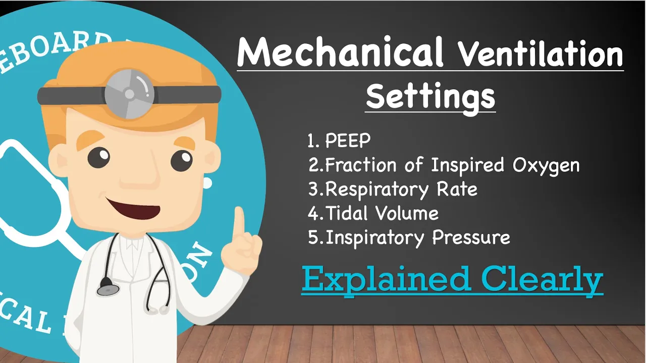
Mechanical ventilation requires four foundational settings: PEEP (Positive End Expiratory Pressure, 5-20 mmHg) maintains constant pressure to improve oxygenation; FiO2 (Fraction of Inspired Oxygen, 21-100%) controls oxygen concentration; Respiratory Rate (8-32 breaths/minute) determines ventilation frequency; and Tidal Volume (6-8 cc/kg ideal body weight) or Inspiratory Pressure (above PEEP) controls volume delivery. These settings operate in either Volume Control (setting tidal volume) or Pressure Control (setting pressure above PEEP) modes, with Respiratory Rate × Tidal Volume calculating Minute Ventilation for carbon dioxide elimination.
Click to watch →
Mechanical Ventilation - Medical-Surgical - Respiratory System | @LevelUpRN
226.7K
Key ventilator settings include: Respiratory rate (breaths per minute delivered to the patient), Tidal volume (VT) which is the gas volume per breath, FiO2 (fraction of inspired oxygen ranging from 21% to 100%), I:E ratio (inspiration to expiration duration, typically 1:2 or 1:1.5), and PEEP (positive end-expiratory pressure) which applies pressure at end-expiration to keep alveoli open and prevent collapse.
Click to watch →
Mechanical Ventilator Settings Explained | PEEP, FiO₂, RR & Tidal Volume Made Simple
497
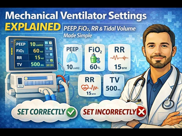
Mechanical ventilation settings are divided into oxygenation (FiO₂ and PEEP) and ventilation (respiratory rate and tidal volume) parameters. FiO₂ (fraction of inspired oxygen) ranges from 21% to 100% and directly increases alveolar oxygen concentration to improve oxygenation, with titration to target saturation of 88-96% to avoid oxygen toxicity. PEEP (positive end-expiratory pressure) maintains alveolar recruitment and improves oxygenation by preventing alveolar collapse, typically starting at 5 cm H₂O and titrated based on pathology. Respiratory rate (16-20 breaths/min) and tidal volume (6-8 cc/kg ideal body weight) together determine minute ventilation for CO₂ clearance, with respiratory rate being the primary determinant of CO₂ elimination. Inspiratory pressure controls tidal volume in pressure control modes and should not exceed 30 cm H₂O to prevent lung injury.
Click to watch →
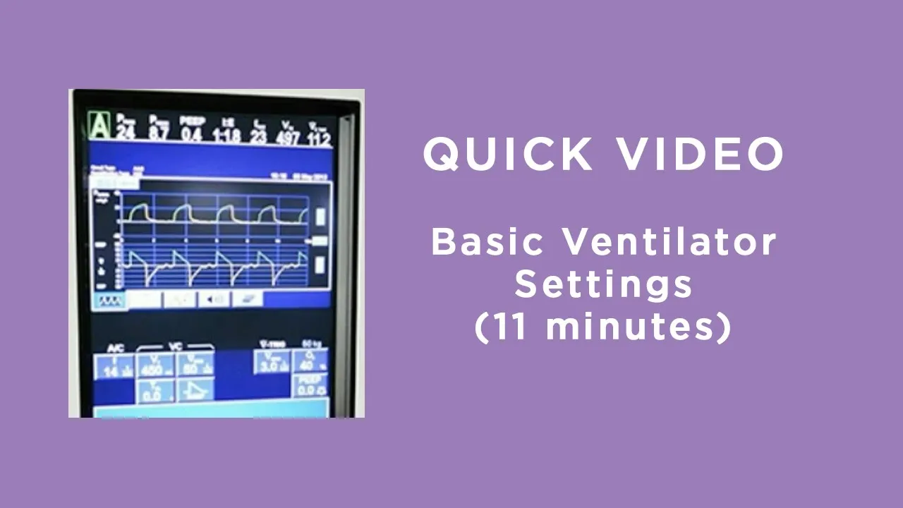
Basic Ventilator Settings
43.5K
Four essential ventilator settings must be understood for respiratory failure management. FIO2 (Fraction of Inspired Oxygen) ranges from 21% to 100%, determining oxygen delivery concentration. Respiratory rate controls breaths per minute, potentially increased in ARDS using permissive hypercapnia. Tidal volume (4-8 mL/kg predicted body weight) sets breath depth, calculated using predicted body weight rather than actual weight since lung size remains constant. PEEP (Positive End-Expiratory Pressure) maintains alveolar pressure at 5-25 cm H2O to improve oxygenation while preventing alveolar collapse. Critical monitoring includes peak airway pressure, exhaled tidal volume, and emergency 100% O2 capability.
Click to watch →

Ventilation settings in ICU - Ventilator settings explained - Hindi / Urdu
8.8K
Mechanical ventilation requires four primary settings: respiratory frequency (breaths per minute), tidal volume (air per breath), fraction of inspired oxygen (FiO2), and positive end-expiratory pressure (PEEP). Frequency is adjusted based on patient's respiratory status and acid-base balance. Tidal volume is calculated at 6-8 ml/kg body weight to prevent lung injury. FiO2 normally starts at 21% and is increased based on oxygenation needs. PEEP maintains alveolar recruitment, with normal settings around 5 cm H2O; values below this increase collapse risk. All settings are dynamically adjusted based on patient condition, with changes typically evaluated every 10 minutes.
Click to watch →
Deep Dive
Interpretation of Arterial Blood Gas (ABG) values and basic oxygenation indices, such as the PaO2/FiO2 ratio.

P/F Ratio | PaO2/FiO2 Ratio
14.5K
The P/F ratio, also called the Horovitz index or Caro index, is calculated by dividing the arterial oxygen tension (PaO2) by the fraction of inspired oxygen (FiO2), and it is used to evaluate oxygenation status and classify the severity of acute respiratory distress syndrome (ARDS) according to the Berlin definition: mild ARDS (P/F ratio 200-300 mmHg), moderate ARDS (100-200 mmHg), and severe ARDS (<100 mmHg with PEEP >5 cm H2O).
Click to watch →

Gasometria Arterial Fácil
94.5K
PaO2 should be 80-100 mmHg; FiO2 is the percentage of inspired oxygen (normally 21%). The P/F ratio (PaO2/FiO2) assesses oxygenation efficiency: normal values above 400 mmHg indicate adequate function. Values below 400 indicate respiratory dysfunction: mild (300-400), moderate (200-300), severe (less than 200). In ARDS, P/F ratios are typically around 60 mmHg. Oxygen toxicity occurs when FiO2 exceeds 60% (some studies suggest 50% is already harmful). PEEP maintains alveolar patency, improving oxygenation but potentially causing barotrauma.
Click to watch →
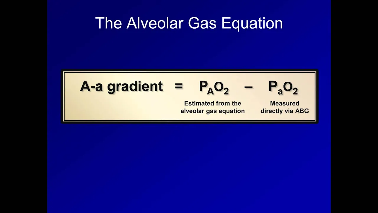
The A-a Gradient (ABG Interpretation - Lesson 16)
156.3K
The PaO2/FiO2 ratio measures severity of hypoxemia in ICU patients. For a healthy young adult breathing room air, PaO2 ≈ 100 mmHg and FiO2 = 0.21, giving a ratio of approximately 476. Values of 200-300 indicate mild to moderate gas exchange abnormality (acute lung injury), while values under 200 indicate severe gas exchange abnormality (acute respiratory distress syndrome/ARDS). These cutoffs are somewhat arbitrary but clinically useful for classification.
Click to watch →

PaO2 to FiO2 Ratio - PF Ratio
129.2K
The PaO2/FiO2 ratio (PF ratio) is a clinical tool used to quantify the severity of respiratory failure by comparing the partial pressure of arterial oxygen (PaO2) to the fraction of inspired oxygen (FiO2 expressed as a decimal), calculated as PaO2 ÷ FiO2; a normal PF ratio is greater than 400, while values below 200 indicate significant respiratory impairment, allowing healthcare providers to trend patient deterioration, determine appropriate oxygen delivery strategies, and guide decisions regarding intubation and respiratory support.
Click to watch →

02/04/20. Principios Básicos de Ventilación Mecánica Intensiva
15.4K
Arterial blood gases are essential for monitoring: PaO2 indicates oxygenation, PaCO2 indicates ventilation. A PaO2 of 108 mmHg on 50% FiO2 gives P/F ratio of 216 (good function), while the same PaO2 on 100% FiO2 indicates poor function. Oxygenation is adjusted by changing FiO2 (direct effect on alveolar oxygen) or PEEP (improves lung function). Hyperoxemia with good function allows FiO2 reduction; poor function requires PEEP adjustment.
Click to watch →
Deep Dive
The clinical indications for initiating mechanical ventilation and the pathophysiology of acute respiratory failure.
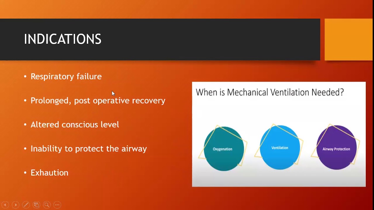
Mechanical Ventilation: Basic concepts -- Dr Vanitha/ Dr Subramanyam
220
This section explains the classification of respiratory failure and criteria for initiating mechanical ventilation. Type 1 (hypoxic) respiratory failure involves alveolar gas exchange pathology with mechanisms including ventilation-perfusion mismatch, intrapulmonary shunting, and decreased functional residual capacity, seen in conditions like pneumonia and pulmonary edema. Type 2 (hypercapnic) respiratory failure involves ventilatory pump failure from decreased respiratory drive (post-anesthesia, drug overdose), chest wall abnormalities (flail chest), respiratory muscle fatigue, or neural disorders. Primary indications for mechanical ventilation include respiratory failure, while prophylactic ventilation is needed for airway protection in altered consciousness patients, post-surgical recovery, and impending ventilatory failure. Goals of mechanical ventilation include improving oxygenation and ventilation to reverse hypoxemia and acidosis, reducing work of breathing, improving pulmonary mechanics, managing secretions, and permitting lung healing. Clinical parameters indicating need include respiratory rates >35 or <5 breaths/min, hypoxia (SpO2 <88-90%, PaO2 <55-60 mmHg), hypercapnia (PaCO2 >45-50 mmHg), Glasgow Coma Scale <8, tidal volume <5 ml/kg, and vital capacity <15 ml/kg.
Click to watch →

Respiratory Failure EXPLAINED!
272.7K
Mechanical ventilation becomes necessary when non-invasive support fails or when specific clinical criteria are met. Indications include respiratory arrest, apnea, respiratory rate exceeding 30 breaths per minute sustained, altered level of consciousness, respiratory muscle fatigue (tiring out), hemodynamic instability, refractory hypoxemia despite non-invasive support, and refractory hypercapnia with pH <7.25. Goals of mechanical ventilation include correcting hypoxemia through controlled FiO2 and PEEP, correcting respiratory acidosis by adjusting minute ventilation (tidal volume × respiratory rate), and allowing ventilatory muscles to rest.
Click to watch →

Invasive Pediatric Mechanical Ventilation,by Dr. D. K. Jha, M. D. Pediatric Pulmonologist ,Delhi
440
Mechanical ventilation is indicated when other oxygen therapy modalities fail, including respiratory failure, prevention of respiratory muscle exhaustion, airway protection in unconscious patients, support of left ventricular function in cardiogenic shock, and treatment of intracranial hypertension. Respiratory failure can occur at multiple levels: central drive failure, nerve pathway lesions, neuromuscular junction dysfunction, respiratory muscle failure, airway obstruction, or ventilation-perfusion mismatch. Respiratory failure is classified as hypoxemic (PaO2 <60 mmHg) or hypercapnic (PaCO2 >60 mmHg). Mechanical ventilation should be initiated when PaO2 <60 mmHg on supplemental oxygen, PaCO2 >60 mmHg, or pH <7.25. The alveolar gas equation determines alveolar oxygen concentration based on atmospheric pressure, water vapor pressure, FiO2, and CO2 pressure. The A-a gradient (normal 12-15 mmHg on room air, <70 mmHg on 100% O2) indicates gas exchange impairment.
Click to watch →

Pneumologia Intensiva - Podcast EMED - Aula para Residência Médica
173
Acute respiratory failure is the inability of the respiratory system to maintain adequate gas exchange, classified into Type 1 (hypoxemic) involving V/Q mismatch, shunt, and diffusion impairment, and Type 2 (hypercapnic) resulting from pump failure or hypoventilation. Mechanical ventilation supports patients through invasive (endotracheal intubation) or non-invasive (mask interfaces) methods. Non-invasive ventilation is indicated for COPD exacerbations, cardiogenic pulmonary edema, and postoperative respiratory failure, with evidence of reduced mortality. Absolute contraindications include obvious need for intubation and cardiac arrest. Mechanical ventilation operates through four phases: trigger, inspiratory phase, cycling, and expiratory phase. Key parameters include tidal volume (6 ml/kg predicted body weight), minute volume, PIP, plateau pressure, PEEP, and driving pressure. The I:E ratio is adjusted based on pathology: shorter for restrictive disease, longer for obstructive disease.
Click to watch →

ENFERMERIA EN ALTO RIESGO TEMA 4 - ASISTENCIA RESPIRATORIA MECANICA (ARM)
3.2K
Mechanical ventilation is indicated when respiratory insufficiency becomes unsustainable and all compensatory mechanisms fail. The fundamental purpose of respiration is to oxygenate cells for metabolic processes, with carbon dioxide playing a crucial role in pH balance. Indications include hypoxia (PaO2 <50 mmHg despite FiO2 >60%), respiratory rate abnormalities (below 10 or above 35 breaths/minute), decreased depth, cyanosis, metabolic acidosis (pH <7.25), and PaCO2 >50 mmHg. When oxygen is critically low, the body switches to anaerobic metabolism, producing lactic acid and causing metabolic acidosis. Negative Inspiratory Force (NIF) measures respiratory muscle strength (normal 60-120 cm H2O), with values below 25 cm H2O indicating insufficient strength for independent ventilation.
Click to watch →
Prerequisite Knowledge
- Concept 01Basic respiratory anatomy and physiology, including the mechanics of breathing, gas exchange, and lung compliance.
- Concept 02Fundamental concepts of mechanical ventilation, specifically the key settings such as Tidal Volume (Vt), Positive End-Expiratory Pressure (PEEP), and Fraction of Inspired Oxygen (FiO2).
- Concept 03Interpretation of Arterial Blood Gas (ABG) values and basic oxygenation indices, such as the PaO2/FiO2 ratio.
- Concept 04The clinical indications for initiating mechanical ventilation and the pathophysiology of acute respiratory failure.
Subsequent Learning
- Step 01The clinical protocol for conducting, monitoring, and evaluating a Spontaneous Breathing Trial (SBT).
- Step 02Extubation techniques, immediate post-extubation care, and the prevention of post-extubation airway complications.
- Step 03Strategies for utilizing non-invasive ventilation (NIV) and High-Flow Nasal Cannula (HFNC) therapy as post-extubation support.
- Step 04Algorithms for managing 'difficult-to-wean' patients and tracheostomy-related weaning protocols.
Weaning Criteria
Assessment Indices
Overall Readiness
Weaning Criteria
0:00Playing Section
- 1
Primary criterion is resolving the original reason for ventilation.
- 2
Assess four key areas: respiratory, cardiovascular, neurologic, and psychologic status.
The Overreliance on Predictive Weaning Indices vs. Direct Spontaneous Breathing Trials
While traditional weaning parameters like the Rapid Shallow Breathing Index (RSBI) and maximal inspiratory pressure have long been used to predict extubation success, a significant body of contemporary critical care research challenges their utility. Critics argue that these static physiological measurements have low predictive accuracy, often yielding false positives or negatives that can lead to premature extubation or, conversely, unnecessary delays in liberation from mechanical ventilation. Modern clinical guidelines increasingly advocate for minimizing the use of these traditional parameters. Instead, they favor a simplified, protocolized approach centered on daily Spontaneous Breathing Trials (SBTs) combined with holistic clinical assessment. This counterpoint suggests that a patient's actual performance during an active, trial-of-breathing challenge is a far more reliable indicator of extubation readiness than rigid numerical indices, which can overcomplicate the weaning process and prolong ventilator days.
Deep Dive
The clinical protocol for conducting, monitoring, and evaluating a Spontaneous Breathing Trial (SBT).

Weaning Failure in ICU: Causes, Assessment & Management | Critical Care Webinar
170
This section covers the complete protocol for conducting and interpreting spontaneous breathing trials. The SBT can be performed using T-piece or pressure support modes, with the RSBI (Rapid Shallow Breathing Index) serving as the primary predictor of success/failure. A cut-off of 105 indicates successful weaning. The duration of SBT can vary from 30 to 120 minutes depending on patient factors, though evidence shows no significant difference in outcomes between durations. Weaning failure is classified as simple (single failure), difficult (up to 3 attempts or 7 days), or prolonged (more than 3 attempts or 7 days). Five categories of parameters are used to assess failure: hemodynamic (heart rate, blood pressure), respiratory (rate, accessory muscles), gas exchange (PaCO2, pH, oxygenation), neurological (delirium, agitation), and others.
Click to watch →

9) Discontinuation of MV - Case based discussion - Dr Mohamed Torayah
728
The Spontaneous Breathing Trial (SBT) is performed after meeting readiness criteria to assess weaning tolerance. The patient is placed on spontaneous breathing mode with minimal support (PEEP 5 cmH2O, FiO2 0.21, or PEEP 5-8 cmH2O). The trial lasts 30 minutes to 2 hours. Critical monitoring includes: heart rate changes not exceeding 20% from baseline, blood pressure changes not exceeding 25%, respiratory rate not increasing by more than 50%, and absence of increased work of breathing signs. Successful completion indicates readiness for extubation.
Click to watch →

Heuer_Spontaneous Breathing Trails SBTs and Ventilator Weaning
118
The SBT algorithm begins with assessing weanability: clinical stability, mental status, oxygenation, and pulmonary mechanics. If ready, patients proceed to SBT for at least 3 minutes to calculate RSBI. If RSBI is poor (>105), return to full support and rest for 24 hours. Exception: sedated but stable patients may have sedation lightened for later assessment. RSBI interpretation requires context - values of 75 are better than 95 unless caused by agitation or pain.
Click to watch →

Respiratory Therapy: Spontaneous Breathing Trials & Weaning Parameters
46.1K
A spontaneous breathing trial (SBT) is the gold standard for assessing readiness to wean patients from mechanical ventilation, involving placing the patient in a spontaneous mode with minimal support (such as CPAP 5 cm H2O with pressure support 5 cm H2O or proportional assist ventilation at 30%) for approximately 30 minutes; successful completion requires meeting multiple criteria including a rapid shallow breathing index (RSBI) less than 105, negative inspiratory force (NIF) greater than -20 cm H2O, vital capacity greater than 10 mL/kg, stable hemodynamics, normal ABG values, adequate cough, and neurological responsiveness, with the goal of achieving an extubation failure rate around 15% rather than zero to optimize patient outcomes and reduce hospital stays.
Click to watch →

Weaning from mechanical ventilation
127
SBT protocol involves attaching patients to heated nebulizers with 10% higher oxygen concentration than during ventilation, with PEEP exceeding peak inspiratory pressure. Duration is typically 30 minutes, extendable to 120 minutes for COPD, heart failure, or neuromuscular disorders. Successful SBT requires respiratory rate <35, heart rate <130, heart rate variability <20%, maintained P/F ratio, and no distress. Failed SBT shows increased muscle workload, asynchrony, tachycardia, diaphoresis, cyanosis, tachypnea, or abdominal paradox. Cough reflex and cuff leak (<110 ml) must be assessed.
Click to watch →
Deep Dive
Extubation techniques, immediate post-extubation care, and the prevention of post-extubation airway complications.

Ventilator NCLEX Review: Respiratory Nursing Concepts & Questions
9.4K
Post-extubation care requires careful monitoring and intervention. Patients should remain NPO until demonstrating adequate gag reflex, with humidified oxygen via face mask and upright positioning. Stridor is the priority assessment finding requiring immediate intervention as it indicates airway edema and potential obstruction. Essential emergency equipment includes an Ambu bag with high-flow oxygen (15 L/min) at the bedside. Tracheostomy tube security is assessed by placing one finger under the tube—if more than one finger fits, the tube is not secure. For dislodgement within 7 days, cover with occlusive dressing and apply non-rebreather mask before notifying provider.
Click to watch →
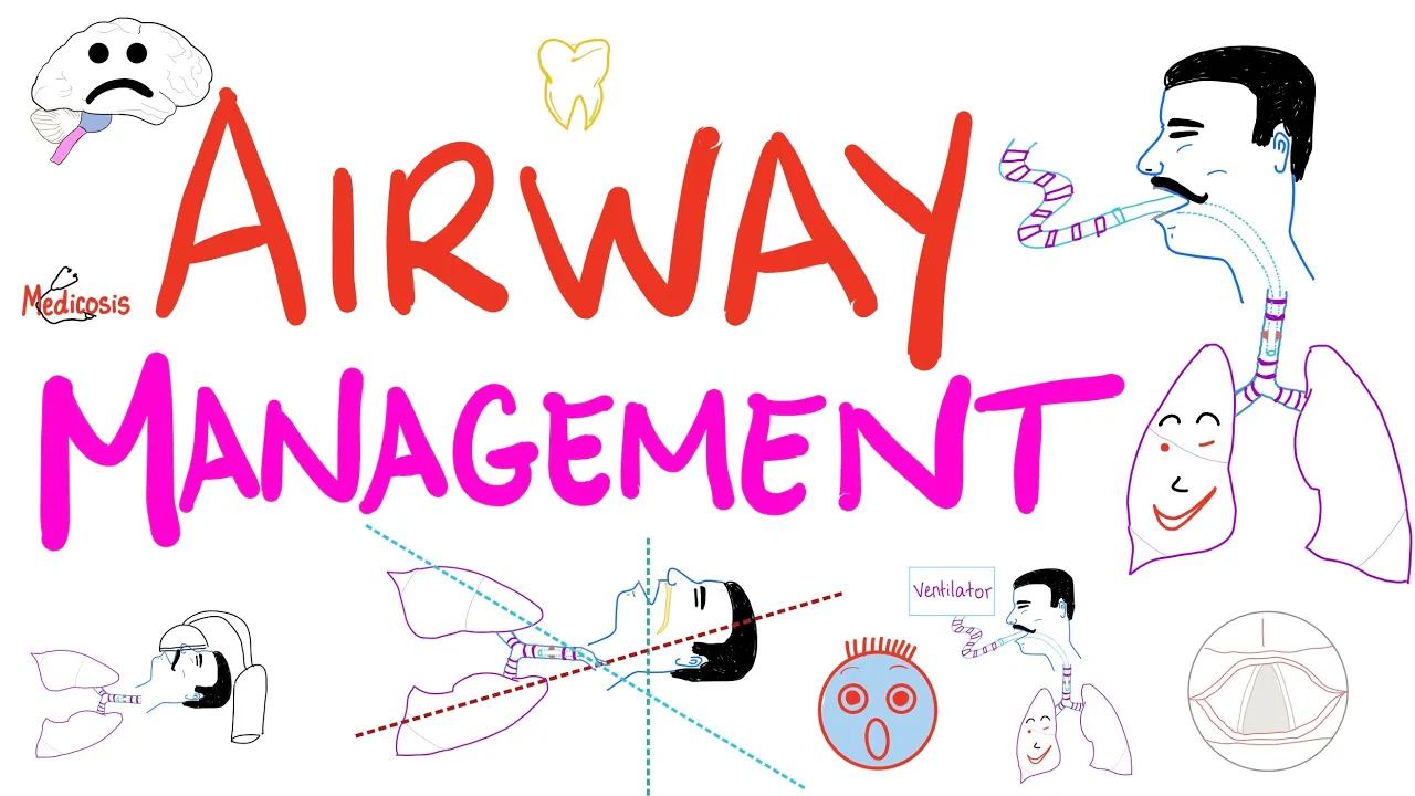
Airway Management - Tracheal Intubation - Anesthesiology Series
77.1K
During intubation: oral/dental trauma, aspiration, hypertension, tachycardia, myocardial infarction. While tube in place: esophageal/bronchial intubation, tube obstruction, cuff leak, pulmonary barotrauma, nasogastric distension, accidental disconnection, accidental extubation. After extubation: aspiration, pharyngitis, laryngitis, laryngospasm, laryngeal ulceration, tracheitis, tracheal stenosis, vocal cord dysfunction, arytenoid dislocation. Post-extubation stridor results from tracheal mucosal edema from large tubes. According to Poiseuille's law, decreased radius increases resistance to the fourth power. Treatment: mild cases receive humidified oxygen and PACU observation; severe cases require racemic epinephrine and ICU monitoring. Reintubation may be necessary if patient does not improve.
Click to watch →

The Art and Science of Extubation
7.9K
Staged extubation uses wire encased in plastic jacket to reduce irritation; reintubation catheter passes over wire, then ETT railroads over. Post-extubation care includes keeping patient fasted, upright, and awake, with antiemetics and humidified oxygen. Laryngospasm responds to PEEP at laryngospasm notch and propofol/suxamethonium. Negative pressure pulmonary edema (pink frothy sputum, pulmonary edema on CXR) requires relieving obstruction, oxygen, and PEEP—no evidence supports diuretics or steroids. Vocal cord dysfunction (stridor, paradoxical motion) responds to oxygen, ventilation, and benzodiazepines. Laryngeal edema is universal after >4 days intubation; steroids help, but nebulized adrenaline lacks evidence. Comprehensive assessment of both airway and weaning factors is essential, accepting that some extubations will fail while optimizing for success.
Click to watch →

Extubation Guidelines & Aids: Dr Manpreet Singh, Chandigarh
5K
Extubation is the safe removal of an endotracheal tube, transitioning from artificial to natural airway. Difficult extubation occurs when removal proves challenging or when clinicians fear complications. Michigan data (1985-2003) showed 0.12-0.25% ICU incidence with devastating outcomes. NEJM/Lancet 2013 identified all anesthesia-related deaths occurred during emergence/recovery, not induction. One-third of major airway complications occur at extubation, with airway obstruction, laryngospasm, and edema being most common. Risk factors include restricted mouth opening, obesity, full stomach, pregnancy, restricted airway access, and surgeries requiring smooth extubation. The Difficult Airway Society provides a four-step framework: Plan based on patient assessment; Prepare by optimizing systems and ensuring skilled help; Perform using appropriate techniques; Provide post-extubation care. Risk stratification divides patients into low-risk and at-risk categories. Second-generation supraglottic airway devices (LMA Supreme, ProSeal, i-gel) revolutionized extubation by providing superior emergence profiles and allowing vocal cord visualization. Bury's maneuver places an LMA behind the endotracheal tube, preventing epiglottis down-folding. The airway exchange catheter (85-100 cm) serves as a guide for reintubation, reducing failure rates from 14% to 87%. Hemodynamic responses during extubation (10-30% BP increase) can be suppressed using esmolol, lidocaine, fentanyl, or dexmedetomidine. The cuff leak test predicts post-extubation stridor by measuring tidal volume difference with and without deflated cuff.
Click to watch →

Intubação Traqueal: materiais necessários e como preparar
89.6K
Post-intubation care includes: maintaining the patient in semi-recumbent position (30 degrees) to prevent aspiration, performing suctioning when secretions are observed or when ventilator waveforms indicate turbulence, and performing routine oral hygiene. These measures help prevent ventilator-associated pneumonia and other complications.
Click to watch →
Deep Dive
Strategies for utilizing non-invasive ventilation (NIV) and High-Flow Nasal Cannula (HFNC) therapy as post-extubation support.

Weaning from mechanical ventilation : Update and Experience
146
Post-extubation respiratory failure occurs in 10-20% of properly assessed patients and carries high mortality risk. Prevention strategies include: (1) Facilitative NIV for COPD/hypercapnic patients transitioning from pressure support to NIV, (2) Prophylactic NIV or HFNC for high-risk patients, and (3) Therapeutic trials for those developing acute respiratory failure. A multicenter RCT comparing HFNC versus NIV in hypercapnic patients showed both non-inferior in preventing reintubation within 72 hours. Meta-analysis demonstrates HFNC reduces weaning failure by up to 50% versus conventional oxygen therapy. For high-risk patients, NIV is superior to HFNC for reducing reintubation. For surgical patients, HFNC fairs better than conventional oxygen. Both modalities are similarly effective overall, but selection should consider patient-specific risk factors. High-risk patients require close monitoring and low threshold for reintubation if respiratory failure develops.
Click to watch →
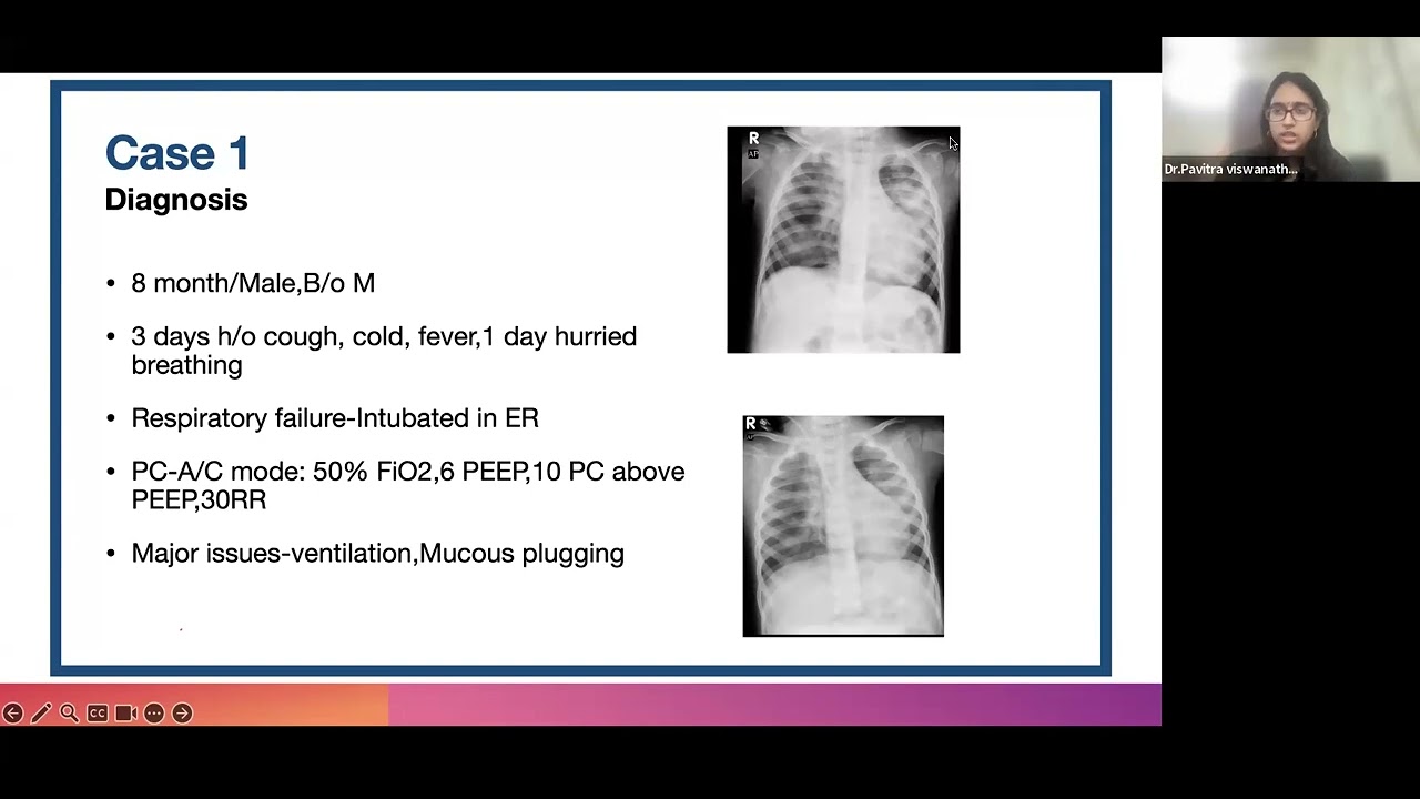
Liberation from Mechanical Ventilation-Step by step guide by Dr Pavitra
389
Post-extubation support includes planned (elective) and rescue NIV. Elective NIV reduces reintubation risk by anticipating pathology. Selection depends on underlying condition: CPAP for prior CPAP requirement, positive pressure for cardiac dysfunction (assists left ventricle), HFNC for simple bronchitis. Children under 1 year at high risk prefer CPAP. For neuromuscular patients (GBS), assess respiratory muscle strength: P0.1 (inspiratory drive), cough strength, shoulder/neck lifting ability. Inspiratory muscle strength: negative inspiratory force below -30 to -40 cm H2O indicates good strength.
Click to watch →

Non-Invasive Respiratory Support
511
Non-invasive respiratory support (NIV) and high-flow nasal cannula (HFNC) are essential tools for managing respiratory failure by avoiding endotracheal intubation; NIV is primarily indicated for acute hypercapnic COPD exacerbations and cardiogenic pulmonary edema, while HFNC is preferred for hypoxemic respiratory failure, with both modalities reducing intubation rates but lacking mortality benefits, and post-extubation support using either modality can reduce reintubation rates in high-risk patients.
Click to watch →
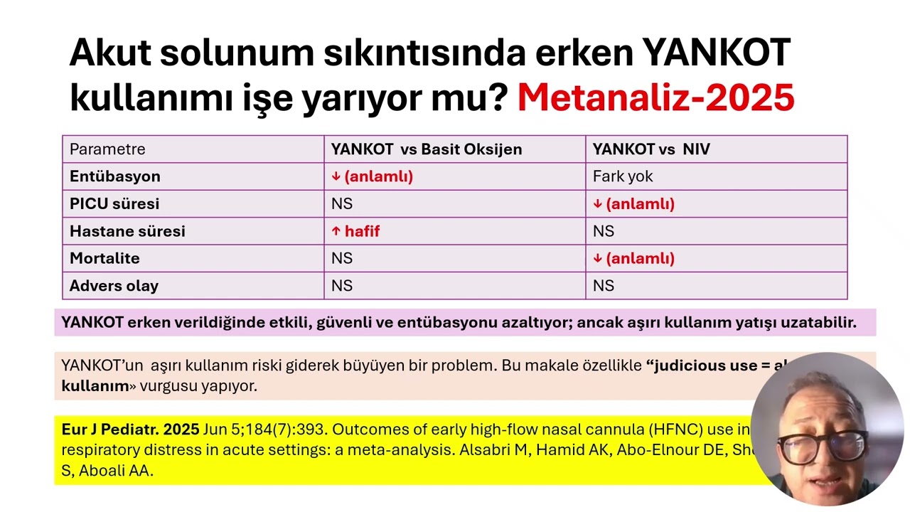
ÇOCUKLARDA AKILCI OKSİJEN TEDAVİSİ= YANKOT ile ilgili kanıtlar
297
HFNC shows benefits in neonatal and post-extubation care. A 2022 NEJM study of 202 premature infants showed HFNC significantly improved intubation success rates (83% vs standard care), providing effective pre-oxygenation and reducing desaturation risk. A 2023 Cochrane review in term neonates showed HFNC and NIV had similar outcomes but HFNC had fewer adverse effects (less nasal trauma, less abdominal distension). A 2022 Hacettepe University study showed HFNC and NIV had similar weaning success rates, ICU stays, and mortality, concluding HFNC is a safe and effective alternative. A 2023 JAMA systematic review showed NIV was most effective for post-extubation support, followed by HFNC, then simple oxygen methods. HFNC is a good alternative to NIV for post-extubation support with easier tolerability and lower sedation requirements.
Click to watch →

Weaning from mechanical ventilation - ผศ.พญ.พิชญาภา รุจิวิชชญ์
3K
Post-extubation support preparation depends on patient risk level: low-risk patients may only need mask with bag, while high-risk patients (history of reintubation, age >65, poor cough, COPD, or CO2 retention) may need NIV or HFNC. NIV reduces reintubation by approximately 7%. NIV contraindications include: inability to cooperate with mask, excessive secretions, airway obstruction, inability to protect airway, high intracranial pressure, recent upper GI surgery, or undiagnosed GI bleeding. For prolonged ventilation (>2 weeks), tracheostomy should be strongly considered as early tracheostomy (within 21 days) reduces mortality, VAP, and ICU stay duration.
Click to watch →
Deep Dive
Algorithms for managing 'difficult-to-wean' patients and tracheostomy-related weaning protocols.

WEANING 1 ( MECHANICAL VENTILATION) : DR CHANDRASHISH CHAKRAVARTY
3K
Mechanical ventilation weaning classification guides clinical expectations: simple weaning discontinues after first assessment; difficult weaning discontinues 2-7 days after initial assessment (most common concern); prolonged weaning cannot be discontinued beyond 7 days or requires >6 hours daily support. Prolonged weaning commonly results from neuromuscular weakness, neurological impairment, and severe lung disease. Early tracheostomy (<4 days) in medical patients lacks evidence of benefit; wait at least 7-10 days before considering tracheostomy for difficult cases. Tracheostomy is indicated for patients unlikely to be liberated within the expected timeframe. Neurological patient weaning requires special criteria beyond GCS: ability to follow simple commands, secretion clearance within 2 hours, good cough reflex (FEF ~-25 cm H2O), and gag reflex. Common causes of weaning failure in neurological patients include hypoventilation, secretions retention, and inadequate respiratory drive despite seemingly adequate GCS scores.
Click to watch →

Weaning from MV ( Tracheostomy weaning, difficult to wean and weaning protocol) . Dr. Jack khalil
342
Tracheostomy weaning requires a systematic protocol that includes cuff deflation, cuff dilation, and cuff inflation to assess the patient's ability to maintain airway patency and adequate ventilation, with the goal of determining readiness for decannulation while preventing complications such as airway obstruction or respiratory distress.
Click to watch →
9) Discontinuation of MV - Case based discussion - Dr Mohamed Torayah
728
Difficult weaning is defined as failed weaning attempts over 7 days. Prolonged ventilation (>7 days) carries significantly higher mortality and requires long-term mechanical ventilation or tracheostomy. Management requires: (1) Comprehensive review of underlying causes; (2) Optimization of comorbidities including thyroid function, electrolytes, and infection; (3) Minimization of sedative use (midazolam, propofol, ketamine); (4) Assessment of neuromuscular blockade; (5) Daily reassessment of weaning readiness. The prognosis depends on addressing reversible factors and optimizing the patient's overall condition.
Click to watch →

ISCCM Vodcast On Difficult & Prolonged Weaning from Mechanical Ventilation
538
A systematic approach to difficult weaning begins after the first SBT failure. The subsequent 24 hours are critical for recovery without repeating SBTs. Key interventions include correcting metabolic derangements (electrolytes, hemoglobin >7, glucose control), treating pulmonary edema with diuretics, and addressing ventilator dyssynchrony. After 24 hours, decremental pressure support trials may be attempted. Cuff leak testing guides extubation readiness, with post-extubation NIV or high-flow oxygen supporting vulnerable patients. Individualized care tailored to each patient's specific barriers is essential.
Click to watch →

Tracheostomy weaning
531
The tracheostomy weaning protocol progresses through stages: starting with spontaneous breathing trials (SBTs) on a T-piece, transitioning to a Passy-Muir valve (a one-way valve that allows air in but not out, enabling breathing and speaking simultaneously), followed by a cap that seals the tracheostomy to simulate decannulation; patients typically wear the cap for 24 hours to confirm they can breathe independently before actual removal, with the goal being decannulation prior to discharge for most patients, though some with extensive head/neck cancer requiring chemoradiation may need long-term tracheostomies.
Click to watch →
Weaning Criteria
Assessment Indices
Overall Readiness
Weaning Criteria
0:00- 1
Primary criterion is resolving the original reason for ventilation.
- 2
Assess four key areas: respiratory, cardiovascular, neurologic, and psychologic status.
The Overreliance on Predictive Weaning Indices vs. Direct Spontaneous Breathing Trials
While traditional weaning parameters like the Rapid Shallow Breathing Index (RSBI) and maximal inspiratory pressure have long been used to predict extubation success, a significant body of contemporary critical care research challenges their utility. Critics argue that these static physiological measurements have low predictive accuracy, often yielding false positives or negatives that can lead to premature extubation or, conversely, unnecessary delays in liberation from mechanical ventilation. Modern clinical guidelines increasingly advocate for minimizing the use of these traditional parameters. Instead, they favor a simplified, protocolized approach centered on daily Spontaneous Breathing Trials (SBTs) combined with holistic clinical assessment. This counterpoint suggests that a patient's actual performance during an active, trial-of-breathing challenge is a far more reliable indicator of extubation readiness than rigid numerical indices, which can overcomplicate the weaning process and prolong ventilator days.
Basic respiratory anatomy and physiology, including the mechanics of breathing, gas exchange, and lung compliance.
Fundamental concepts of mechanical ventilation, specifically the key settings such as Tidal Volume (Vt), Positive End-Expiratory Pressure (PEEP), and Fraction of Inspired Oxygen (FiO2).
Interpretation of Arterial Blood Gas (ABG) values and basic oxygenation indices, such as the PaO2/FiO2 ratio.
The clinical indications for initiating mechanical ventilation and the pathophysiology of acute respiratory failure.
The clinical protocol for conducting, monitoring, and evaluating a Spontaneous Breathing Trial (SBT).
Extubation techniques, immediate post-extubation care, and the prevention of post-extubation airway complications.
Strategies for utilizing non-invasive ventilation (NIV) and High-Flow Nasal Cannula (HFNC) therapy as post-extubation support.
Algorithms for managing 'difficult-to-wean' patients and tracheostomy-related weaning protocols.
92K views1.4Klikes4:23@MedMasteryOriginal Release: 2017-02-23
once our patient is on a spontaneous breathing trial for 30 minutes to 2 hours it's time to monitor weaning parameters that will help guide the decision of exavation now before moving forward according to evidence-based practice we should always note that the single most important Criterion to consider when evaluating for exavation is whether there has been significant alleviation or reversal of the primary cause for mechanical ventilation okay so success with exavation is related to assessing four key areas the assessment of the respiratory cardiovascular neurologic and psychologic status of the patient each of these areas can be assessed using a variety of indices but the challenge is sometimes choosing which indices to use for each key area for example to evaluate the respiratory status during a spontaneous breathing trial you can use the PF ratio aa gradient MIP vital capacity minute ventilation mvv rsbi the area occlusion pressure p0.1 and on and on and on in fact a comprehensive evidence-based review identified a possible role for 66 specific measurements as predictors of wiing success yikes that's a lot of predictors and unfortunately there are enormous discrepancies in the literature regarding their accuracy with regard to the prediction of successful excavation so let's see if we can bring some Simplicity to at least give us a general overview of how to assess for exavation first the respiratory status the clinician should evaluate the ventilatory work of breathing and the oxygenation status during the spontaneous breathing trial the ventilation status can be assessed using two generally good observations the first is the rsbi or the rapid shallow breathing Index this is assessed during 1 minute of unsupported spontaneous breathing with no pressure support and it is calculated using the patient's respiratory rate divided by the spontaneous title volume an rsbi of less than 105 May indicate successful excavation an rsbi less than 80 is associated with an almost 95% probability of successful discontinuation now another good observation is the absence of dnia and a respiratory rate less than 35 breaths per minute if the patient has an increased respiratory rate greater than 35 breaths per minute for more than 5 minutes the patient most likely will have a low probability of successful exavation now in assessing the oxygenation status the patient should tolerate an fi2 less than 40% and a peep of 5 to8 or less additionally if performing an ABG to assess the oxygenation status the clinician can calculate the PF ratio generally a PF ratio of greater than 150 on 40% of oxygen or less is acceptable next is evaluating the cardiovascular status during weaning the cardiovascular status usually is a hemodynamic assessment ensuring that the patient is stable with minimum or no pressures now this varies from institution to institution and from case to case next is the evaluation of the neurologic status generally the patient should should be awaken alert free of seizures and able to follow instructions and lastly evaluating the psychologic status fear anxiety and stress should all be minimized since these non-respiratory contributing factors may actually lead to prolonged ventilator dependence so with these four key areas in mind hopefully you'll be better equipped to monitor patients during weaning and ensure the best probability for successful exavation so I hope you like this video absolutely make sure to check out the course this video was taken from and to register for a free trial account which will give you access to selected chapters of the course if you want to learn how met Mastery can help you become a great clinician make sure to watch the about met Mastery video so thanks for watching and I hope to see you again soon
Up Next
7:07
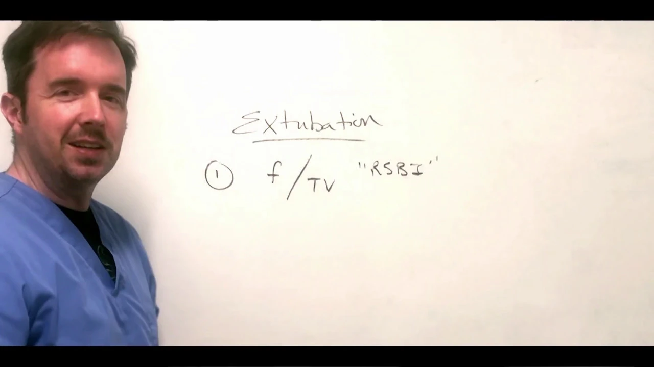
ICU Extubation Criteria: The 5 Key Factors for Safe Decisions
@IUCriticalCare
49.2K views•2020-04-11

48:47
Graphic Medicine: Comics for Collaborative Healthcare Communication
@nationalpatientadvocate
189 views•2023-12-04
3:43
Beta Blocker & Calcium Channel Blocker Toxicity Treatment
@MedMastery
8.9K views•2022-07-25

3:05
Stages of Labor and Vaginal Birth | Childbirth Animation
@nucleusmedicalmedia
52.1M views•2017-08-18
Related Study Plans & Knowledge Roadmaps
Structured learning paths in Medicine
medicine
Abnormal LFTs: Diagnostic Workup & Pathology
01Liver Anatomy and Basic Physiology
02Introduction to Liver Function Tests (LFTs)
03Hepatocellular vs. Cholestatic Patterns
+ 3 more learning modules
12h6 modules
Explore Roadmap
medicine
Analyzing Blood Smears: RBC, WBC & Platelets
01Introduction to Blood Components & Smear Preparation
02Normal Morphology of Blood Cells
03Erythrocyte Pathologies & Morphological Variants
+ 2 more learning modules
12h5 modules
Explore Roadmap
medicine
Mechanical Ventilation: Waveforms & Alarms
01Anatomy and Physiology of Breathing
02Introduction to Mechanical Ventilation & Core Modes
03Ventilator Settings and Waveform Interpretation
+ 2 more learning modules
12h5 modules
Explore Roadmap
medicine
Neuro Exam: Localization & Stroke Syndromes
01Foundations of Neuroanatomy and Vascular Supply
02The Neurological Examination: Step-by-Step
03Principles of Neurological Localization
+ 3 more learning modules
18h6 modules
Explore Roadmap