
















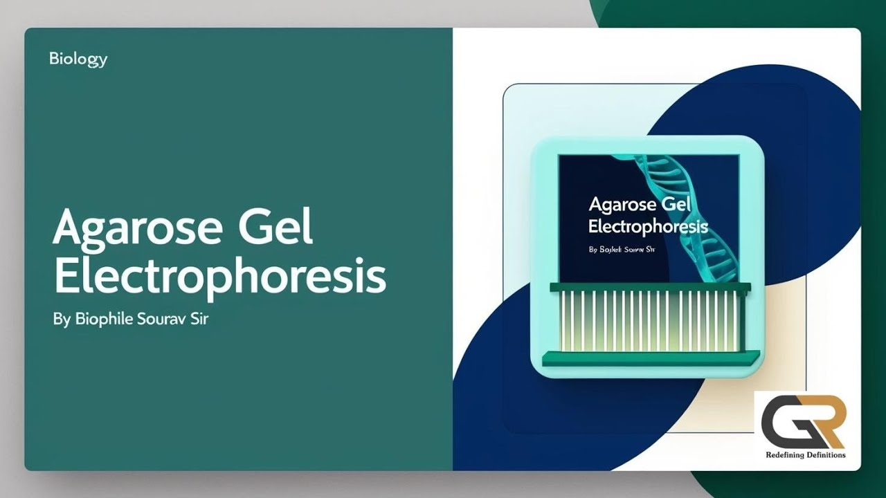





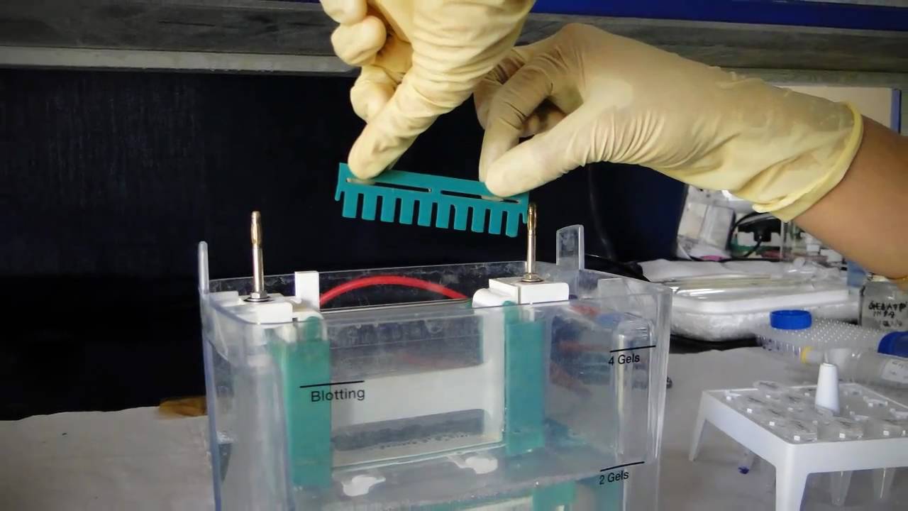


Agarose gel electrophoresis separates DNA fragments based on their size (length) because DNA is uniformly negatively charged due to its phosphate backbone, allowing smaller fragments to migrate faster through the gel matrix pores while larger fragments move more slowly; the gel is prepared by dissolving agarose powder in buffer solution, heating to 40°C, and cooling to form a semi-solid matrix with pores whose size depends on agarose concentration, and DNA bands are visualized after staining with DNA-binding dyes like ethidium bromide under UV light.
Gel Electrophoresis of DNA: Agarose Gel Procedure Explained
Added:Understanding the chemical structure of DNA, specifically its negatively charged phosphate backbone.
Basic principles of electrochemistry, including how charged molecules behave in an electric field.
The concept of molecular weight and how polymer structures, like agarose, can act as a physical sieve.
Fundamental laboratory concepts such as preparation of buffers and the importance of maintaining pH.
Methods for DNA visualization, including the use of fluorescent dyes like Ethidium Bromide or SYBR Safe and UV transillumination.
Downstream applications of gel electrophoresis, such as DNA gel extraction, plasmid mapping, and PCR product analysis.
How to use a DNA ladder (molecular weight marker) to quantitatively estimate the size of unknown DNA fragments.
Advanced separation techniques, such as Polyacrylamide Gel Electrophoresis (PAGE) for high-resolution protein or RNA analysis.
264.4K views8.8Klikes23:17Original Release: 2019-12-28
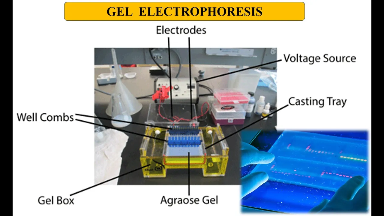
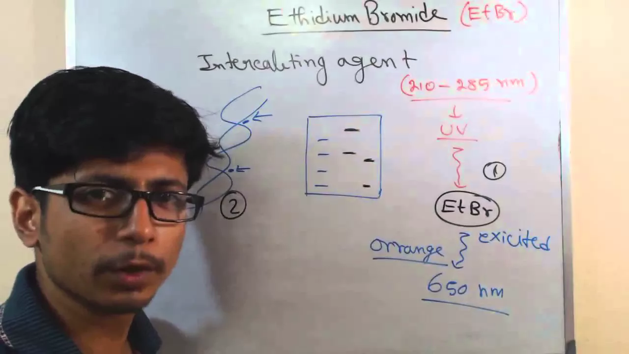
hello friends welcome back to another video tutorial from source value G and in this video lecture we want to talk about the gel electrophoresis now when I use this term gel electrophoresis it's a broad term can be used for many different purpose right but the gel electrophoresis of what the gel electrophoresis of a DNA the gel electrophoresis of a protein it can be up many different samples so in very simple terms I can tell you the gel electrophoresis is a process is a molecule ER technique to separate samples which can be DNA or proteins based on their charge and size that's the very simplest form of the definition ok so when we are saying that we can separate DNA and protein based on their charge or size now work things matter the most now think about it I told you that the electrophoresis can be done for the DNA or for the protein now in this series of video we are going to talk about two videos in one video we'll be talking about the DNA electrophoresis the second one will be talking about the protein gel electrophoresis but when we are using a gel for separating DNA from a mixture of DNA sample that case we use a separate gel that is known as agarose agarose gel and when we are separating proteins from mixture of proteins using a gel then we are using a different gel known as poly acrylamide polyacrylamide gel simply actor i am i D gel poly because multiple acrylamide are fused together with the help of hydrogen bonds they are connected together so two different types of gels are used to separate two different types of sample but the major gel electrophoresis principle remains the same now what is the gel electrophoresis principle okay let us talk about the basic first you probably know the basics so I'll just start with the basic very fast then I'll tell you the actual unique important features that you need to know for the exams so when we are talking about the electrophoresis as a whole the idea of electrophoresis is that separating a mixture of sample based on this charge and size so I write it down here so we are separating them based on charge and all size in this case it can be molecular weight as well right now let's say we have a mixture of proteins those different proteins they have different molecular weight so we need to find a way to separate them based on their molecular weight that means for the smaller molecular weight and then medium then large we can separate that for DNA the idea is different for DNA it's the length of the DNA based on which we can separate them right why because think about DNA the backbone of DNA is negatively charged DNA is always negatively charged all the DNA molecules are negatively charged so the only way we can separate DNA is with the help of with the based on their size means the based on their length okay this is the property that we use for for DNA for proteins they are of different shots you know the surface of the proteins they're made up with different types of amino acids there may be positively charged amino acids negatively charged amino acids there are hydrophobic amino acids and to fill each amino acids so many types of amino acids are there and we are not talking about a single amino acid to be separated we are talking about whole protein and the whole protein when it's folded form does not have any kind of charge distribution like that on the surface right so in order to separate proteins with the help of gel electrophoresis we need to use we need to unfold them this is another very important thing unfolding is needed and then after unfolding we use the process of genetic references to separate them again based on size itself okay we'll see those process in details later on so what happens in electrophoresis particularly in this to talk about the DNA in gel electrophoresis which is also known as the agarose gel electrophoresis now to separate DNA from a mixture of different lengths of the DNA we can separate them using agarose gel electrophoresis method that means we need to create a gel matrix okay through which the DNA samples or the fragments of the DNA can be transferred can be moved like they can move through that matrix okay and based on their length their movement varies so for example let me draw the idea of the gel let's say this is how the gel looks like you know you probably have seen this picture a lot earlier and there are this solid structure now gel we always a jello like structure we all know gee Latino jello like structure solid substance semi solid kind of substance so normally the agarose that we get we get it is it as a powder flakes format of the powder format of white white format and then we put it into the buffer solution we generally don't use water for that we use buffer solution we we heat it up to 40 degree Celsius remember that we heat it to till 40 degree Celsius then we cool it down and when we cool it down it forms a semi solid jello like structure that is known as that or Rose gel okay so gel is done while we you know there is a chamber chamber like this where we put the liquid version of the agarose which is dissolved in the buffer and then what we do here is we also put a comb the comb looks something like this this is how the comb looks like okay so let's say you put this comb like this while we add the liquid and then when it solidifies we take the comb up so three small wells are created which is known as wells see small wells are created three four ten twelve twenty how many number of wells you can create with the help of the comb you can create that depending upon the sample that you want to run for the gel electrophoresis so these are the waves so wells are created and once wells are created then what we do the jelly solidified we put this gel under a chamber we put it under a chamber and this chamber is connected with electrodes remember that okay so this chamber is connected with electrodes one here and one from this side okay so let's say in this side the electrode is negatively charged in this side the electrode is positively charged here we are talking about a gel which is placed like this vertically okay vertically in some other occasion most of the time actually the chamber is placed something like horizontally and let's say this is where the well is present and this is negatively charged this is positively charged this is known as horizontal gel electrophoresis where the gel electrophoresis chamber the electrophoresis chamber is lying down in the horizontal plane and the direction of movement will be horizontal from the negative side to the positive side that means from from the cathode towards the anode okay that will be the movement same thing here this is an old and this is cathode from the cathode towards anode but this is a vertical gel when we put the gel block vertically that's a vertical gel so vertical gel electrophoresis horizontal gel electrophoresis now the thing is in both these cases they are placed inside a chamber obviously there will be a chamber inside okay and in the chamber is filled with buffer again they're filled with buffer now what is a buffer you all know what buffer is buffer can maintain pH a constant pH throughout it because it's pH is very very important in terms of maintaining the structure of the DNA as it is of the protein as it is because you are not going to change them during and while we are running the experiment so it's some salts comes the solutions under some salts under the distilled water that's how we prepare the buffer Tris buffer zdt different kinds of buffers are used which is glycine buffer if you've seen many other buffers are used now generally the buffer pH varies okay for for DNA for protein there are different ideas of the buffer that we use now the thing is here we filled it with buffer and then we load our target then let's a mixture of DNA where we have small fragment as well as large fragment okay so we just load our DNA's in the chamber and why we load them in the chamber along with that we load like one chamber we keep one chamber as what is known as a ladder or yardstick we can call it as a ladder ladder means where we put the fragments of the DNA of known length okay so that we can compare it with the length of the target DNA and can find out the approximate length of our target DNA by comparing it with the ladder okay so this is where we put the ladder and then what we do with applying electric field we apply the electric field so minus plus why because you know DNA as I told you earlier the bad mode contains phosphate phosphodiester bond is there so phosphate are negatively charged so DNA as a whole is negatively charged so the DNA will always migrate towards up to end so the DNA will always migrate towards Catford I mean sorry now anode DNA will always my migrate towards the anode because DNA is negatively charged so as the DNA is migrating towards the anode why you know what happens in the matrix is that why the gel solidifies the gel is heated to 40 degree Celsius then solidifies in a cooler temperature while the gel is solidifying the components inside the jail for example in this case a agarose they are forming what is known as Network or mesh like structure like a knit mesh like structures so I can draw the structure something like this let's say let's say like this miss like structures so when we form the Miss like structures what we have here we have pores you can see these are pores okay so this pores are present in the network okay now that is the beauty that is the real principle behind any electrophoresis is to use a material to form the matrix that can create pores of specific size now as the smaller DNA can easily move through the pore so they will reach the end faster but the large or linear DNA longer DNA cannot move through the pore quite easily so they will move slowly so the large DNA will move slowly the small DNA will move fast as a result of this differential ability of their movement we can we can easily find out their differential position in the agarose gel plate that is the main principle behind agarose gel electrophoresis now you may ask me a question now while forming this network or mesh-like structure with agarose how we denote like how do you know what will be the pore size it depends on the concentration of agar that we are using we should not use a very high concentration normally five six seven percent eight percent ten quill percent depends on what kind of DNA we are sampling what kind of DNA we are separating based on that we separate we create the concentration in maintain the concentration of the agarose in the matrix formation okay sometimes we need very high concentration of agarose because the more concentration you provide the better and very fine pores you can produce so if your DNA samples are very very very very tiny all of them are very tiny then you need to use very concentrated version of the agarose and if your DNA is bigger one you need to use lower concentration of agarose but your concentration of agarose must not drop below certain critical concentration beyond which no polymerization will be formed okay that's another very interesting point okay this is another very interesting fact so now think about it the network is created we load the DNA and now we run the gel what we know is that in the ladder one we have no sequence okay so we have what is known as the DNA they move from different places and what they give us is what is known as the banks now what we mean by banned while we apply the voltage the DNA start migrating from minus to the plus end and as they are moving we continue it to somehow because you know how many like how many hours what is the time it depends on again what kind of concentration of agarose gel you are using okay how many what kind of sample you are using depending upon that but we always keep a track of the final dye because what we put there we do not only put the DNA we also put us die in the DNA so that we can see the movement we can see the attract the movement of the DNA head from the top to the bottom it is very important to monitor the movement of the DNA because if the DNA you know if we keep this voltage on for too long time for a long time then what happened that this DNA can run out of the gel and that will not be good for the experiment the whole time and all the effort will be wasted for that reason we add some dye and as the dye moves we can easily see the tracking of the movement and when the dye reaches almost like little bit of we keep a little bit of buffer zone from the bottom part of the gel so ideally a gel should end near about like 1 inch 2 inch 1 inch track to the end of the gel so that's where when the dye reaches we stop the electric current we stop the current DNA will stay at their own places but that will not give us the band remember that color is already pre applied color just to see that and track the movement that that does not mean that DNA only came here because you know only see the DNA one one colored band in that case only here at the end that's not the band that is the only the colored copy of the DNA then what you need to do you need to stain this deal will to take this agarose gel out of from the chamber and to open the chamber and release this slab of gel out with the help of a spatula and then you tag it with a dye when you tag a DNA binding agent you can use any kind of DNA intercalating agent in there okay when we use that that that agent will bind with this for example in the-- diem bromide that binds to the DNA okay so III diem bromide is one example of such kind of dye that binds to the DNA which is the intercalating agent and once i attach to that then you can put this dyed slab of gel under UV and you are going to see the position of the DNA that will be known as the bank so then you can see the bands okay so do not think the band at the end of the running jail okay that's not the band but at the end when you see under UV chamber or gelled off chamber then you can see the band and let's say the band is somewhere and what we can see is that normally the band that we can also observe for the ladder we already know the sequence we know the the smallest one will go here so one hundred let's say two hundred three hundred four hundred five hundred six hundred base pairs for example for simplicity okay generally it's more than that but let's keep it like that so the other way let's say you got a band here and here and in this case you get a band here and you get a very big man very broadband in here so this kind of data you can achieve now once you see this data what you can get to know approximately what will be the length of this band you see this band is very close to 300 base pairs so near about 280 290 some sort of like that and this one is you can see near about 400 so you can think about it I mean sorry if this is not 290 this can be 310 320 is something like that and this is again C 80 or something like that okay so these are the type of band let's say you got another band in here then you can say this is a 500 base pair brand by comparing it with the ladder sequence the known sequence and here we have another band again 500 this is 500 base pair because it's exactly the same length travel the same length like the known 500 base pair fragment so this is 500 base pair length of the DNA fragment when we see a broader band what does that mean that means a lot of the DNA that are present in the mixture meaning of the DNA they belong to the similar range they belong to 320 to 350 base pair range that's why you get a very very thick band very thin band means less number of DNA thick band means more number of DNA now you may have one more question then when we see the banks the number of band corresponds with the number of DNA now answer is no not at all for thousands of DNA you may get one band for hundreds of DNA you may get one band so for example like this thick band means there are many DNA of similar fragments lengths are present together okay thin band means there are less DNA but it does not mean the only one DNA fragment no single DNA can not be visualized as a band in diagonal gel that kind of resolution is very very hard to achieve so generally when we see a band generally there are multiple fragment of the similar length present together okay so that is about the agarose gel electrophoresis for the DNA now the question is one more question that I want to ask an again answer we load this chamber with buffer you know the salt solution so the question is why we load this chamber with buffer why not we use water simple water or distill what I even now the answer to it is that there is a conductance of this okay because the DNA need to move from the minus end to the plus end there is a conductance required for the process and for the conductance electrolytes play very vital role and salts continue electrolytes electrolytes play a vital role of bringing the DNA or any other sample that is present in your proteins from the cathode towards anode in this case so if you use only water it's not going the dad gel with water is not going to be here exactly like the way it will behave if you use a buffer okay that's why I always need to use buffer for that same process it's very very important to use it so this is the idea of agarose gel electrophoresis and DNA gel electrophoresis as a whole because for DNA gel electrophoresis we can easily separate DNA based on their length the size without thinking of their charge because they are all negatively charged that's quite easy to do with the help of agarose gel okay one very important question that many times you may receive during bye-bye is that in other is gel electrophoresis we always use for the DNA separation and why we should not use the polyacrylamide gel electrophoresis for DNA because acrylamide is kind of universal matrix creating polymer that can form and as a result of which we can get a very good resolution for the protein separation so protein separation is well done with poly acrylamide but for DNA separation why we cannot use poly acrylamide and why we always use agarose gel now what is the answer to it the answer to it is that the poly acrylamide you know there is a particular concentration there is a particular concentration of these monomers needed and to better help separating the sample or for a better resolution of the separation of the sample and that concentration is the effective concentration of either agarose or poly acrylamide whatever matrix molecule be used so there is a fixed concentration of critical concentration needed that is enough to be solidified and to be able to separate the sample but if we use poly acrylamide for DNA separation dealing has a large molecular weight and that is near about over 1 million dalton 1.5 million dalton so for this high molecular weight components the poly acrylamide needs to be of a very less concentration so for that less concentration of poly acrylamide the poly acrylamide can never become solid so poly acrylamide will remain liquid which is needed at the concentration needed to separate molecules like DNA that is the reason polyacrylamide is never used for the DNA separation it is only used for the protein separation even if you take a protein which II which has the molecular weight more than that one media and I'll turn polyacrylate actor element cannot separate it that's all the only reason we should not use poly acrylamide for the DNA separation because it simply cannot do that because if you take even very tiny concentration of poly acrylamide which is solidified you load DNA into it you'll see DNA no movement across the poly at relevant at all okay so that is another bonus tip that I give you if they ask you any question from that particularly in the PhD viber you can answer that so if you liked this video please hit the like button hit the subscribe button and also click the bell icon so that you get notified whenever we upload a video and also subscribe to our channel and share this video with your friends thank you bye [Music]
Up Next

3:46
Polymerase Chain Reaction (PCR) Animation: Step-by-Step DNA Amplification Technique
@shomusbiologyofficial
347K views•2016-06-11

50:12
Bacterial Communication: Quorum Sensing and Biofilm Formation Explained
@JHUAAP
12.6K views•2011-06-28

14:34
RNA Interference Mechanism: siRNA and shRNA Pathways Explained
@shomusbiologyofficial
379.7K views•2015-02-21

7:08
Bacteriophages: Earth's Deadliest Killers and Future Antibiotics
@kurzgesagt
34.6M views•2018-05-13
Related Study Plans & Knowledge Roadmaps
Structured learning paths in Biology
biology
RNAi: Biogenesis, RISC & Gene Silencing
01Molecular Biology Foundations: DNA to Protein
02Introduction to RNA Interference (RNAi)
03Molecular Biogenesis of miRNA and siRNA
+ 2 more learning modules
10h5 modules
Explore Roadmap
biology
Mycorrhizae: Signaling, Biophysics & Ecology
01Foundations of Mycorrhizal Symbiosis
02Molecular Signaling and Root Colonization
03Biophysics and Bioenergetics of Nutrient Exchange
+ 1 more learning modules
11h4 modules
Explore Roadmap
biology
Circadian Clocks: TTFL, SCN & Periphery
01Foundations of Molecular Biology & Gene Expression
02Introduction to Chronobiology and Actograms
03The Core TTFL: Molecular Architecture of the Clock
+ 3 more learning modules
14h6 modules
Explore Roadmap
biology
Population Genetics: Math, Drift & Selection
01Genetic & Probability Foundations
02The Hardy-Weinberg Principle and Baseline Models
03Mathematical Modeling of Natural Selection
+ 3 more learning modules
18h6 modules
Explore Roadmap