
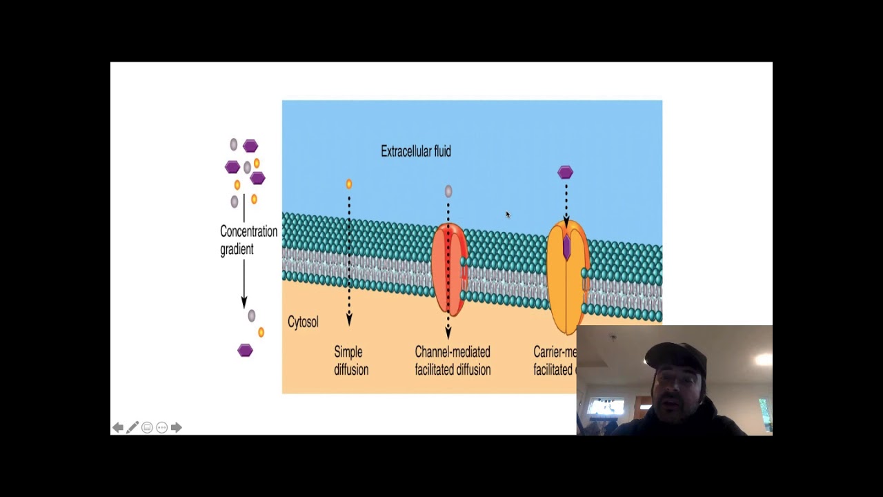
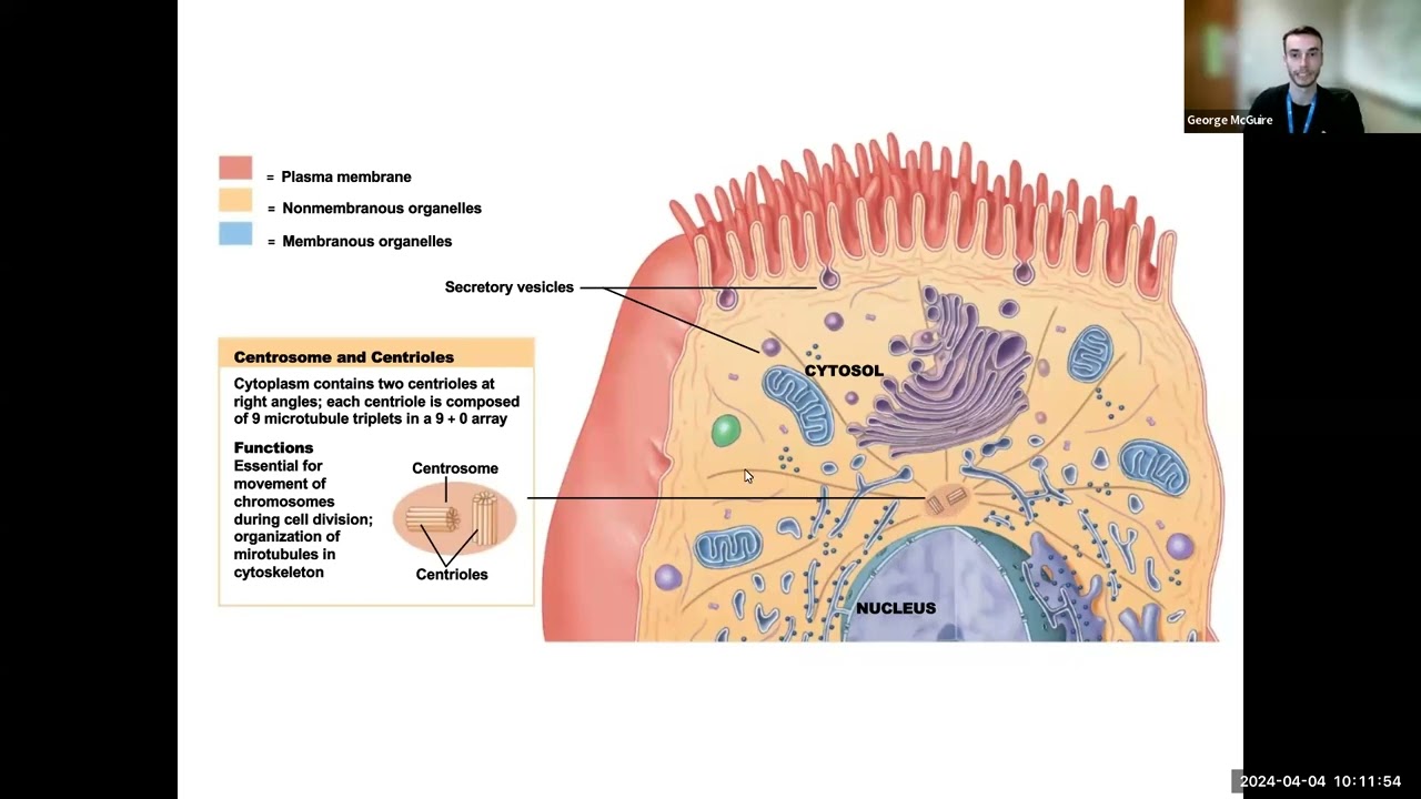

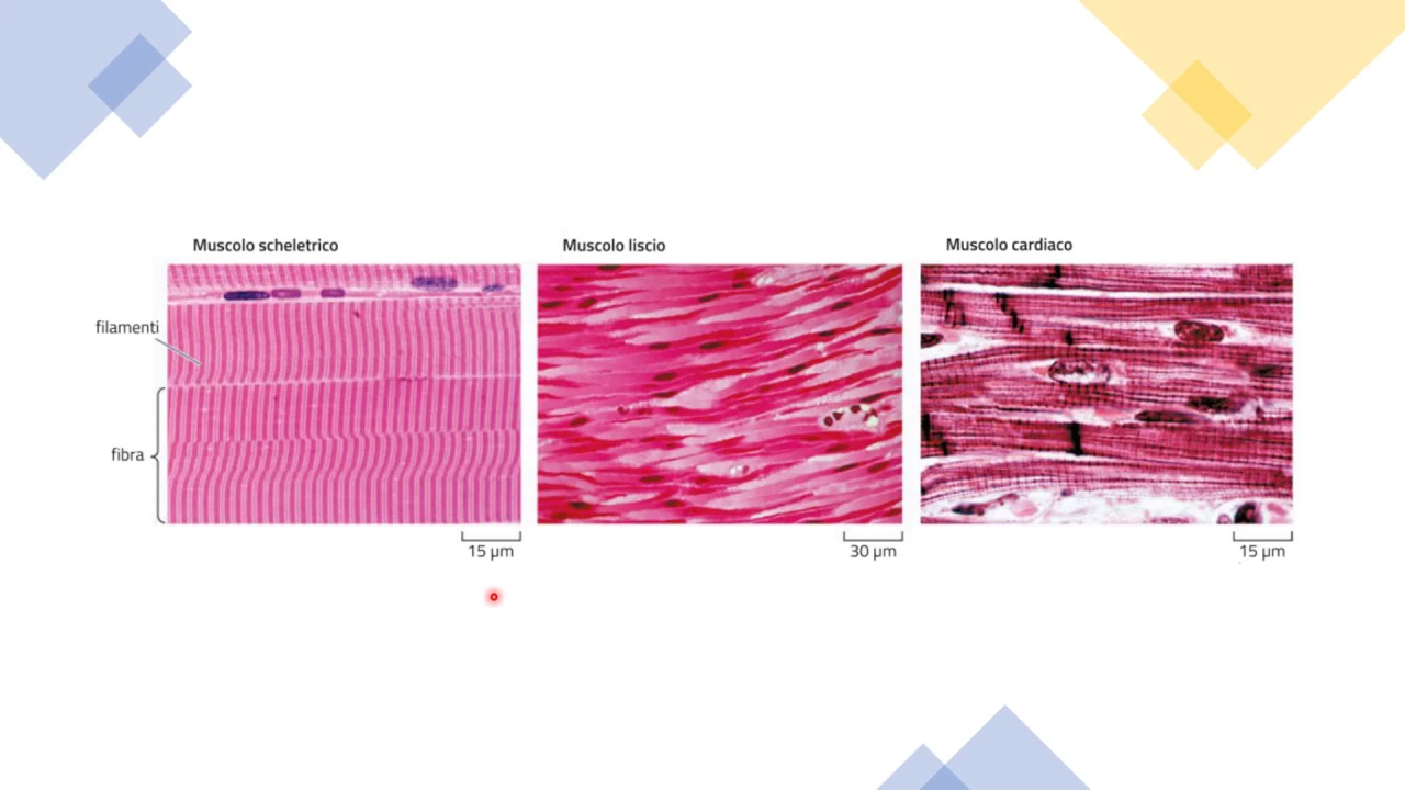

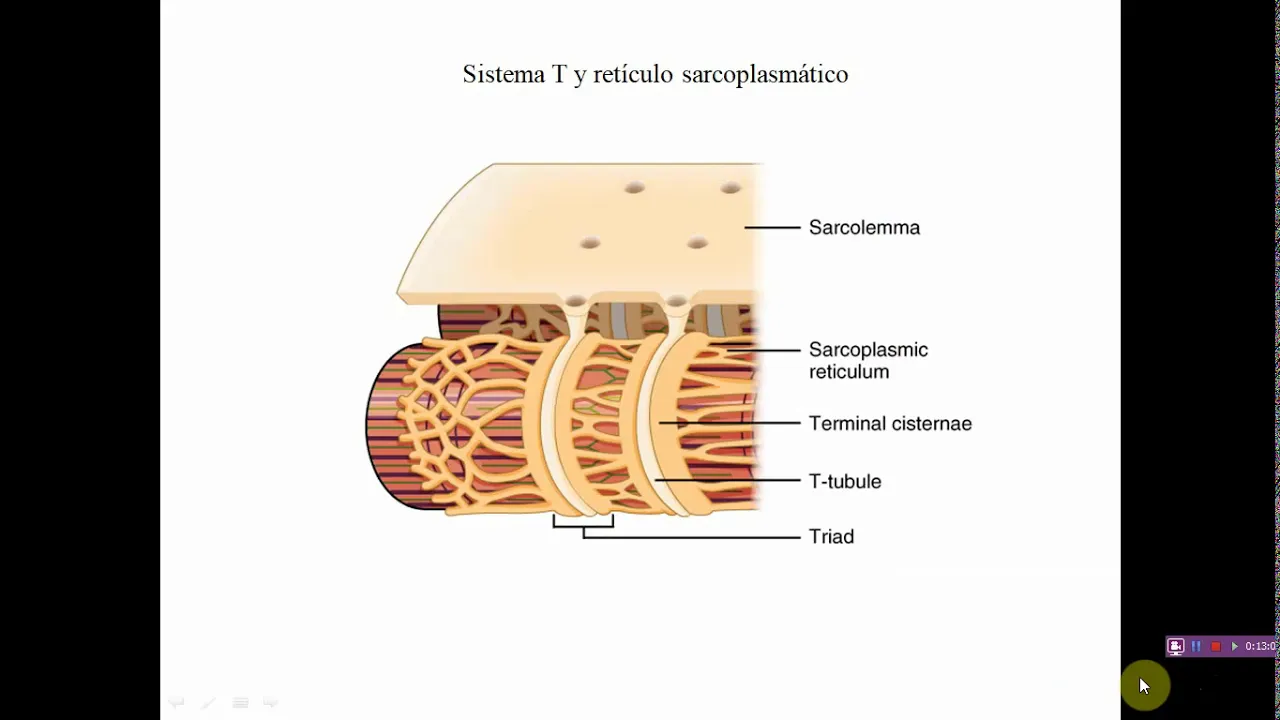















This comprehensive NEET PG revision session by Dr. Soumen Manna covers essential physiology topics including cell signaling types (paracrine, autocrine, endocrine, juxtacrine), membrane transport mechanisms (primary active, secondary active, facilitated diffusion, simple diffusion), thermoregulation and fever mechanisms, muscle contraction physiology (skeletal vs cardiac muscle, electro-mechanical coupling, tetanus phenomena), sarcomere proteins (titin, nebulin, alpha-actinin), touch receptor physiology (Merkel cells, Meissner's corpuscles, Pacinian corpuscles), receptor adaptation and accommodation, Darrow diagram for fluid balance, nephron physiology (substance profiles, tubular fluid tonicity), cardiac response in transplanted hearts, heart sounds and aortic pressure, PV loop interpretation, lung compliance, and GI smooth muscle physiology.
NEET PG Physiology Rapid Revision Marathon with MCQs | PrepLadder
Added:[Music] thank you thank you foreign [Music] [Music] [Music] [Music] I hope everything is Audible and visible at your end so this is Dr showman Mana your physiology teacher good evening to all of you yes I think everything is perfect at your end audio and video right good evening to all of you I'm not taking names but I can find out many joining very good right thank you so much so without wasting much of the time let's start the session right perfect okay shallow so ah we are going to discuss the important images of physiology so this is an image based question session I'll discuss few images which are which has already been asked in the mcqs or they may be asked in future right but before starting the session first of all I will give you a glimpse that what are the important topics you should concentrate in physiology because you know key I know that there is a crucial time you do not have much time you have to revise many subject all the 19 subject you have to revise so in between that 19 subject revision for physiology uh is a very important part but you cannot devote much of the time for physiology I know right so what are the important topics that you should concentrate for physiology so these are the first thing right so General physiology I have written some topic okay so these are most important topic in general physio for upcoming neat entrance examinations you have to revise at least this topic similarly the normal muscle physio see I have written only few topics in numbers and physiology so at least please revise this topic before going into examinations similarly in central nervous system epsp that is the excitatory postsynaptic potential right inhibitory postsynaptic potential motor in potential and rinser cell inhibition a very very important topic and all of you know the muscle spindle and Golgi tendon operators apparatus along with the tactile receptor and the adaptation phenomena of the receptor all this thing you have to read and in hypothalamus particularly the nuclei of the hypothalamus and their functions these are the important topic similarly in git at least you read migratory Motor complex and basal electrical Rhythm right and for CBS C very least a very less than number of topic I have noted here at least you read jvp preload after load and PV Loop okay so these are the important area from where the questions have been asked not only in Need for ini site examination also similarly you have to read about the baroreceptor Cushing reflex Bainbridge reflex and visual Jerry's reflex okay and in renal physiology the list of topics are here in respiratory the list you can find out here and in endocrine only the hormone receptor is the most important thing that you have to revise before going into examination right so I think right so I think they will check and the volume will be increased soon but otherwise video and audio is taken up right okay PDF of this okay obviously it will be provided don't worry so now after discussing this important topics we are entering into the direct discussion of the mcqs so I will give you McQ first you will look at and try to answer as first as possible because you know there is a time lag and we have only two hour session right so first McQ is coming in front of you this is a repeat McQ in net entrance examinations I think all of you know the answer of this so here you can find out that two cells has been shown and in one of the cell is secreting certain product and that product is working or acting on the nearby cell adjacent cell so obviously this kind of signaling is known as paracrine type of signaling okay all of you are giving great good answer right and obviously this is the correct answer paragraph signaling so what we have to read in this topic we have to read that there are four types of signaling I have to remember one is endocrine one is autocrine you know this and one is paracrine and one is juxtagrine so basically if you look at this endocrine and autocrine signaling from the name you can easily make out the what is the meaning of the signaling say endocrine signaling means if the gland is here the gland is secreting some product the product will travel through blood and it will work on a distant organ so it is traveling through blood that is the most important thing in endocrine type of signaling right in autocrine you can easily understand Auto means it is acting on the cell same cell right so the product is secreted from the cell and it is acting on the same cell onto the receptor the typical example of this autocrine type of signaling is paf okay so platelet activating Factor this is one of the important autocrane signaling example that you have to remember now look at this two important thing one is paracrine and one is juxta crime okay according to the meaning both are almost same but there is a little difference between these two paracrine means when a cell is secreting some product and the product is going to the nearby cell by the process of diffusion right two cells are located near to each other cells secretion from one cell is acting on the nearby cell by the process of diffusion so it has to struggle the product has to travel but not through Blood by the process of diffusion only but in case of juxta crime the cells are too close with one another so there's two cells they are so closed with one another that the ligand from one cells is directly interacting with the receptor on other cells getting my point so here the cells are in close contact so juxta crime means the cells should be in very close contact so that the surface receptor from one cell is interacting with the ligand from other cells the typical example of this kind of thing is that ah the transforming growth factor beta and Alpha right so transforming growth factor Alpha is one of the important examples of this juxta current type of signaling now paracrine signaling examples many examples are there all of you know that somatostatin if you think of somatostatin at the level of so somatostatin by the process of diffusion goes towards the parietal cells and it inhibits the secretion from the parietal cell so that is a typical example of paracrine signaling this much is important in this question I am going to the next question so next question is here ok look at the questions and try to answer right so I'm waiting for answer the question stem of the question is a little linear but I think this is ok ok so three different types of Transporter has been given in this question one is X one is y and one is z right and the questions is saying that the cells are cells contain the concentration of the iron which is similar to the mammalian cell concentration means you have not changed the composition of the cell intracellular and extracellular means sodium concentration calcium concentration everything is same ah like that of the normal mammalian cell yes so as you can find out here the first the transporter X it is throwing sodium out of the cell and taking back potassium inside the cell so this is typically the sodium potassium a t pH pump sodium potassium atph bomb right getting my point so sodium potassium atph pump this is an primary active transporter but this is not your question the question is regarding y so y here is throwing calcium out of the cell this is against the concentration gradient and taking in the sodium along the concentration gradient so this then typically the antipor system right so all simple and antipor they are which type of Transporter secondary active transporter so please note down that this y transporter is nothing but the secondary active transporter because all the simple are an anti-porter they are secondary active transporter if the molecule name is attached with atph remember if the molecule name is attached with atph suppose hydrogen potency phase hydrogenity phase all of them are primary active transporter right now looking at these two facilitated diffusion and simple diffusion all of you know that diffusion is a passive process it will occur from high concentration to the low solute concentration but if the diffusion is occurring with the help of a carrier protein then that type of division is known as facilitated diffusion what I told facilitated diffusion means it is a passive process but you require the help of what you require the help of carrier protein right you require the help of carrier protein but whenever you look at this trans Z here Z what is z what do you think if I ask rather than y what is z then what will be your answer within this ABCD um yes what will be Z is a z is a carrier protein no Z is not a carrier protein what I can find out here yeah very good very good so what you can find out here the Z content are lumen through this Lumen or you can say there is a pore through this pore one molecule can goes in bi-directional manner right so this is typically of a channel this is typically of a channel and you know channels are nothing but they are simple Channel through if the something is moving through channel that is a simple diffusion process right so look at here if you do not believe me then you look at here this is the diagram I have taken from diaton so here you can find out that this is channel protein channel proteins are put into the category of simple diffusion right so basically you have to understand that there is a difference between carrier protein right there is a difference between carrier protein and the channel protein right so in case of channel protein there will be an internal pore inside the protein and through this internal pore the molecule can move without any actual binding with the protein but if you look at the carrier protein see here if you look at the carrier protein suppose it is transporting sodium and potassium or suppose it is transporting sodium and calcium so first the sodium will binds right then the carrier protein will change its shape right then after just changing in shape the sodium of the calcium which are attached with it it will be released on the other side getting my point suppose this is calcium same way if it is transporting sodium first the sodium has to attach to this carrier protein and then the carrier protein will change its shape and after changing the shape it will release the sodium from other side so the bottom line is that carrier protein means it is actually binding with the substances which has to be transported and then it is changing its shape and then it is releasing the molecule but in case of channel protein neither it is changing its shape neither it is actually binding with the substances which has to be transported so all channel protein mediator transport they are simple diffusion process simple diffusion can also occur without protein also because you know if the substances is lipophilic substances like oxygen carbon dioxide gases here they can easily move without any channel without any carry protein that is also a simple division so basically whenever a carrier protein mediated transport is occurring it can be energy dependent also it can be without energy also non energy dependent transport also so carrier protein can be involved in energy dependent process also and non energy dependent process also if it is energy dependent process then it is an active and if it is non-energy dependent process or belages and passive but this passive is nothing but the facilitated diffusion we say that all the glutes all the glutes are facilitated diffusion but all the supporter and anti-potal like for example if I say sglt these are the secondary active transporter at least you remember this much of information for this kind of question this is a diagram which has been given in Guyton so you can find out here here the carrier protein is responsible for facilitated diffusion also as well as active transport process also am I clear right so we are moving on to the next question so the next question is the repeat questions of neet 2022 examinations try to answer the question so here ah whenever you get any graph in your exam OK physio foreign image based question means most of the questions will be graphs ok so whenever you are looking at any graph in physiology what you are going to look at first you look at x axis and then you look at y axis and then you try to understand don't look at what is happening inside the graph because here you can find out one broken line blue color line one red color line whenever you are seeing this kind of images first time in your exam you will get confused oh my God I have not seen this kind of image in my life one is broken line one is red line so do not do like that first you look at x axis look at Y axis so Y axis here is the body temperature so if I follow the blue color line blue color line is saying to me that body temperature is rising suddenly and if I look at the red color line on the first half it indicating the body temperature is rising but slowly this is the meaning of this two graph if I have to explain For the First Time OK suppose I do not know anything about this graph so what I will understand from this blue blue color line it is indicating that yes Blue Line indicating that the temperature is suddenly rising to a higher value and red line indicating that the temperature is rising to a a slow manner ok so let us look at how it happens so all of you know that hypothalamus is the main Center which is responsible for thermoregulation right so I am writing here hypothalamus hypothalamus OK it contain various nucleus one of such nucleus control our thermoregulation process now hypothalamus has a set point what do you mean by set point set point means hypothalamus will say that the body temperature will remain at this temperature now that is known as the set point set point can increase decrease depending on the environmental temperature or depending on difference parameter but set point means hypothalamus is setting a temperature as a particular value so when the body temperature is normal all of you know the body temperature is in and around 98.8 degree Fahrenheit okay suppose hypothalamus has already set the body temperature to 98.8 what will happen in this condition the whole body whole body will remain at the same temperature that is 98.8 degree Fahrenheit ok now suppose due to release of various pyrogens you know pyrogens obviously you know so pyrogens are the substances which can causes fever like for example interleukin one is one of the important pyrogens so whenever this Pi regions are released right what will happen this pyrogens will act on the hypothalamus and it will it will suddenly rise the set point of hypothalamus suppose now the set point is set at one zero three degree Fahrenheit so who is rising the set point who is changing the set point here the pyrogens it can be changing other means also but here the pyrogens is rising the set point of the hypothalamus but look at your body temperature your body temperature is still at 98.8 degree so hydrogens has suddenly rise the set point but your body temperature is still at 98.8 now what the body tissue will try to do body tissue will also try to rise in temperature to match with the set point means body temperature will also rise and it will try to match with 103 degree Fahrenheit but how to increase the body temperature body temperature increment cannot be sudden it has slow process why slow process now you either have to increase heat production into your body so either you have to increase heat production into your body or you have to decrease the heat loss from the body so that's why whenever your set point is suddenly rise to a higher value at the level of hypothalamus body tissue will try to generate more and more heat and it will try to prevent heat loss as much as possible so what is going to happen how can you do that you know that all the mechanisms which happens in your body when you expose to cold environment like your curling of the body okay shivering of the body OK pilo erection of the body all this thing will happen and that will generate heat slowly and whenever the heat production is sufficient it will match with the hypothalamic set point temperature that is why if you look at this curve OK the a indicating that the body tissue is gradually generating heat to match with the set point change so this blue color Point already given in the question this is the set point temperature has been raised and now the body is try to reach to the set point by producing more and more heat production so now you look at even if you don't understand the other part I will come to that but even if you don't look at the other part what will be your answer which of the following a b c d may say which of the following is ah ok is a heat production mechanism right so obviously answer foreign so that is why my answer here is savoring right then what will happen you know that this is the thing which is happening when the fever is coming right now what happens when the pyrogens are not there in the body hypothalamus again will decrease the set point ok now suppose the pyrogens 8.103 method now this Set Point again come back to 98.8 degree Fahrenheit normal temperature but your body tissue already it is at one zero three degree Fahrenheit but set point has suddenly decreased to 98.8 again your body temperature is 103 degree Fahrenheit so what the body tissue will try to do to match the set point now the body tissue has to lose the heat from the body so that's why there will be loss of heat from the body OK and whenever you want to lose your heat from the body yes there will be sweating there will be vasodilation so there will be vasodilation the skin blood flow will be increased your skin will looks like warm okay there will be huge amount of profuse amount of sweating ok so whenever this kind of thing is happening was a as a doctor you know now the fever is subsiding right so this is the part first part there will be chill OK chill and riger and this is the second part when the fever is finishing that time there will be flash reaction there will be flash reaction just come to both the crisis reactions so flush or Price is Right so you know whenever your fever is coming for the first time this is the first thing which is happening and when you fever is subsiding from the body the second thing is happening that is the flush or crisis your body vasodilation will be there your body will be profusely sweat so that is why you look at this is the exact graph which is taken from chitin and this has been put into the question so on the first half side this is the a in your McQ okay what is happening there is chills right so the body is trying to generating heat vasoconstriction pile erection epinephrine secretion and savoring right and when the crisis reaction is happening the body is try to lose heat by vasodilation and sweating something mayor so this is the thing that you have to remember okay okay so I think discussions for giving a rest to your eye for a few seconds so as you know he pre-bladder is ready for next preparation right and all of you know that next is round the corner so our team has um has is already prepared the question Banks as well as the video lecture and there is an elite plan for next entrance examinations right in this elite plan you know what you are going to get you are going to get all the video lectures the updated video lecture of all 19 subject not only in the visual lecture there will be question Banks which are Yeah question Banks which are completely next type of question bank which contains 70 percent of the clinical waste question along with that you know there will be next Edition test series also there as well as there will be the rapid revision course in this plan so if you are opting for this plan that is the elite plan for next entrance examinations so you can book here from 21st January okay so this is a short brick now again the next question so now we look at this question ok so looking at this question it is a detailed question but read the question first and try to answer okay answer yeah okay so there is a depolarization ok depolarization means this is an action potential after the action potential muscle is Contracting and relaxing okay all of your thinking if you look at the options now this question is easy but apart from options I want to understand that what is the meaning of this graph right so first of all all of you look at so the first this green color curve this is the depolarization depolarization means suppose you are generating one action potential on the muscle membrane what will happen the muscle will contract and relax so this blue color and red color curve both are indicating that there is a contraction so the contraction is going on here and followed by after the end of contraction there will be a relaxation this is the relaxation part so this is relaxation part and this upper part is the contraction part OK hold on OK I will tell you the answer so A and B both are muscle contraction and relaxation to understand this thing suppose this is my muscle suppose this is my muscle skeletal muscle and this is a neuron okay this is the neuron which is connected to the muscle if I stimulate the neuron now don't try to answer look at the explanation very important question so if I give a stimulus here if I give a stimulus here there will be Action Potential on this neuron then that action potential will travel down the neuron and the action potential will cross the neuromuscular Junction then after crossing the neuromuscular Junction the muscles membrane will produce another action potential and that action potential will causes contraction of the muscle obviously after contraction there will be relaxation of the muscles if I record this contraction and relaxation of the muscle then in graphically how it looks like it will looks like that there is a phase of contraction where the tension where the force generated by the muscle is rising so this pink color curve is the tension generated by the muscle or the force generated by the muscle that is the phase of contraction and then there will be a phase of relaxation there will be a phase of relaxation where tension is decreasing so this is phase of contraction this phase of relaxation so I have generated a action potential at this point but I can find out that there is a Time gap between this action potential and the onset of contraction this time gap between the action potential and the onset of contraction is known as the latent period latent period of contraction and the contraction and relaxation this contraction and relaxation of the muscles as a whole this is known as simple muscle twitch simple muscle t w i t c h simple muscle muscle so this is known as smt we say this is smt simple muscle which means muscle will contract and then relax only one contraction relaxation simple muscle twitch getting my point so this A and B if I ask you whoa what are these These are muscle contraction relaxation or you can say they are the twitch of the muscle they are the muscle twitch simple muscle twitch now skeletal muscle are of two type do you know what are the types of skeletal muscle there is something called red muscles and there is something called white muscles all of you know that but if I ask you red muscles are which type type 1 or type two anybody red muscles and white muscles skeleton muscle are of two type red and white red is which type type one or type 2 type 2 detail I am not going to discuss here because time is less but tell me red one is type 1 or type two red one is type one muscle and white is the type 2 muscle now which one is slow to each muscle if I ask you type 1 or type two which one is the slow twitch muscle yes which one is the slow to each muscle exactly very good very good the new era in physics okay so this type one is known as the slow twitch muscle okay so this is known as the slow muscles but iska metabolism is oxidity type of metabolism and it is a fatigue resistance muscle right and this type 2 is the is the fast twitch muscle fast twitch muscles so although it is fast switch but it is easily fatigable but the type one is the slow twitch muscles but this is fatigue resistance now this is Twitch slow twitch muscle what do you mean by what do you mean by that means the contraction and relaxation process that means the simple muscle twitch will be slow slow means what will happen to the time duration time duration will be prolonged and this white muscle is Type 2 that is the first which means contraction and relaxation both will be rapid so if I give this graph and rather than asking this question if I ask you which one is the type one muscle and which one is the type 2 muscle your easy answer will be type A okay sorry graph a is the fast twitch muscle that is the type 2 muscle and grab B is the slow twitch muscle that is the type 1 muscle first you have to understand this first you have to understand this right second thing the question here is asking what is the cause of this latent period getting a point what is the cause of this latent period so if I give a stimulus at the level of neuron right the action potential is traveling down the neuron then it is crossing the neuromuscular Junction then it is stimulating the muscle so latent periodical cause is age of neurons then it is crossing the neuromuscular Junction all this will come into the latent period okay but but even if I give a direct stimulus on this muscle membrane direct stimulus on muscle rather than giving stimulus to neuron even if I give direct stimulus on the muscle membrane then also muscle will contract but again there will be a latent period and that is the question they are asking here if you give a depolarizing stimulus directly on the muscle although it is not given but the question is related to that that you have given a stimulus directly on the muscle membrane muscle will contract but it will take some time why question is if I give a stimulus on the muscle that is the electrical activity how the electrical activity leads to contraction of the muscle you have to understand electrical activity leads to contraction of the muscles by releasing calcium by release a calcium now in which organelle calcium is stored sarcoplasmic reticulum so if you think of a skeletal muscle you know that if this is a skeletal muscle membrane all of you know that there will be a t tubules on the muscle membrane so this pink color line that I have drawn here this is a circle muscle membrane okay and there will be presence of a sarcoplasmic reticulum all of you know this now the channels which are present on this T table membrane this channel is known as the dhpr receptor dihydropyridine receptor block this is the calcium channel okay dhp are dihydropyridine receptor this is a kind of voltage-gated calcium channel is calcium b stands for voltage gated cab 1.1 this is the recent naming of dhpr Channel cab 1.1 right we have to remember it okay you have to remember it and the channel which is present on this Sarco plasmic reticulum membrane this is r y r rhinodin receptor Channel but in skeletal muscle the subtype of the rhinodin receptor which is present is R wire one TK now if I generate our Action Potential on this muscle membrane what will happen there will be depolarization of this muscle membrane there will be depolarization of this muscle membrane right this depolarization positive sign depolarization rmp change okay that is depolarization this depolarization will activate this dhpr channel after the activation of the dhpr channel look at concentrate this dhpr channel will change its conformation and one part of the dhpr channel is going to interact with this ryr channel so this is basically simple this is basically dhpr and Ro whenever the dhpr channel is activated how it will be activated by depolarization whenever it is activated the protein of the dhpr channel will change its conformation and because of the conformation change it is going to interact with the r wire and then it will causes twisting of the r wire now whenever the r y r is Twisted because of the dhpr interaction it will open up and through this open R wire Channel calcium will be released from sarcoplasmic reticulum and this calcium will causes contraction right so as you can find out here that even if I generate an action potential on the muscle membrane it is taking some time why time because this dhpr and ryr they are interacting with one another interacting with one another this is known as dhpr R wire coupling dhpr okay r y r coupling this is known as dhpr rir coupling this coupling will take some time and then the calcium will release and then the contraction is going to happen so obviously you require some time so basically if I go back to this question the answer of this question will be C option number c that calcium to accumulate in the sarcoplasm this is the answer of this question am I clear right now okay am I clear Titanic stimuli coming hold on Titanic is coming right so first of all you tell me whether you understood this or not so this phenomena where the dhpr and ryr is interacting with one another what type of interaction this is see dhpr and r y are their couple but this coupling is happening through physical interaction mechanical interaction and when these two receptor are mechanically interacting with one another in presence of electrical activity in presence of action potential that's why this coupling is known as Electro exactly Electro mechanical coupling okay Electro mechanical coupling why electrode is used because in absence of electrical activity the coupling between dhpr rir is not going to happen that's why the complete name is Electro mechanical coupling Maya so in case of skeletal muscle contraction hundred percent calcium sarcoplasmic reticulums is a hundred percent source of calcium is sarcoplasmic reticulum and the coupling between dhpr and rir is electromechanical coupling always choose electromechanical word if it is given in the options otherwise mechanical coupling is okay but without electrical activity mechanical coupling cannot happens right but you have to understand one step ahead here because this is typical of skeletal muscle in case of cardiac muscles this mechanism is a little bit different okay so that difference you have to make out otherwise you will not be able to answer in your McQ so suppose this is cardiac muscles in cardiac muscle similar kind of diagram is seen OK there will be T tables and obviously just opposite to this T tables the sarcoplasmic reticulum will be there and it is said that the distance between these two t tables and sarcoplasmic reticulum is very very less only 15 nanometer easily Whenever there is change in conformation there will be interaction but the thing is that in case of cardiac muscle the dhpr channel is different in nature okay although the name is dhpr but in case of skeletal muscle I told you what is the name CA V 1.1 in case of cardiac muscle this is Cab 1.2 see so why 1.1.2 why I am giving different name although both of them are dhp or diode apparent receptor because the property of these two channels are different in skeletal muscle in cardiac muscle so in cardiac muscle this is Cav 1.2 right and the ryr channel which is present on the sarcoplasmic reticulum this is also different this is R wire type 2. foreign muscle two smooth muscle three and each of calcium one point smooth muscle maybe this is also present in the smooth muscle cardiac and smooth muscle in both cab 1.2 am I clear okay all this thing you have to remember believe me okay these are the recent thing you have to remember sign okay so if I produce a action potential directly on cardiac muscles again the same thing is going to happen there will be depolarization of the membrane listen carefully but whenever there is depolarization of the dhpr in cardiac muscle so this DH square is different CIB 1.2 if you activate this cab 1.2 the channel will open up the channel will open up this is one of the major difference in case of skeletal muscle dhpr although it's a channel but it was not opening up here it is opening up because it's a separate sub variety so whenever the dhpr channel is opening up it's a calcium channel so calcium has to enter through this channel so where from this calcium is coming ECF calcium that is the blood calcium is coming no mechanical interactions so outside calcium ECF calcium blood calcium is entering in the cell in skeletal muscle we have not this seen this in skeletal muscle blood calcium was not entering the cell for contraction only sarcoplasmic that's why I told you hundred percent source of calcium in case of cardiac muscle outside calcium will enter the cell but this calcium which is entering from ECA amount is less amount is less means it cannot produce full contraction of the cardiac muscles so it has to take help of sarcoplasmic reticulum but how it will take help see the r wire type 2 Channel which is present in the cardiac muscle this channel property is again different from the skeletal muscle this is known as calcium gated calcium channel this is known as calcium gated calcium channel what is the meaning calcium gated calcium channel means the Gate of the channel will open in presence of calcium that's why the name of the channel is calcium gated calcium channel TK so whenever the extracellular calcium is entering in the cell this calcium is a stimulus for r y r Channel opening gate opening and whenever it is stimulating the opening of the ryr gate the sarcoplasmi reticulum calcium will also enter the cytoplasm and then the combination of these two calcium oil causes interaction getting my point so in this cases the outside calcium which is coming from ECF this is responsible for 20 percent and the sarcoplasmic reticulum calcium is responsible for 80 percent so if the question is like this indirect action of digital is is not here that is the transporter that is a Blocker of the sodium potassium 80 phase pump OK right look at here so what I say in cardiac muscle suppose the question is like this what is the major source of calcium your answer is sarcoplasmic reticulum but if the question is what is the essential source of calcium essential means if you remove that calcium cardiac muscle contraction will stop then your answer will be e c f calcium blood calcium because if this ECF calcium is not entering then the r wire channel will also not open up cardiac muscle contractions the scientists have proven this in experiment that in hard if you if your extracellular solution does not contain okay if it does not contain the calcium then cardiac muscle stop contraction will stop so blood calcium muscle contraction will also stop getting my point so these two things and here the d h p r d h p r and r y are coupling is it a mechanical coupling are you any mechanical interaction physical interaction between these two receptor answer is no there is no physical interaction but who is interacting who is communicating between these two channel calcium calcium is a chemical substances that's why this is known as Electro chemical coupling this is known as Electro chemical coupling in case of cardiac muscle okay electrochemical coupling sometimes is also known as c i c r calcium induced calcium release again I'm repeating calcium is inducing release of calcium from sarcoplasm a reticulum that is why this outside calcium is inducing the release of sarcoplasmic reticulum this is known as calcium induced calcium and so electromechanical coupling is skeletal muscle and electrochemical coupling in cardiac muscle electrochemical coupling is also known as cicer ok so I hope this is clear this is one of the important topic I am telling you OK which has already been asked and it will be asked in the future also okay we are going to the next question so someone of your asking and the Titanic so this is the question okay regarding your Titanic contraction laughs foreign examination so here again ah the diagram which is given here is a simple muscle twitch I explained what is simple muscle to each right so you remember simple muscle twitch now second thing that they are asking here the tetanizing frequency so I have to first understand what is tetanization right now everybody look at my board so if this is the graph x axis and y axis okay are absolutely correct Dr dreamer your answer is correct but look at the understanding so if I give a stimulus okay so if I give a stimulus so there will be an action potential after this action potential the muscle contraction and relaxation will happen this is nothing but known as the simple muscle twitch we have already understood this right now if I give multiple stimulus okay suppose I have given a stimulus here now I am giving another stimulus here this is the first stimulus this is the second stimulus what will happen the second contraction will start here and it will go like this but look at the height of the second contraction is much much more than that of the first contraction you may ask that why this is happening you know whenever you have stimulated one muscles the calcium which has been released into the cytoplasm OK it will again go back to the sarcoplasmic reticulum it will again go back to the sarcoplasmid reticulum by a transporter protein here there is a transporter body which is known as the serca sarcoplasmic endoplasmic reticulum calcium antiph right but it will take some time now so the calcium which has already entered into the cytoplasm it will be mop up by the sarcoplasmid reticulum with the help of this Circa pump but it will take some time by the time if you give a second stimulus so already some calcium is still there in the cytoplasm and you have given a second calcium so further calcium will enter in the cytoplasm from sarcoplasmic reticulum so available calcium will be more and that will rise the height of contraction or force of contraction this is known as the beneficial effect beneficial effect this is known as a beneficial effect what do you mean by that means second contraction is getting benefited by the first contraction that is the meaning of beneficial effect second contraction is getting benefited by the first contraction that is beneficial level right ok so if you continue this kind of stimulation rather than giving second stimulation if I go on giving multiple stimulation like this what will happen there will be gradual increase in height of contraction if I go on giving this multiple stimulation so the contraction relaxation contraction full relaxation contraction full relaxation contraction full relaxation height will increase up to the fourth but after that no further height will increment means this will be like this because there is a limit now circular calcium there is a limit so obviously after this fourth there will be no further increment of height the height of first contraction yoga and the maximum height will be four times that of the first contraction height right but look at so if you look at this contraction and relaxation I can find out that there is a step-by-step increment of height of contraction this step by step phenomena where the increment of height is occurring this is known as the staircase phenomena or this is also known as Dre PPE trapy phenomena staircase or trippy right staircase or trapy phenomena Maya okay so question the other that is so basic thing is that you are giving multiple stimulation but multiple stimulation is coming after the full relaxation after the full relaxation of the muscle but suppose you are giving multiple stimulation but the relaxation is not complete relaxation is not complete like home so contraction relaxation is going on but relaxation is not complete before that you have given the second stimulation so here is the first contraction stimulus this is the second stimulus so now the second contraction will start from here again it is going to relax but before that you have given the third stimulus again it is going up like this so what is the difference between this and the staircase or trapy the difference is that in staircase or trapy every contraction is getting fully relaxed and then I am giving the next stimulus but here I am not allowing the muscle to relax completely in complete relaxation so this is known as incomplete tetanus generation in complete tetanus generation so tetanuska meaning with a sustained contraction here there is sustained contraction but there is partial relaxation in between getting my point this is incomplete tetiness so whenever I am saying incomplete Fitness if I say complete tetanus what will happen to the diagram complete tetanus means you will give no relaxation time so whenever one contraction is going on it is just about to relax but before starting of relaxation I have given the second stimulus here means all this contraction will fuse up with one another so whenever it is trying to relax I have given the third stimulus so means all the contraction are getting fused with one another without any relaxation in between this is known as complete tetanus generation and this complete tetanus generation requires a high frequency stimulus why high frequency stimulus because you have to give multiple stimulus and all this stimuli Should Fall In the period of contraction getting my point here incomplete details means the stimulus is falling in the period of relaxation OK entropy also the stimulus is falling after relaxation but in complete tetanus each stimulus is falling in the period of contraction means I have given second stimulus within the period of the first contraction third stimulus within the period of the second contraction fourth stimulus within the period of the third contraction right so the basic bottom line is that if you produce if you want to produce a complete tetanus if you want to produce a complete Fitness your stimulus frequencies should be so high that each of the stimulus should fall within the period of contraction means in this simple muscle twitch if I want to produce a complete stiffness I have to give a first stimulus here second stimulus must be within the period of contraction that is why the formula for calculating tetanizing frequency detonizing frequency is 1 upon contraction period duration what is mean once one means one second one second means 1000 millisecond upon your contraction period duration in millisecond so my basic bottom line which I have to understand from here sorry for the disturbance so I have to understand from here is in this simple muscle which you find out what is the period of contraction what is the duration of contraction OK and you can find out here that it is starting almost from 0 and the contraction is up to this level so if I drop a perpendicular from here the duration of the contraction period is 40 millisecond so if I divide this thousand millisecond right upon this is 40 millisecond that is the construction period duration so this will be 25 Hertz and that is why the answer here is 25 hours so everybody tell me whether you got it or not so meghal God I know what is your confusion adaptation and accommodation question is coming hold on okay question is coming but you have understood these are not okay so contraction period duration you have to find out you have to divide one second period upon the contraction period duration whatever is the frequency all this frequency all this stimuli will fall within the period of contraction correct okay shallow got it okay so Avi says this is a very easy question you know easy question but your ah understanding should be this whatever I discussed here okay now next question so this is a repeat question as per I remember so tell me the answer of this question read the question the answer is within the stream of the question even if you don't understand the image even if you don't understand the image the answer is within the stem of the question okay cello so electron micrograph of human gastronomers muscle right so tighten tight in ah Titan a little different answer so what I can find out here I can find out here that this is basically a something which is attached on this Z line I am very good okay so little bits ups and down but overall I am good okay concentrate on the question so look at here this is a molecule if you look at the description protein Mark in a is in the Z test following diagram helps in binding of actin to Z line okay Titan is a protein which is extended from one part to other part but it's not a binding protein for acting the answer here is a alpha actinine alpha actinine so look at for quick understanding I have made this diagram so you can find out that this is the diagram of the sarcomere all of you know the bands dark band light band is right I hope all of you know that the myosin is the dark OK a band okay forget about that so this is my Z line right this is my Z line so the distance between two Z line is a sarcomere okay and this white color structure is the myosin right wow so this white color structure is a myosin and this pink color structure that you are finding out this is acting so this pink color this is acting schedule length of the one micrometer one micrometer is called length right and is a myosinical length this is 1.6 micrometer is the length of this ok so here also this is one point six micrometer and here also this is one micrometer forget about the length even if you do not remember no problem but look at ah few molecules I have to remember for McQ what is that at the middle of the myosin there is a little elevated region there is a little elevated region like this this elevated region okay when it touches with one another why touches because this is the diagram of only one circumvent but there will be thousands of sarcomere which will be packed inside the muscles so whenever one myosin is touching with another myosin the center of the myosin that is a little elevated area and this elevated area will touch us with one another and it will gives the appearance of a line which is known as the M line M line right so M coming from a German word Mitchell's capable forget about that so M line say Lake a z line from this m line up to the level of Z line there is a elastic molecule connected this molecule is known as titin this molecule is known as Titan getting my point so what I have to remember about Titan very very important information t i t i n titin Titanic so Titan is a elastic molecule questions that elasticity of the cardiac muscle due to which protein your answer is tight in whether it is skeletal muscle or cardiac muscle elasticity if you stretch the muscle muscle will relax a little bit this is elasticity elasticity is due to this Titan so it is an elastic protein okay connected from connected from M to Z Line This is the typical description of the titin protein and it is said that Titan protein the molecular weight is very very huge one of the largest protein in human cell it contain around 25000 amine acid okay so this is one of the largest protein in human cell elastic protein M two Z line protein now see this function of this Titan is that it holds myosin it hold myosin at the center of of sarcomere at Center of sarcomere OK OK see myosin always we draw at the center of sarcomere but we have not think of that how the myosin is there at the center of sarcomere white is not moving here and there what is the attachment with this myosin and Z line there must be some attachment and that attachment is done by titin so Titan is fixing the myosin at the center of sarcomere by connecting the m to Z line right the third information that you have to remember that if there is a mutation of this protein OK could you tell me what disease is going to happen what is going to happen in case of heart if this protein is mutated Titan protein then hypertrophic and dilated cardiomyopathy so you remember cardiomyopathies right myopathy is hypertrophic and dilated cardiometric one of the causes mutation of this Titan protein and second if there is mutation of this title in skeletal muscle there will be myopathy but typically the myopathy that has been found out is the tibialis muscle spypathis tibialis muscles mypathies has already been found out so tibialis my path is and cardiomyopathy particularly hypertrophic and dielect cardiopathy you have to remember very good very good right ok so this is first protein Titan second the actin remain attached with Z line with the help of a protein this protein is known as Alpha actinin protein Alpha actinin protein or it is also known as cap cap Z protein so in this question this Alpha actinine is there even if the actinine is not there suppose cap Z is also there then also going to be your answer so this alpha actinine or cap Z it is a z-line protein it is present only on the Z line and the function of this protein is to attach the actin with the Z line right so this is Cap Z Now actin molecule you know that actin molecule I told you what is the length one micrometer now there is a possibility that the length of the actin myosin May decrease or increase so who prevent the change in length of the actin myosin you have to remember there is a molecule which is connected from this Z line up to the tip of the actin molecule and this molecule is known as the nebulin molecule this is known as the nebulin molecule so nebulin from where to where it is connected look at again I am drawing so from the tip of the actin it is connected up to the level of Z line it's not elastic protein although I am making like elastic protein but it is not an elastic protein its known as a fencing protein barrier protein surrounding the actin means acting it will form a barrier so that it will prevent the loss of length of actin right so it is known as nebulin so this protein is known as nebulin we said that nebulin is attached from tip of actin tip of actin to Z line tip of actin to Z line and what is the function regulation of length of regulation of length of actin molecule regulation of length of actin molecule right so again nebulene protein I hope you understood so Titan nebulin myosin Bucky you know okay so nebulin protein is attached from the tip of the actin up to the level of Z line if there is mutation of this nebulin protein then again there will be an myopathy the name of this myopathy is known as nemaline myopathies nimaline myopathy is right so the mammalian hypothesis can affect any of the muscles in the body it can be congenital also it can affect the childhood also there will be weakness of the muscles and this disease is known as nemaline myopathies right fine so these are the basic information that you have to remember for this information now again okay so this is the next question try to answer look at this question try to answer so the figure here as you can find out here the motor neuron and motor neur is connected with the yeah trying okay so in this question you can find out a motor neuron and motor neuron is connected with an inhibitory interneuron you know the name of this inhibitory interneuron is nothing but arenza cell ransa cell and rensa cell is a typical example of negative feedback animation very good very good ok so basic diagram that has been given in this diagram is that if this is a motor neuron okay so motor neuron is supplying the skeletal muscle skeletal muscle okay there will be a branching from this motor neuron OK this branch is going to act on an inhibitory interneuron and this Innovative interneuron is inhibiting the same motor neuron is a white color cell this is known as renza cell rensa cell inhibition is a negative feedback inhibition negative feedback inhibition it is also known as recurrent inhibition it is also known as recurrent inhibition okay yes feed forward mechanism is there the perk initial inhibition is a example of feed forward so this is negative feedback or rinse cell inhibition and second question that can be asked what is the neurotransmitter of this rain Circle obviously inhibited neurotransmitter but here the neurotransmitter is glycine right this much of information you should remember here ok so this is typical of rensa cell inhibition diagram cello now this is a little bit difficult question little bit difficult question okay let's see how you answer the receptive field okay so I can find out that there is a large receptive field here and there is a smallest receptor field here okay so ah among these four diagram I can find out that this is the smallest receptive field okay very good very good my dear very good Dr manika excellent so this question you have to answer based on your concept that which touch receptor which mechanoreceptor which are present on your fingertips has the smallest receptive field and the answer is directly to Miracles right so that's why the answer of this question is merkels the smallest receptor field is merkels now I hope all of you know the what is the meaning of receptive field receptive field means that area of the receptor from where if you give a stimulus the stimulus will be perceived in the brain right so if the receptor field is large then if I give a stimulus here if I give a stimulus here both will be perceived by the brain but if the reciprocal is small then if I give a stimulus here it is not going to perceive by the brain but if I give a stimulus here that is going to be perceived by the brain right that is the meaning of receptor field that from where the from where the the reception is happening that is the receptive field okay F A 1 f a two s a one and sa2 what is the meaning of that if a stands for fast adapting fast adapting receptor fast adapting maybe you have already read that is known as a rapidly adapting receptor rapidly adapting receptor and S A stands for a stands for slow adapting receptor slow adapting receptor now of answer key rapidly adapting rapidly adapting touch receptor so rapidly adapting receptor is the passenian and measners measners right these two other rapidly adapting receptor now rapidly adapting receptor is r a or f a first adapting receptor right but if a one whether it will be f a one or F A two that one or two is determined by the receptive field if the receptor field is smallest then it will be one if the receptor field is large then it will be two soles may say if you look at then passinion corpuscles as larger receptive field and misnuts are small so easily Miss nuts is known as F A okay one and passinion is fa first adapting or rapidly adapting receptor two similarly slow adapting receptor hamara body major receptor that is The Mortals okay markles and Roofing is right and Roofing is similarly markles has the smallest receptor field so that is why this is sa1 and Raffin is the largest recipient in comparison to Markle so that is why this is a set two this is just to make you confuse simple just to make you confused otherwise there is no meaning of this right so in this question only information that I have to remember is the smallest receptive field touch receptor is the markers please please note down smallest receptor field touch receptor is the markers right because of this property that markles has the smallest receptor field this is the best receptor for two point discrimination are you getting right so if I give two point on this receptor Suppose there is a gap of two millimeter between this touch so this is one touch this is another touch point there is a gap of two millimeter and it is falling on the same receptive field of a single receptor the brain will perceive them as a single touch brain is not going to differentiate but if I give a stimulus here and if I give a stimulus here ok One Touch is here and another touch is here so these two touch are falling on two different receptor field brain will perceive them as separate brain will percept them as two touch so this is nothing but your two point discrimination property and this is best perceived by which receptor Mortals so please please note down two point discrimination two point discrimination is best done by merkels am I clear because of the same reason the reading of Braille there is a confusing area the Braille reading is also best done by Merkel so b-r-a-i-l-l-e Braille what you know the Braille content dot like this dot like this okay in different different direction different different direction right ok different different direction right so first you have to perceive this dot as a separate one the best receptor for perceiving the dot point is the markers so that is why the best receptor for reading Braille is miracles but rather than perceiving the dot you also have to understand that whether the dots are in the horizontal direction or in the vertical direction that means you have to move your finger on this dot blind person has to move your finger on this Dot and they have to perceive whether these dots are in horizontal or vertical Direction what is the direction so whenever you are moving your finger moving touch is best detected by Miss nuts that is why I also require misners listeners but without markers without merkels Braille cannot be read because the dots the elevated point you cannot perceive so if I have to answer only one that what is the best receptor for reading Braille here I have taken this from the candles principle of neural science where clearly it is written that Markle cell has the best receptor for breeding Braille is your answer will be done hi okay now next question is here okay look at here look at here question is diagram C so what is this diagram which receptor this is so first of all you have to understand that in this diagram what is this and what is this so stimulus is given and the action potential is also given stimulus and action potential both are given so look at the explanation OK look at the explanation so suppose I am giving a stimulus like this okay what is the meaning of this diagram if I say that this is my stimulus what is the meaning of this means suppose I am giving a touch on your skin touch means there will be an indentation of the Skin So if I give a touch here there will be indentation of the skin suppose I am measuring how much indentation is happening happening maybe one millimeter two millimeter three millimeter so I am Rising this indentation I am giving more and more touch more and more touch more and more touch right whenever I am giving more and more touch if I record the Action Potential from neuron of the receptor if I record the Action Potential from the neuron of the receptor right what will happen the action potential will appear like this so this single single line this single single lines are action potential is your depolarization and repolarization this is a single line denoted here as a single line right so the single single line is the action potential okay then you look at then if I keep this indentation for long duration I am holding this touch for long duration I am holding this touch for a long duration I select this that when you holding your when you holding the hand of your girlfriend or boyfriend for the first time you holding the hand of your girlfriend or boyfriend for the first time you will feel that touch and that's a very electrical kind of sensation but if you hold the same hand for one hour do you feel the same touch answer is no why because although the touch is still there but the receptor is modified receptor is adapted and why the brain is not perceiving the touch because brain can understand only the action potential if I hold the stimulus for long duration so this is sustain sustained touch for long duration what will happen the action potential will disappear gradually action potential will disappear gradually and whenever the action potential are disappearing gradually then you are not going to perceive the sensation this is the typical this is the typical understanding of which phenomena adaptation adaptation phenomena I'm getting my point and this is the typical example of adaptation phenomena so what is adaptation if you keep a sustained touch for long duration the action potential is disappearing Vanishing right and that's why you are not perceiving the Sensation that is known as adaptation so adaptation is occurring at the level of receptor receptor is modifying in a way your your action potential is not coming right so if the action potential is continuously coming in the same manner then what I will say the receptor has been adapted or not adapted adapted or not adapted receptor is not adapting that's why the action potential is coming and coming in coming in getting my point so if the receptor is continuously coming up then receptor adaptation is not happening so this is typical of slow adapting receptor diagram this is typical of slow adapting receptor diagram and if the action potential are disappearing quickly if the action potential are disappearing quickly like this then the receptor is getting adapted quickly then that will be the example of rapidly adapting receptor so now if I go back to this diagram ok so in the x axis I can find out this is stimulus this is stimulus this is stimulus this is stimulus same kind of stimulus is given all four types of receptor are you understanding but if I count the number of action potential here I can find out one two three action potential and then there is no action potential for a long time means this is adapted if I count here one two three four five and then adaptation so here five then adaptation here three then adaptation so which one is a more rapidly adapting receptor C or a what will be your answer which is the most rapidly adapting in this receptor in this diagram you just count the action potential number okay if it is disappearing quickly then that is a rapidly adapting receptor so I can find out here that diagram C okay diagram C is a rapidly adapting receptor diagram a is also a rapid reduction receptor and here in the action potential is noise disappearing it is continuously coming it is continuously coming continuously coming means SS2 adapting receptor maybe it will adapt maybe later on but here I am not getting adaptation adaptation over the action potential ironing right so if I have to differentiate between C and A which one is the most rapidly adapting receptor your answer is C you know most rapidly adapting touch receptor in our body is passinian corpuscles that's why the answer here is passing in are you understanding okay are you understanding so that's why C is the most rapidly adapting receptor and C is what we know from our information that e s b and d this is also slow adapting this is also slow adapting b and d s slab pattern very good right so this is the answer passion and corpuscles I am getting are you getting right so here two things you have to understand one is the adaptation just now we have discussed see why see more that's why I was explaining that count the number of action potential okay in both the cases action potential are disappearing but before disappearance of the action potential how much action potential the receptor has generated here only one two uses right and here it is disappearing but before that it has generated few action potential so little bit slower adapting than that of the C okay that is why C is the most rapidly adapting receptor passinion receptor is best for vibration the best stimulus for passing is a vibration that question has been asked already okay so now you look at so adaptation phenomena is if you continue a stimulus for long duration that stimulus should be suprasal okay yes a will be the answer so if you keep a Supra threshold stimulus for long duration right then receptor will be modified and because of the modification of the receptor you are not going to perceive the sensation this is known as adaptation phenomena right adaptation phenomena but accommodation means what is written refer to a gradual increasing stimulus gradual referred to a gradual increase in threshold caused by prolonged slow Rising sub threshold stimulus very important if you are stimulating a neuron you are giving the stimulation in a slow manner stimulation so what the neuron will do neuron will be very angry why you are doing like this you give me a sudden stimulus I will generate action potential but you are giving me slow stimulus for long duration neuron will be very very angry and whoever neuron is angry it will raise its threshold okay so you are giving stimulus neuron is supposed to generate action potential but now the neuron is very angry because you are giving slowly Rising stimulus stimulation there are so neuron will raise its thresholder now now you do now you do I am not going to generate action potential because my threshold is raised this is the phenomena which is known as accommodation phenomena this is the phenomena which is known as accommodation phenomena getting my point right so this accommodation and adaptation is very important for your McQ please try to understand right ok next question so next question here in the following diagram which best illustrate the state of a person who receive one liter of drinking water one liter of drinking water which of the following is the best so this diagram which has been given here okay this is d y diagram this is known as Darrow unit diagram unit diagram d y diagram ok so now here so d y diagram look at this diagram first x axis is the volume axis this is also volume axis and y axis is the osmolality axis osmolality axis right now in this diagram you are finding out this white color box you are finding out this white color box so this is the volume total volume of water which is present in your body so this white box indicate this is the total volume of water which is present in the body we know the total body water has been divided into two part one is the ECF which has volume of one third so this is the ECA volume and this is the IC volume which is two third of the total volume two third of the total body order so left side indicator I say volume right side ECA volume in steady state condition in steady state condition both ECF and ICF has same osmolality equilibrium is maintained same osmolality now in this picture if you give a isotonic saline solution suppose 0.9 percent NaCl infusion you have given 0.9 percent initial solution you have given so you are giving this infusion to blood directly the fluid is going into your blood means your ECF volume will increase so EC volume increase means this box will look like this this box will look like this there is increment of ECF volume but will there be any change in osmolality of the ECF answer is no because this is a kind of isotonic fluid same ok so if you give ECF solution to a person only the ECF volume will increase there will be no change in osmolality of the body fluid but if you give hypotonic solution suppose you are giving hypotonic solution to a person hypotonic means is your drinking water foreign after absorption it will reach to the blood means again there will be increase in ECA volume first okay so ECA volume increase means it will increase like this direction but tell me if you ingest a hypotonic solution what will happen to the tonicity or osmolality of your ECF hypotonic solution even you have injected so what will happen there will be decrease in tonicity of the ECM means this diagram this ECF now it will look like this so look at this pink color box look at this pink color box so what is the meaning of this box this Paints the osmolality of this new ECF is lesser than that of the original ecl and the volume of the ECF has been increased so volume has been increased in this direction and osmolality has been decreased but this is not a steady state condition you know this is not a steady state condition it cannot remain like this whenever the tonicity of the ICF is higher than ECF tonicity ECF ICF ICF tonicity is higher than ECF in this condition what will happen there will be osmosis osmosis means water will move from low solute concentration to high solute concentration so this is the moment of water whenever the ECF is hypotonic now the water will move from ECF to ICF only water whenever water is moving into the ICA what will happen to the volume of the ICF the ice a volume start increasing like this IC volume will start increasing and this increment will continue continue continue until and unless a new equilibrium developed so now the ICF will looks like this so what is happening if you are taking a hypotonic water there is increment of volume of the ECF also there is increment of volume of the ICF also and there is decrease in the tonicity of the ICF also there is decrease in the tonicity of the ECF also meaning okay now after understanding this if you look at this question what will be the answer okay so here only you have to look at solid line represent the original condition OK and dash line indicate the final steady state so I can understand that here this is the dash line This is the dash line and this is the solid line so obviously the answer is going to be C no not b b is means solid and broken line you have confused dozen otherwise look at solid line indicate steady state conditions so steady state condition is this and then the new condition will look like this new condition will look like this right so this is best here in C okay so that is why the answer is C here ok so this is the new condition this is the original condition okay next question so next question is here very easy question very easy question answer I am absolutely sorry for the disturbing noise in between that hi which curve describe the alanine profile along the nephron alanine profile along the nephron so what is alanine it's an amino acid what we know we know that amino acids and glucose they have similar kind of property all of them will be completely absorbed at the level of PCT at the level of PCT hundred percent reabsorption will occur so if you look at this curve Y axis is the percentage of filter load remaining in the nephron percentage of filter load remaining in the nephron you first have to look at the Y axis very carefully so during filtration suppose during filtration the load is hundred percent right suppose the load is hundred percent amino acid or glucose we know that from proximal convoluted tables hundred percent is going to be reabsorbed so what will happen to the luminal concentration or percentage of filter load it will be near equal to zero so that is why if we follow this curve a b c OK A and B both of them are completely reabsorbed in the nephron eighth meaning A and B dome in both the cases the concentration the percentage of filtrate load percentage of filtered load is getting near to zero a is completely zero and B is near equal to zero so this B curve is probably most of the cases it may be sodium or it may be potassium because we know in case of sodium 99.9 percent is reabsorbed so this B curve is probably sodium and this a curve is typical of glucose or amino acid so obviously the answer of this question is curve a so this C C curve indicate that whatever is the amount of filtration here hundred percent filtration there is no change in the amount throughout the nephron what is the substances this is typical of inulin this is typical of innovative because it is neither reabsorbed neither secreted in the nephron am I clear what is D that we will discuss in the next question so the same kind of curve is given but here the question is para amino heparic acid okay inulin yes absolutely very good very good so already answered so same curve okay so what I am showing the same question because you know key is the same curve can be given for a different question here para amino hyperic acid we know that it is a freely filterable substances freely filterable substances right as well as its substances which is hugely secreted at the level of at the level of PCT hugely secreted at the level of PCT so secretion means there will be increase in concentration so D answer here is the D am I clear am I clear so these two I think this is clear very simple question okay so this is the diagram I have taken from genome okay here you can find out the different substances the discussion that we are doing here okay you can find out the same thing here see this is creatinine what is the property of creatinine it is like that of the pH but pH secretion is very very high creatine secretion is very less so that is why this curve is above the inulin curve so this is the inulin curve right okay and this is the glucose curve you can find out here proximal convolution and the sodium curve is getting near equal to 100 reabsorption but at the end of the Nephron ok so that is why this curve is important to understand ok next question is here at which nephron site is the tubular fluid upon plasma osmolality is lowest in a person who has deprived of water a little difficult question ok yeah cut for glucose and amino acid is same both of them are reabsorbed hundred percent in the Nephron proximity will be absorbed at 100 right so now in this question what do you mean by this tubular fluid upon plasma concentration what do you mean by this you know the tubular fluid upon plasma concentration if I say that this is one indicating the osmolality of the tubular fluid is equal to plasmos molality means this is what kind of solution isotonic isotonic urine tubular fluid tonicity is same with the plasma chronicity that's why the ratio is one what is the meaning of this means the tubular fluid is isotonic to plasma if the tubular fluid upon plasma concentration is greater than one then tubular fluid is hypertonic in nature that is my point and obviously if the tubular fluid upon plasma tonicity is less than one then this is the hypotonic getting my point okay now the question is saying that the person is deprived of water why this statement is given when the person is deprived of water for some times there will be increase in osmo La Leyte there will be increase in osmolality increase osmolality is a stimulus for secretion of one hormone that is ADH that is ADH right now with this concept we are going to discuss this question so we know from our basic knowledge that if this is the Bowman's capsule proximal table then this is the nephron I sorry Loop of N and then this is the DCT and this is the collecting duct this is schematic diagram of nephron what you know what kind of fluid is present at the level of proximal convoluted tables it is isotonic isotonic screen is cleared check your network okay thank you for helping so PCT is isotonic isotony means what will happen TF upon P will be equal to 1 right then you know that when the fluid is going through this Loop of Handler water will be absorbed but solute is not going to be absorbed that's why at the tip of loop of the solution will be hypertonic right then when it is going through the ascending part of the loop of Henley all of you know that solute is going to be reabsorbed here so when the solute is reabsorbed then what will happen to the tonicity of the tubular fluid first it will be isotonic followed by it will be hypotonic means at the level of DCd the tonicity of the fluid will be hypotonic right now this hypotonic solution which is present in the DCT ok this will move through this collecting duct and jitna Sarah why because the actions of ADH where the ADH reabsorb water it reabsorb water at the level of collecting duct mainly collecting duct mainly all the later part of the DCd is also there but mainly collecting duct so up to the level of DCT ADH has no direct action means isotonic hypertonic hypotonic this will happen whether the ADH is there or not but if the ADH is present then it is going to reabsorb huge amount of water when the ADH is positive so if it is reabsorbing water from a hypotonic solution which is flowing through the collecting duct ultimately the urine which is going to release from here that will be hypertonic again right means in presence of ADH answer my question in presence of ADH maximum tonicity of urine or tubular fluid is at which level at the level of collecting duct and minimum tonicity hoga at the level of d c t getting my point TCT which is in presence of ADH so this pink color writing that I have drawn here this is in presence of ADH but if the ADH is absent then water reabsorption here is not going to happen there will be very little percentage two percentage as per so the hypotonic solution which was flowing through this collecting duct you are not able to reabsorb water from this hypotonic solution or you can say you are reabsorbing solute continuously even at the level of collecting duct but you are not reabsorbing water radically so what will happen the final tonicity of the urine will be further hypotonic further hypotonic decent image this is in absence of ADH ADH negative that means the question is in absence of ADH OK ADH minus if my question is maximum tonicity of urine foreign adequate amount of water in respect to solute but if the question is in absence of ADH the minimum tonicity will be where then you are collecting duct end see same question I am asking maximum minimum tonicity but just depending on this Ada the question answer is different right so if the ADH is absent then urine will be very very hypotonic at the end of this collecting duct and if the ADH is present urine will be very very hypertonic at the end of the collecting duct right so in this question they are asking that the person is deprived of water so ADH concentration is very high ADH concentration is very high mean lowest tonicity ADH is present lowest 20 City copy lowest 26 ADH is present low is 26 DCT so in this diagram which known as the GCT side number D so that's why the answer here is side number D right thank you so much cello are you understanding okay I think now look at the next question next question is similar next very good publicity right now next question is similar at which nephron site does the amount of potassium in the tubular fluid exceed the amount of filtered potassium in a person on a high potassium diet a typical question high potassium diet foreign I hope you understood okay now here you look at when a person is taking high potassium diet potassium is the only electrolyte mark my word potassium is the only electrolyte which is reabsorbed as well as secreted in the Nephron or as a Kobe electroline is now secretion of potassium occurs here this is the site where secretion of potassium occurs right means DCT mainly the ending part of the DCd is the most important site Japan potassium secretion although there is a little secretion of potassium at the level of collecting that so DCT and collecting Dogma potassium secretion means but secretion of potassium occurs at the level of DCd the ending part of the DCd which is known as the connecting tables as well as collecting duct remember remember okay now when the person is taking high potassium diet body will try to get rid of potassium not required to land here okay when you are taking high potassium diet means your blood contain huge amount of potassium the body will try to get rid of potassium how to get rid you have to secrete more potassium so where from the secretion will happen the secretion is going to happen here so if the secretion is happening in this Zone then maximum potassium concentration will be in which part of the Nephron that's why the answer here is d right getting my point all of you are getting my point or not right okay so secretion of potassium is happening here that's why secretion maximum concentration is e ok now when your potassium is low in the diet in condition when you are taking less amount of potassium in your day-to-day diet then body will not try to secrete potassium obviously your blood potassium is less body is not going to secrete potassium but here reabsorption so everywhere the potassium is getting reabsorbed when your body potassium is less right now when your potassium is high in the diet now the body has to get rid of potassium now where the body is secreting potassium in which part of the Nephron so this is the area where the secretion of potassium is happening and look at the percentage of potassium secretion the secretion amount can be 180 percent also 180 percent means suppose your filtered amount of potassium is hundred percent secretion can be even more that of the filtrate amount so if I ask a question that a person who is high in potassium diet the major source of urinary potassium the major source of urinary potassium is filtration or secretion the person who is high in potassium diet the major source of urine potassium is coming from filtration or secretion what is your answer secretion can be more than that of the more than that of the filtration so that is why it is coming from the process of secretion and that has been asked here in this question so answer is I hope this is clear to all of you okay cello next question okay so next question is this is a repeat question in neet I hope all of you know the answer of this question all of you will answer I am hundred percent sure okay so yes so this is basically your okay LaPlace law okay law of Laplace OK lob LaPlace is saying yes that P is equal to twice T by R okay so T is the tension whenever more and more urine is entering in your bladder okay the tension at the level of this wall tension at the level of this wall will increase and proportionally there will be expansion of this bladder so radius will also increase so basically the bladder tension is also Rising radius is also rising in a ratio both if denominator and numerator both are rising proportionally then P will remain constant P will remain constant so that is why each part this part is the application of the LaPlace reflex whenever your bladder urine volume is more than 400. okay now your micturition reflex will start and whenever micturition reflects start sudden increase in intravasic blood pressure will be there and that is shown here in this curve ok so this is simple answer is 1B lap does law okay cello next question ok so little difficult question but I will say that this question can be given without this image also without this image also only one information you have to take from this image is this transport maximum that is 300 that you have to take from this image okay otherwise this question can be given without the image also so this image is explaining that reabsorption of the glucose is happening happening happening then there is a plateau of reabsorption means glucose reabsorptions occurred by sglt so there is a limitation that how much glucose can be absorbed by sgld transporter Whenever there is saturation no further increment of glucose is absorption is possible and that point where all the sglt are saturated that point is known as the transport maximum of glucose ok so now equation 55 year old overweight patient estimated GFR is yes yes estimated GFR is 150 and glucose is 400 Milli gram per DL very good ok so look at so if this is the afferent arterial this is the glomerular capillary it is said that the glucose concentration is 400 milligram per DL so first of all I will convert this into 4 milligram per ml DL is the 100 ml so I am converting the blood glucose into ml right it indicate that one ml of plasma contain four milligram of glucose and in one minute what is the GFR GFR is 150 ml is the GFR in one minute so I can understand that one ml of Plasma gluc 1 ml of plasma contain four milligram of glucose if there is filtration of 150 ml of plasma how much glucose will be entering in the Bowman's capsule that is 4 multiplied by 150. credible Point means If I multiply the blood glucose multiplied by g f r then you are going to get a amount that how much amount of glucose is going to be filtered here this is known as the tubular load or filter load of glucose filter load or tubular load of glucose very good very good ok so filter load of glucose here is 4 into 150 that is 600 milligram within the 600 milligram the transport maximum T Max is given how much that is 300 milligram per minute means within this 600 milligram of glucose which has reached here within the 600 you are maximum you can absorb maximum how much glucose 300 milligram so if you absorb 300 milligram of glucose from here the remaining glucose that will appear in the urine is 300 right so that is why the answer of this question is D that is the 300 milligram foreign next question in a scientific research okay what you are finding out that large number of subject has been followed okay and you are finding out that the systolic BP is rising and diastolic BP is falling ok systolic BP is rising and diastolic BP is falling so this typically happen after a age of 60 or more and that is due to increased stiffness of the arterial wall increase stiffness of the arterial wall whenever your arterial wall is stiff your systolic blood pressure will rise up ok so it is said that increased stiffness of the arterial wall means there is decreased compliance of arterial wall decrease compliance so whenever there is decreased compliance or increased stiffness of the artery systolic blood pressure will rise and diastolic blood pressure will fall right so that has been asked in this question the detailed discussion is not possible in this session it will be there in the lecture okay okay now next cardiac response to moderate supine exercise in normal human and a patient with a transplanted heart are recorded ok so now here you look at the Curve one curve is from normal person one is from transplanted person right the mark difference that you can find out here that the heart rate is rising gradually in transplanted person okay but in case of normal person the heart rate is rising very rapidly OK so you know that in case of normal person heart rate increases immediately after starting of exercise sometimes even before the starting of exercise what is the reasons for this increased heart rate in normal person this is due to sympathetic stimulation what I told after you start the exercise sometimes even before the starting of exercise heart rate increment can also occur even before the starting of exercise and this is due to psychic stimulation psychic stimulation means you are just thinking that I have to start the exercise OK and this thinking will increase your heart rate that is known as psychic stimulation ok so sympathetic stimulation will be activated but before that psychic stimulation will increase the heart rate but a person with a transplanted heart with a transplanted heart okay the sympathetic parasympathetic system has already been cut right and you have transplanted the heart to a different person sympathetic parasympathetic re-innervation is not possible so in this transplanted heart patient heart rate will increase answer is it should not increase because sympathic is not there but still the heart rate can be increased because of epinephrine and not epinephrine what will happen when the transplanted heart person is doing exercise there will be stimulation of the sympathetic system look at this sympathetic system is not going to affect the heart directly no but this sympathetic system will stimulate the Adrenal medulla and this Adrenal medulla will secrete epinephrine and norepinephrine that epinephrine and not epinephrine is going to act on the beton receptor which is present on the heart cell membrane sympathic parasympathetic is not there but interna and membrane major receptor beton that is also intact so this epinephrine and not epinephrine will act on this beta1 receptor and then the heart rate will start increasing that's why in case of transplanted subject heart rate will rise but not immediately immediately nervous systems are possible later on heart rate will increase and that later on increment of heart rate is due to epinephrine and Nod epinephrine which is secreted from Adrenal medulla okay so please remember this thing is the slow rise of heart rate in a transplanted person is due to circulating catecholamines from adrenal Metallica please note down okay so few information you have to remember that in case of transplanted heart person how the cardiac output is rising heart rate Rising slowly but cardiac output cases it is said that mainly because of the Frank Sterling law cardiac output increases even in transplanted heart person also cardiac output increases even in transplant and heart person also due to Frank Sterling law okay okay I will tell you I will tell you so three things you remember increase in industrial volume will increase the length of the heart muscles that will increase the contractility of the heart OK this is known as the Frank Sterling law okay increase in heart rate can also increases the contractility of the heart this is known as bow d i t c h bodies effect increase in after load means outtake pressure can also increases the contractility of the heart this is your and rep effect so remember this three effect okay so increase length increasing the contractility okay thank you so much so this is increase in contractility increase heart rate can also rise the contractility that is biode effect increase in after load can also increase the contractility that is Android effect okay so I think this is clear to all of you this question transplanted Hardware question so please remember that sympathic innervation reunervation is not possible but it is the catecholamines which increases the heart rate slowly okay cello so each point this point we have already discussed that what happens in case of transplanted heart you can read it later on also ok now look at here next question so here you have to remember certain important information that fast heart sound correspond with r wave in the ECG so this heart sound is corresponding with the r web this is my first art sound so this B is the first heart sound right then second heart sound occurs at the beginning of the relaxation of the heart at the beginning of the relaxation of the heart relaxation means after the depot repolarization of the heart so first there will be repolarization then there will be relaxation of the heart or diastole of the heart will happen so whenever there is diastole the second Hudson will happen and repolarization after repolarization diastole will start repolarization in the ECG is denoted by T wave so at the end of the to the heart sound which is appearing that is the second Earth sound so this C is secondary sound right second heart sound then how to understand the third and fourth third sound remember that the fourth heart sound correspond with the atrium contraction Atrium contraction four third sound correspond with the atrium contraction tell me Atrium contraction means you require atrium depolarization Atrium depolarization in the ECG is denoted by which web atrial depolarization in the ECG is denoted by P wave so if a heart sound is corresponding with the P wave or appearing just after the POF then that is the four third sound so this is the four third sound in between the heart sound that you are getting here this is the third sound so D here is the third sound so obviously if the question is asking second that sound your answer is point number c take it okay so I think we can do or one or two questions and then we have to end the session at seven o'clock okay so let's see okay corresponding with the r wave is one correspond with the RO pick R wave picking right now this diagram have you seen this diagram this is the aortic pressure curve okay look at I am drawing the diagram try to understand suppose this is the left ventricle this is the arch operator okay this is the Earth if there is ejection of a stroke volume right if there is ejection of a stroke volume what will happen there will be increase in aortic pressure there will be increase in outtake pressure right so this pressurize if I plotted on a curve if I plotted on a curve what will happen ok so if I plotted on a curve it will look like this it will look like this ok so this is happening during ejection of the blood after that what will happen at the end part of the ejection the aortic pressure will start fall down like this fall down like this suddenly there will be closure of this semilunar valves after the closure of the semilunar bulb missile most carefully after the closure of the semilunar bulbs what will happen the outer which was expanded due to ejection of the blood the out was expanded now this outer will recall back this outer will recall back so your valves are closed your outer is recoiling back so what will happen it will put some pressure at the level of this blood so there will be increase in outtake pressure once again so that's why Whenever there is a closure of this semilunar valves after the closure there will be little increase in iotic pressure once again and then the outtake pressure will Falls to a very low level like this Maya this is the closure of the semilonal bulbs semilionable so this is my second heart sound point after the closure of the semolina balls the positive wave that you have received here this is due to elastic recoiling of the outer this is known as the dichotic wave ok so this dichrotic wave is due to elastic recoiling of the outer and the dichrotic wave is appearing just after the closure of this second valves semilunar valves second heart sound so the point where these valves is closed there is a notch this Notch is known as the dichrotic notch so after the notch the wave is appearing Notch is corresponding with the second valve second heart sound the closure of the semilion novels after the notch you are getting a wave that is a dichrotic wave and the wave which is created by this ejection of the stroke volume this way which is happening during ejection of the blood this is known as the percussion wave in aortic pressure percussion wave in aortic pressure somebody so if you record the pressure from the outer you are getting a maximum pressure point so this is the outtake pressure yeah yeah the reflection wave sometimes and if you go towards the periphery this wave character will change okay that is due to increased stiffness and increased reflection right but at the level of outer we are discussing here so at the level of outer the maximum pressure that you that you are getting here during ejection this is known as the systolic blood pressure because this pressure will be transmitted throughout your blood vessels so the maximum pressure that you are getting in the outtake pressure curve is the systolic blood pressure and the minimum pressure is the diastolic blood pressure ok maximum pressure point is the systolic blood pressure and the West Point is the dashed liquid pressure and the difference between this systolic and diastolic blood pressure is obviously the pulse pressure the pulse pressure we know now you tell me baroreceptor is most sensitive to which pressure no down Barrow receptor is most sensitive to pulse pressure followed by meanaterial pressure I am not saying this is completely written in ok so baroreceptor is most sensitive to pulse pressure so in this diagram at which point pulse pressure is maximum if I measure pulse pressure continuously so I can easily understand the pulse pressure the difference between systolic and diastolic blood pressure so this is maximum here pulse pressure is gradually reducing reducing reducing so if I have to answer that baroreceptor will be activated baroreceptor firing what do you mean by baroreceptor firing Barrel receptor firing means action potential generation of baroreceptor stimulation that is firing activation firing means Activation so biodeceptor activation is maximum at which point the point where the systole pressure that is the point number a answer okay if you don't believe me obviously you are believing but if you don't believe me look at this diagram which is taken from genome so this single single line this single single line this kind of line we have already seen in receptor adaptation this line indicates these are nothing but action potential more the action potential means more the activation of Barrow receptor more action potential means more activation of Barrow receptor so if I think that there is pressure blood pressure is 75 milligram millimeter of mercury blood pressure is 75 millimeter of mercury at this 75 millimeter mercury in which point you are getting maximum firing of battery sector at this point at this point what is this point this point is corresponding with the highest pulse pressure points again whenever the pulse pressure is rising then you are getting some action potential again you are getting action potential here that means if you look at this diagram carefully you will find out more the pulse pressure more is the number of action potential in baroreceptor ok so that means maximum Barrel receptor firing will happen when the pulse pressure is the highest and West highest pulse pressure is appearing in which point is point am I clear am I clear okay okay next time secure next time secure here so same question PV Loop PV Loop preview okay so whenever the questions is coming about the PV Loop okay you know PV Loop you have to understand which phase indicate what right so this PV Loop from D to a percussion versus tidal wave is a little different tidal obvious tidal wave always your descending partner here is your descending part is sometimes odd extra positive which is due to reflection of the pressure wave from the arterial tree that is sometimes known as the tidal wave ok but most of the cases it is not seen okay great okay so now if you look at this a PV Loop ok so ah from this D to a this is the filling of ventricle ventricular filling is going on and then there will be isovolumic contraction so from this B to a is the isovolumic contraction and then from D to C this is the ejection and then from this C to D this is the relaxation a basically in bottom line the PV Loop when cardiac cycle different phases has been described here so if you look at this question here isovolumic contraction they are asking isovolumic contraction means volume is same but pressure will rise inside The ventricle so that is typically this A to B where the volume is same but the ventricular pressure is rising Rising rising Rising gradually so this A to B is the A to B is the iso volumeic contraction part OK and use of C to D this is the iso volumeic relaxation part ISO bulimic relaxation take a very simple I hope all of you know the PV Loop right now second question is also on PV Loop ok but here they are asking the first hot sound or second notes and first starts when they are asking for start sound so you know keep fast heart sound mean Whenever there is onset of ventricular contraction Whenever there is onset of ventricular contraction that will give you the first heart sound ok so onset of ventricular contraction means yes so Point number B Point number B is the answer here so option A is the answer Point number B is the first third sound so I'm ready here this is the first third sound and this D is the second hard sum this D is the second dot sound okay great great cello so now sum of the mcqs of respiratory system ah these are the mcqs which I am not going to discuss in detail OK because it will take some time maybe in other session if it is possible later on but look at the question compliance so only information I have to remember here is high compliance OK High compliance you have to remember emphysema and little bit in case of asthma okay and low compliance OK all of you know this low compliance is mainly fibrosis or any case of restrictive lung disease ok so obviously answer here is a because as you can find out here this a indicate High compliance curve rather than normal okay High compliance cup not all COPD OK because bronchitis in bronchitis compliance is absolutely normal in bronchitis compliance is absolutely normal compliance is only high in case of emphysema and little bit high in case of asthma OK not bronchitis bronchitis compliance is almost normal okay okay second question okay so look at the question static expiratory PV Loop is given in a healthy person with severe emphysema already we have discussed but here the question is different question they are asking you to calculate to calculate the compliance of this person okay so look at how to calculate ok quickly Okay so yeah yeah so this is a normal person so in normal person first you look at this exponential part of the curve first you note down this exponential part of the curve okay and then on this exponential curve you take two point one point I am taking here one point I am taking here look at what I am doing okay what I am doing I have taken two point blue color Point code at which part of the curve I am taking I am taking this point one is here another is here why I am taking here okay so this two point should be should fall on the exponential part of the curve exponential part of the curve here here okay now you drop on perpendicular from this five and drop on perpendicular from three okay now you have to extend this perpendicular line on x axis and from here on x axis okay now if I take a difference between 5 and 3 this is the volume change of the lung this volume change is due to this pressure change P 1 minus P2 so if I change this pressure P one to P two then volume is increasing from 3 liter to 5 liter means this three liter to 5 liter this is change in volume Delta V and this P one to P2 this is Delta p compliance formula is Delta V upon Delta p compliance formula is Delta V upon Delta P so Delta V here is the volume change 3 liter to 5 liter means 2 liter and pressure change if you measure so this is around 10 centimeter of water 10 centimeter of water right so 2 liter means 2000 ml per 10 centimeter of water so obviously this is 200 mL per centimeter of water this way you have to calculate the compliance if the given in like this right so in this question the answer here is C uh okay how to calculate the compliance curve okay obviously there will be some question so now you look at this question two questions I will show same type of Curve will be there okay but don't think that this is the oxygen hemoglobin dissociation curve not it may be difference also so in one question first question here suppose this is McQ number one and another McQ here this is McQ number two ok so in McQ number one y axis is oxygen saturation in case of anemic person oxygen saturation decreases or it remain normal okay answer is so always remember anemic person means enemy person means hemoglobin is reduce but that does not mean saturation will reduce saturation sao2 depends on depends on partial pressure of oxygen in the blood in case of anemia partial pressure of oxygen in the arterial blood is normal arterial blood I am talking about right so saturation of the oxygen will also be normal hemoglobin saturation normal so please please note down in case of anemic person saturation is not change so that's why answer of this question is point number D so this is the saturation hundred percent saturation like that of the normal person but if the y-axis rather than saturation if the y axis is oxygen content look at the question number two here the y axis is oxygen content arterial blood how much oxygen it's contain amount of oxygen in artery that is oxygen content if your hemoglobin is less so saturation could be normal hemoglobin is less now so how much is the total oxygen in arterial blood obviously the content of oxygen in the arterial blood will also reduce that is why this e point will denote that this is the anemic person right so that's why please note down if it is saturation then it will be normal like that of the normal person hundred percent saturation but anyway that is like here hundred percent almost but if it is anemic person and if the y axis is oxygen content then obviously it is going to be reduced because hemoglobin is less hmm yeah what is the doubt sir compliance is inverse of elasticity compliance is inverse of elasticity right so when the outer is hard it will not be able to expand if it is not able to expand it will not be able to recoil that's why the elastic recoiling force will also decrease although if you expand it it will require with high forces but the ejected blood will not be able to expand the outer that is why elastic recurring force will be less now here now in this question this is the expiratory curve look at please note down so this is the starting point of expiration Point number a okay then this is the mid part and this C Milo point this is the ending point of expiration so during expiration the maximum air that you expire is known as the force vital capacity and within one second is one second Point within one second the volume that you expect that is the Feb one so what I have to do I have to drop on perpendicular from this if ev1 and I have to drop on perpendicular from this ABC point Y axis ABC is around 5 liter Casper little more than five liter and fpv1 is 4 liter current so if eb1 is around 4 liter or little bit more than four liter but for calculation is 4 liter 5 liter so four upon five is the eighty percent ratio take it I know I am going a little bit faster but anyway I hope you know this thing okay now coming to this question this has been asked multiple time I will wait for your answer I'll wait for your answer so which hormone increases during exercise also sleep also foreign cortisol sleep effects and thyroid hormone they are not affected by sleep much estrogen estrogen increases sleep but sleep cannot increase estrogen cortisol sleeve has not much role insulin is same only the growth hormone is affected by non Ram sleep as well as hypoglycemia hypoglycemia hoga so that is a strenuous exercise so this is typical of the growth hormone ok so growth hormones stimulus hypoglycemia strenuous exercise and this non-rem slip is the stimulus for group ok now few more questions are coming concentration of major ions in saliva so this is salivary secretion ok so whenever there is increase in flow of saliva Whenever there is increase in flow of saliva there will be increase in all ions like sodium ion potassium ion bicarbonate ion all will increase all will increase there will be only and only decrease in potassium ion this is the only thing you have to remember what I am saying again I am repeating again I am repeating so whenever there is increase in flow rate of saliva Whenever there is increase in flow rate of saliva there will be increase in sodium concentration potassium concentration bicarbonate concentration in the salivary secretion but there will be decrease in potassium concentration so is opposite hoga when the salivary flow is low so because salivary flow May high flow rate means everything is high sodium is high potassium sorry sodium is high chloride is high okay and bicarbonate is also High but potassium is the lowest and when the slice salivary flow rate is very very low then everything will be low sodium is low chloride is low bicarbonate is also low but potassium is conditioned will be highest this is the thing that you have to remember about salivary secretion potassium and bicarbonate duct Celsius and bicarbonate is salivary ducts okay and sodium and chloride secretions now when the flow is high means suppose you are taking some food particle inside your mouth so your salivary flow will be high when your salivary volume is high when your salivary flow is high potassium will be lowest and sodium potassium sodium chloride bicarbonate will be low okay ah here here sorry sorry a chloride this is chlorine absolutely taken taken taken taken taken taken right this is also Rising B is also Rising C is also Rising but D is decreasing so this D indicate potassium okay so this is the way how to identify salivary secretion now coming to the next question or two three questions are remaining just hold on few minutes bus foreign effect of various Ion on membrane potential of the intestinal smooth muscles so look at this diagram this is depolarization and this blue color that I am drawing here this is hyperpolarization right depolarization hyperpolarization which of the following causes hyperpolarization hyper polarization means it will cause a relaxation of the GI smooth muscle and depolarization means there will be appearance of action potential action potential means there will be appearance of contraction so which of the following causes relaxation obviously adrenaline getting my point so this way I have to understand this question OK OK so similar kind of questions so this is all about the discussion that if you give adrenaline epinephrine or sympathetic stimulation so there will be hyper polarization and if you give this acetylcholine or parasympathetic stimulation then there will be contraction ok now another question this is the question which has been asking mspg repeat question answer so if you look at this question this is the bazal electrical Rhythm these are the basal electrical Rhythm ber ok when the basal electrical rhythm is going above this voltage when the ber is going above this voltage there is appearance of the action potential there is appearance of the action potential getting my point diagram whenever the ber is going above this voltage Above This voltage means switch voltage 50 minus 50 millivolt that means this minus 50 millivolt is the threshold voltage absolutely true so this is the true statement frequency of the BR is 6.
this 10 second time scale has been given here 10 second time scale within 10 second I am getting only one b r they go so this is BR 1 BR 1 ber I am getting within 10 second means within one minute what is the rate of beer rate is six so this is also true statement this is also true statement okay tension developed proportionate to the frequency of the spike potential absolutely true more the frequency of this pipe potential means if more action potential numbers are coming then more contraction more stronger contraction will happen so this is also true but Sun potential amplitude has no relation to the force of contraction action potential amplitude follow all or non law okay relationship so this is my fault statement and this is my answer okay all of the following statement are true except which I so this is the answer ok yes 60 upon 60 that is the one minute 60 second upon 10 seconds so that is the 6 is the beer frequency you have to look at the x axis time scale so if you look at the x axis triangle this is the x axis time column okay now the last question I will come to okay next last question so this is the last question OK again the same principle here the question is Del's apparatus what we record in Dell's interest record Del separatus we record the movement of the git movement mechanical movement so contraction relaxation contraction relaxation of the git was happening very strong contraction and relaxation was happening suddenly you have given this drug X there is decrease in contraction and relaxation OK that means git is getting relaxed or you can say because of the presence of this x there is decrease in height of contraction of the GI smooth muscle so again the same question that which of this following agent causes relaxation of the git your answer is adrenaline right so this is all about the today's session I hope you got some of the information ok right so thank you so much please continue no no no two two hours was allotted to me okay I have exceed that time okay you will enjoy you will enjoy physiology I can assure you that okay and and I will also integrate physio with medicine don't worry okay so thank you so much for today okay if you have any question you can contact me later on but today's session I am finishing here thank you thank you thank you so much bye bye take care press the Bell icon so you never miss an update from prep ladder subscribe and press the Bell icon so you never miss an update from preplada subscribe and press the Bell icon so you never miss an update from prep ladder [Music] foreign [Music] foreign foreign [Music] foreign foreign [Music] foreign [Music] thank you [Music] thank you [Music] thank you [Music] thank you [Music] thank you
Up Next

Memory and Long-Term Potentiation | Physiology | MBBS Exam Prep
@doctutorialsmbbs
307 views•2025-08-27

Complex Psychiatric Cases: Diagnosis and Treatment Approaches
@nyphospital
77.3K views•2017-06-20

Psychiatry FMGE 2023 Flashback: Must-Know PYQs
@PrepLaddermedpg
79.1K views•2023-05-26

Stages of Labor and Vaginal Birth | Childbirth Animation
@nucleusmedicalmedia
52.1M views•2017-08-18
Related Study Plans & Knowledge Roadmaps
Structured learning paths in Medicine