Aptamers are engineered DNA sequences that detect target molecules like neuropeptides by undergoing structural changes; when a target binds to the aptamer, its three-dimensional morphology shifts, rearranging the negative charges along its phosphodiester backbone, which can then be precisely measured as changes in electrical current using devices like field-effect transistors, where the conformational change either increases or decreases electron flow through the semiconductor channel depending on whether the aptamer compresses or elongates.
Aptamers Explained: DNA Biosensors for Neuropeptide Detection
Added:Deep Dive
Basic DNA structure and biochemistry, particularly how single-stranded nucleic acids fold into complex three-dimensional conformations.

Protein Folding I Nucleic Acid I CSIR NET Life Science I Biochemistry
3K
This comprehensive section covers nucleic acid structure fundamentals including: (1) Prime numbering system where prime indicates pentose sugar atoms and non-prime indicates nitrogenous base atoms; (2) Sugar puckering around bond number four with four possible conformations (C2' endo and C3' endo); (3) Structures of all four nitrogenous bases: adenine (purine with NH2 at position 6), guanine (purine with NH2 at position 2 and carbonyl at position 6), cytosine (pyrimidine with NH2 at position 4 and double bond at position 3), and thymine (pyrimidine with NH2 at position 4 and carbonyl groups at positions 2 and 6); (4) Lactam formation as an inherent property of nitrogenous bases; (5) Aromatic nature of purines and pyrimidines; (6) Molar extinction coefficients (dGMP > dCMP > dAMP > dTMP); (7) 5-methylcytosine and 5-hydroxymethylcytosine in DNA with TET enzyme involvement; (8) DNA backbone flexibility with six degrees of freedom per deoxyphosphate segment; (9) Glycosidic bond angle of 135 degrees with anti/syn conformations; (10) Three main DNA secondary structures: B-DNA (right-handed, high humidity, 10-base-pair repeat, most common form), A-DNA (dehydrated conditions, central hole, adopted by RNA-DNA hybrids), and Z-DNA (left-handed, alternating purine-pyrimidine sequences, requires high salt concentrations); (11) Sugar pucker differences: A-DNA (C3' endo) versus B-DNA (C2' endo); (12) 2' hydroxyl group preventing RNA from adopting B-DNA conformation; (13) Hydrogen bonding patterns: AT (2 bonds) versus GC (3 bonds); (14) Major groove chemical information for sequence recognition; (15) Groove structures: A-DNA (narrow/deep major groove, broad/shallow minor groove) versus Z-DNA (absent major groove, extremely narrow/deep minor groove); (16) Triplex DNA formation through protonated cytosine and Hoogsteen base pairing involving N7 and N6 positions of purines.
Click to watch →

Single Strand Conformation Polymorphism | SSCP | SSCP Marker
1.2K
Single-stranded DNA molecules spontaneously fold into specific three-dimensional shapes through intramolecular base pairing. Hydrogen bonds form between complementary bases (A-T and G-C) along the same strand, causing the molecule to fold back on itself. Even small changes in the nucleotide sequence can alter these folding patterns, resulting in different conformational structures for mutant versus wild-type DNA.
Click to watch →

Structure and Types of RNA || CH 06 || Molecular Biology || Biology 09 || National Book Foundation
3.4K
Unlike DNA which is double-stranded, RNA is a single-stranded nucleic acid. This single-stranded structure allows RNA to fold into complex three-dimensional shapes that are essential for its various functions in the cell.
Click to watch →

A, B and Z DNA | Different conformation of DNA | Biochemistry #biochemistry
4.5K
DNA exists in three major structural conformations: B-DNA (the natural right-handed form found in cells with 20 Å diameter, 10.5 base pairs per turn, and C2-end sugar puckering), A-DNA (a dehydrated form with 26 Å diameter, 11 base pairs per turn, and C3-end sugar puckering that forms in DNA-RNA hybrids), and Z-DNA (a left-handed zigzag form with 18 Å diameter, 12 base pairs per turn, and alternating C2/C3-end sugar puckering that forms in GC-rich, negatively supercoiled, or methylated regions and plays roles in gene regulation).
Click to watch →

MI Lecture notes 1 - Part 4
136
DNA folding refers to the process by which single-stranded DNA molecules fold into secondary structures: (1) Single-stranded DNA can fold back on itself; (2) Complementary bases pair through hydrogen bonds; (3) This folding creates secondary structures such as hairpins, loops, and stems; (4) The folding process is driven by the thermodynamic stability of the resulting structure.
Click to watch →
Deep Dive
The physiological role of neuropeptides as signaling molecules and chemical messengers in the nervous system.

Reproductive longevity, Aging, R&D, New funding models - Jennifer Garrison | 171
450
Neuropeptides are signaling molecules secreted by neurons that act as neurotransmitters or neuromodulators to transmit messages within the brain and nervous systems, involved in pain sensation, stress responses, feeding behavior, and reproduction. The term 'neuropeptide' is somewhat of a misnomer because these peptides can also be made and sensed by non-neuronal tissue, making them more accurately described as bioactive peptides. Neuropeptide signaling differs fundamentally from classical neurotransmission at synapses, operating on slower timescales (seconds to hours) and signaling over long distances between cells that are not physically connected. Neuropeptide signaling systems are highly complex with multiple regulatory components including the signal, receptors, and enzymes (peptidases) that shape timing and anatomical paths of signaling. The diversity of neuropeptide signaling systems across species is remarkable, with humans and worms having similar numbers of neuropeptides and receptors, suggesting this signaling system evolved early and has been maintained throughout evolution.
Click to watch →
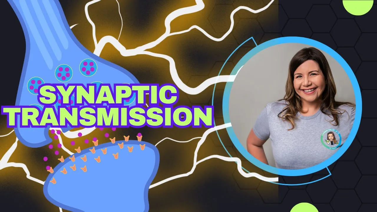
How Neurons Talk: Synaptic Transmission, Neurotransmitters, and Ending the Signal
158
Neuropeptides are a special class of signal molecules in the nervous system. Unlike classical neurotransmitters, which are usually small molecules acting quickly and locally, neuropeptides are larger chains of amino acids that often act more slowly and over longer distances. They are made in the cell body of the neuron and transported down the axon to vesicles in the synapse. Instead of simply turning the next neuron on or off, neuropeptides modulate neural activity, meaning they fine-tune the strength, duration, or sensitivity of signals, often working alongside traditional neurotransmitters to shape how the nervous system responds. For example, they can make excitatory signals stronger, inhibitory signals more effective, or adjust how responsive a neuron is to repeated stimulation. Because of this modulatory role, neuropeptides are involved in complex and lasting processes like pain perception, stress responses, appetite, sleep, mood, and learning. Well-known neuropeptides include substance P (transmits pain signals in the spinal cord and brain) and the body's natural opioids (endorphins and enkephalins, which reduce pain and produce feelings of euphoria).
Click to watch →

From the scientist's view: a conversation with William Schafer
594
Neuropeptides represent the most biochemically complex group of chemical signals in the nervous system. Even in C. elegans, which has only 302 neurons, there are hundreds of different individual neuropeptides, each binding to specific receptors. There are approximately 150 different neuropeptide binding receptors even in this small nervous system. These neuropeptide signaling networks function as 'wireless' communication systems that allow neurons not directly connected by synapses to communicate with each other. This form of communication is distinct from classical synaptic transmission and represents a fundamental mechanism for modulating brain activity and behavior.
Click to watch →
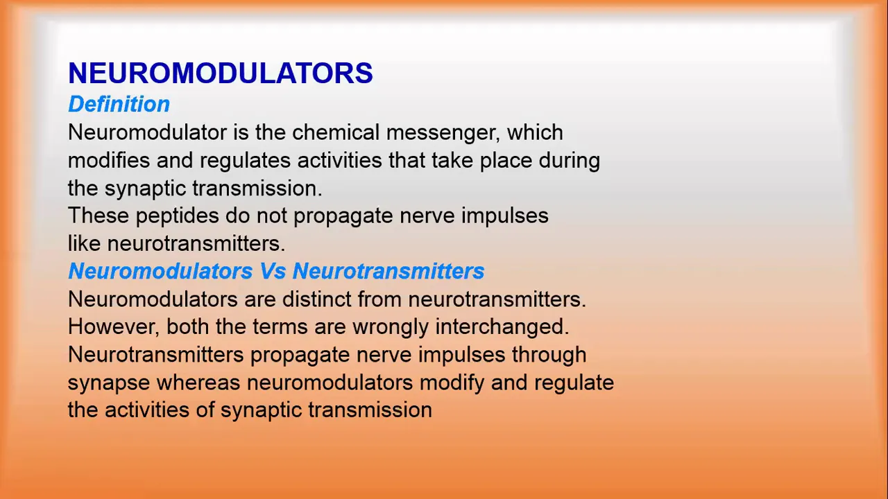
Neuromodulator, what is Neuromodulation
5.2K
The nervous system contains numerous neuropeptides that serve as neuromodulators with diverse physiological functions. Oxytocin and vasopressin are involved in social bonding, stress response, and regulation of water balance and blood pressure. Growth hormone releasing hormone (GHRH) stimulates growth hormone release from the pituitary gland, promoting growth and metabolic processes. Somatostatin inhibits the release of growth hormone and other hormones. Endorphins and enkephalins are endogenous opioid peptides that bind to opioid receptors to produce analgesic effects. Neuropeptide Y is involved in appetite regulation, stress response, and cardiovascular function. These neuropeptides are distributed throughout the nervous system and modulate various physiological processes including pain perception, appetite, stress response, and endocrine function.
Click to watch →

Healing the Mind Battlefield | Thoughts, Brain Chemistry & God’s Word
173
Neuropeptides are small protein-like chemical messengers that function as neurons. They are part of the nervous system's chemical language and play a major role in how we think, feel, and respond. They regulate emotions, pain, stress responses, immune function, hormone release, social bonding, learning, and memory. There are over 100 known neuropeptides in the human body.
Click to watch →
Deep Dive
Fundamental operating principles of Field-Effect Transistors (FETs), specifically how electrical conductivity changes in response to surface charge.
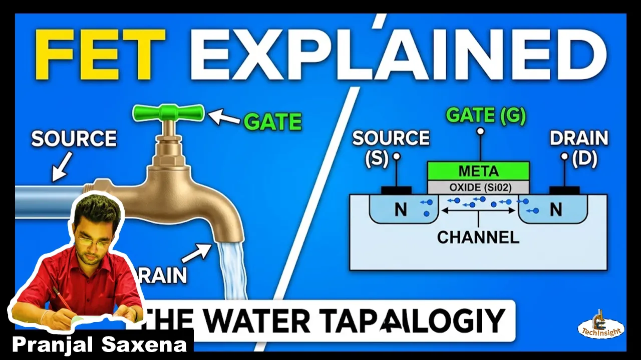
Structure of FETs | Power Electronic Series
210
A Field Effect Transistor (FET) consists of three terminals: Drain, Gate, and Source. The Source supplies majority charge carriers (electrons in N-channel, holes in P-channel) to the channel, while the Drain collects them. Charge carriers flow from Source to Drain through the channel, creating Drain-to-Source current (ID for N-channel, IS for P-channel). FETs are voltage-controlled devices where the Gate terminal applies voltage to create an electric field that controls channel conductivity. N-channel FETs are preferred over P-channel because electrons have higher mobility than holes. The Gate voltage controls the channel width and conductivity without requiring significant current flow into the Gate, making FETs more efficient than current-controlled BJTs.
Click to watch →

#1 | MOSFET Visualization | MOSTOK | MSQ & Quizzes in English for GATE 2021 by Shishir Das Sir
3.4K
A Field Effect Transistor (FET) is a phenomenon where the conductivity of a semiconductor is modulated by an electric field applied normal to the semiconductor surface. The electric field affects how easily charge carriers can move through the semiconductor material, thereby changing its conductive properties without requiring additional doping of the semiconductor material.
Click to watch →
Основы полупроводниковой электроники, Свинцов Д. А. 15.12.2021г.
337
A field effect transistor (FET) is a semiconductor device where resistivity between source and drain terminals is controlled by a third gate terminal. Without gate voltage, the p-doped semiconductor structure blocks current flow like a blocked pn junction. When positive gate voltage is applied, electrons accumulate at the semiconductor-insulator interface, forming an inversion channel that erases the junction barriers and enables current flow. The simple theory relates conductivity to carrier density via Ohm's law, with carrier density controlled by gate voltage through a capacitance relationship. This creates a linear gate-voltage dependence of conductivity, forming the basis of FET operation as a voltage-controlled switch.
Click to watch →
088 Transistores de efeito de campo FET
29.1K
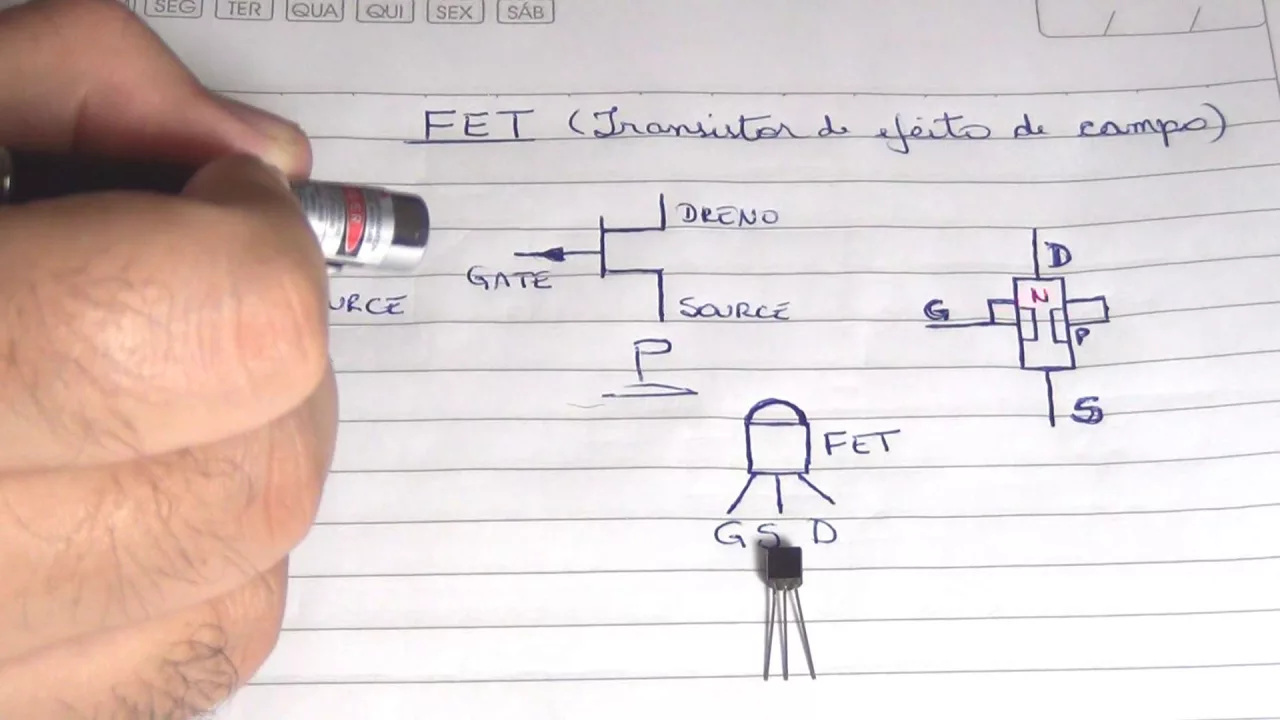
The FET operates based on the gate voltage applied to control current flow between drain and source. When a positive voltage is applied to the gate of an N-channel FET, it attracts positive charges toward the channel, which in turn attracts negative charges from the N-type crystal, allowing current to flow from drain to source. Conversely, a negative gate voltage attracts positive charges to the gate and repels negative charges, preventing current from flowing through the channel. This creates an electric field that controls conductivity.
Click to watch →
Transistores Efecto de Campo #1: Introducción
44.1K
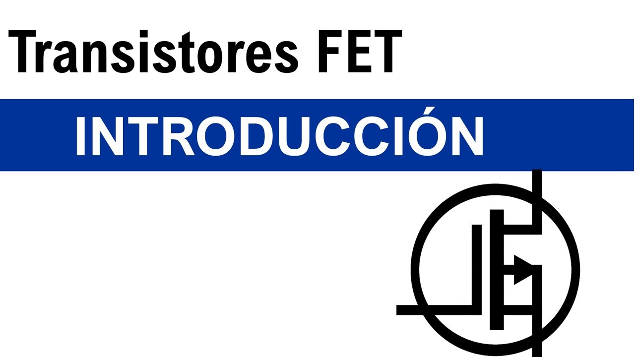
Field Effect Transistors (FETs) revolutionized electronics in the 1970s-1980s, enabling calculators, computers, and modern electronic devices. Theoretical work by Julius von Lilienthal in 1930 led to Bell Labs' development, with William Shockley patenting the FET in 1951 and the first successful fabrication in 1952. FETs are unipolar devices that use electric fields to control single-type charge carrier channels. The electric field is defined as a force field created by charge attraction and repulsion, always drawn from positive to negative charges. FETs exploit this principle to generate conduction channels when sufficient voltage creates strong enough attraction forces between particles.
Click to watch →
Deep Dive
Core biosensor architecture, including the distinct roles of a biorecognition element (receptor) and a transducer.

Biosensors Introduction: From Fabrication To Application
4.2K
Biosensors are integrated receptor-transducer devices that provide selective analytical information using biological recognition elements. Unlike physical sensors like thermometers, biosensors specifically detect biological species through molecular recognition. The three essential components are: (1) biological recognition element for target analyte interaction, (2) transducer converting recognition events to measurable signals, and (3) readout system. Recognition elements require selectivity, stability, and strong binding affinity. Common interactions include DNA hybridization, receptor-ligand binding, antibody-antigen recognition, and enzyme-substrate reactions.
Click to watch →

Short Learning Object on Smart Sensor Molecules for Advance Sensing by Dr.V.Violet Dhayabaran
115
Biosensors consist of two main components: (1) Receptor - the biological component that recognizes the target analyte (such as enzymes, antibodies, or microorganisms); (2) Transducer - converts the biological changes into electrical signals. Semiconductors or electrodes can be used as transducers. The three steps involved in biosensor operation are: (1) Biological recognition takes place; (2) The transducer converts the biological change from one form of energy to another; (3) The result is amplified and useful information is displayed.
Click to watch →

Track 6: Magnetic Based Biosensors
301
This section details how biosensors function as analytical devices converting biological responses into quantifiable signals. The core architecture consists of two interconnected components: a recognition element (receptor) and a transducer. Recognition elements include nucleic acids, antibodies, cells, and enzymes, each offering unique specificity for target analytes. The working principle involves: (1) bio-receptor binding to analytes, (2) biological events at interfaces, (3) signal pickup by transducers, (4) electrical signal conversion, (5) amplification by detector circuits, and (6) computer-based data processing. Two sensing mechanisms are distinguished: direct sensing where targets bind directly to electrode ligands, and indirect sensing where ambient reactions produce detectable products. These principles underpin all subsequent discussions of magnetic biosensor design and application.
Click to watch →

Webinar about electrochemical enzymatic biosensors
3.4K
A biosensor consists of two main components: a biorecognition element and a transducer. The biorecognition element specifically interacts with the target analyte (such as glucose, DNA, or virus particles), while the transducer converts this interaction into an electronic signal. Biosensors can be classified by their biorecognition elements: enzymatic biosensors use enzymes, immunosensors use antibodies or antigens, DNA sensors use single-stranded DNA, aptasensors use artificial antibodies called aptamers, cell sensors use whole cells, microbial sensors use bacteria or algae, and molecular imprint polymer sensors use synthetic polymers shaped to recognize specific molecules.
Click to watch →
Curso Intensivo RIBA - Biotecnología Molecular
149
A biosensor consists of three main components: (1) The analyte to be detected; (2) A biological recognition element (enzyme, antibody, nucleic acid, or whole cell) that specifically binds to the analyte; (3) A transducer that converts the biological recognition event into a measurable signal. The transducer can be electrochemical, optical, thermal, or piezoelectric. The signal is then processed by connected equipment, which can include portable devices that read signals directly or connect via Bluetooth to smartphones or displays.
Click to watch →
Prerequisite Knowledge
- Concept 01Basic DNA structure and biochemistry, particularly how single-stranded nucleic acids fold into complex three-dimensional conformations.
- Concept 02The physiological role of neuropeptides as signaling molecules and chemical messengers in the nervous system.
- Concept 03Fundamental operating principles of Field-Effect Transistors (FETs), specifically how electrical conductivity changes in response to surface charge.
- Concept 04Core biosensor architecture, including the distinct roles of a biorecognition element (receptor) and a transducer.
Subsequent Learning
- Step 01The SELEX (Systematic Evolution of Ligands by Exponential Enrichment) methodology used to screen and engineer custom aptamers.
- Step 02In vivo neurochemical monitoring and the biocompatibility challenges of implanting FET-based biosensors in living brain tissue.
- Step 03The integration of advanced nanomaterials, such as graphene-field effect transistors (GFETs), to enhance biosensor sensitivity and limits of detection.
- Step 04Clinical diagnostics and therapeutic applications of aptamers, comparing their efficacy and stability to traditional antibody-based assays.
Aptamer Basics
Structure Switching
Charge Dynamics
FET Transduction
Signal Readout
Response Patterns
Aptamer Basics
0:00Playing Section
- 1
Aptamers are DNA sequences engineered to change shape.
- 2
Unlike antibodies, they undergo large 3D conformational shifts.
Debye Screening and Stability Limits: The Case for Antibodies and MIPs
While DNA aptamer-based field-effect transistor (FET) biosensors are highly sensitive, critics highlight significant limitations when translating this technology to real-world physiological environments. A primary challenge is the 'Debye screening effect': in high-ionic-strength fluids like blood or cerebrospinal fluid, mobile ions shield the target's charge, severely hindering FET detection unless samples are heavily diluted. Furthermore, unmodified DNA aptamers are prone to rapid degradation by nucleases in vivo. Consequently, many bioengineers advocate for alternative approaches, such as traditional antibodies or synthetic Molecularly Imprinted Polymers (MIPs). These alternatives often provide superior chemical stability, easier scaling, and avoid the electrostatic shielding limitations inherent to FET-based electrical sensing.
Deep Dive
The SELEX (Systematic Evolution of Ligands by Exponential Enrichment) methodology used to screen and engineer custom aptamers.

Aptamers and a quick comparison of small nucleic acid tools/therapeutics (RNAi-based & ASOs)
344
SELEX (Systematic Evolution of Ligands by EXponential Enrichment) is an iterative in vitro selection process for developing high-affinity aptamers. The method involves creating a diverse library of random nucleic acid sequences, incubating with target molecules, isolating sequences that bind the target, amplifying these sequences using PCR, introducing mutations through error-prone PCR, and repeating cycles until optimal binders are obtained. Aptamers feature constant adapter regions at the ends for PCR amplification and variable internal regions that fold into specific shapes. This design enables easy isolation, copying, and modification of selected aptamers, making SELEX a powerful tool for developing customized aptamers for various applications.
Click to watch →

Аптамеры на основе нуклеиновых кислот — Андрей Головин
2.6K
SELEX (Systematic Evolution of Ligands by EXponential enrichment) is an in vitro evolution method that accelerates molecular selection. The process involves: (1) chemical synthesis of diverse DNA libraries, (2) enrichment and amplification of molecules binding to predetermined targets, (3) selection of sequences forming complex 3D structures complementary to targets. Targets range from small molecules to proteins to entire cells. The key distinction is between base complementarity (A-T, G-C matching) and surface complementarity (complex 3D structural matching). Once a binding sequence is identified, it can be chemically synthesized and used therapeutically as an aptamer.
Click to watch →

Build-a-Cell seminar Steve Benner: Biotechnology in Artificial Cells in Fluorinated Media
176
SELEX (Systematic Evolution of Ligands by EXponential enrichment) is a method for selecting functional nucleic acids. In six-letter genetic systems, libraries are synthesized chemically and exposed to target cells (e.g., liver cancer cells). Non-binding sequences are washed away, and bound sequences are extracted. Counter-selection removes sequences that bind to non-target cells. After about 11 selection rounds, specific binders emerge. Six-letter libraries are richer than four-letter libraries, with nitro group functionality providing universal low-affinity binding. Mutations in Z and P bases significantly reduce binding affinity, demonstrating that the expanded genetic alphabet provides richer functional diversity for aptamer evolution.
Click to watch →
Aptamers and SELEX: The Past, The Present, and The Future
37.7K
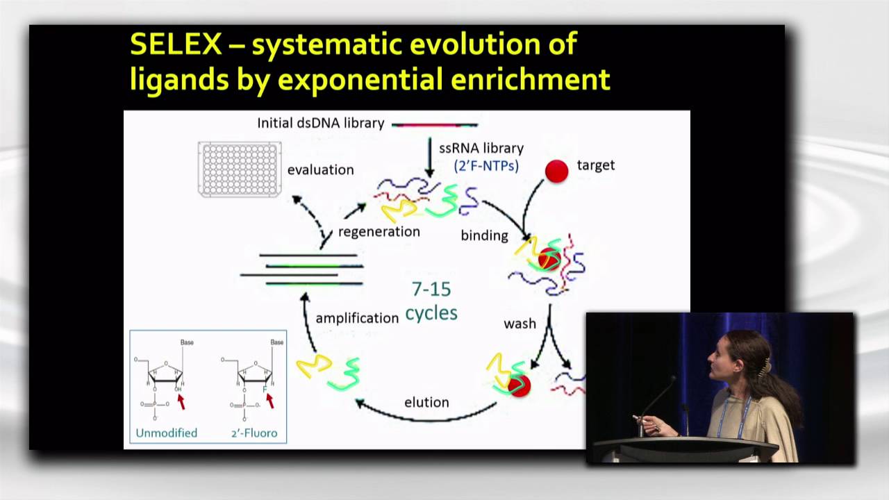
Aptamers are highly structured, short single-stranded DNA or RNA oligonucleotides discovered in 1990, named from Latin 'aptus' (to fit) and Greek 'meis' (a part). They offer advantages over antibodies including complex 3D structures for high affinity/specificity, small size (<5 nm) for tissue penetration, and ease of chemical modification. SELEX (Systematic Evolution of Ligands by Exponential Enrichment) is the iterative selection process: starting with a double-stranded DNA library containing a variable region (10-90 nucleotides), converting to single-stranded RNA with modified nucleotides for stability, combining with targets, and enriching binders through multiple rounds of selection, washing, and amplification. Advances include negative selection for specificity, alternative amplification methods (emulsion PCR, droplet digital PCR), and high-throughput sequencing integration.
Click to watch →
Molecular Therapy Podcast: COVID-19 Diagnostics with Dr. Maxim Berezovski
152
Aptamers are synthetic nucleic acid molecules (DNA or RNA) that can be engineered to bind specifically to target molecules including proteins, cells, and small molecules. Developed since 2002, aptamers offer significant advantages over antibodies: they are easier to develop using iterative selection processes, can be synthesized at low cost, and allow precise control over attachment to sensors. Unlike antibodies, aptamers are considered chemical entities rather than biologicals, resulting in simpler regulatory pathways. The SELEX (Systematic Evolution of Ligands by EXponential enrichment) process enables any graduate student to develop aptamers through iterative rounds of incubation, washing, and amplification. Starting with a library of ~10^15 random-sequence DNA molecules, researchers perform multiple selection rounds to enrich for sequences with high binding affinity to the target. Negative selection steps can remove aptamers binding to unwanted targets. This methodology was applied to develop aptamers targeting SARS-CoV-2 nucleocapsid protein, producing sequences with nanomolar binding affinity.
Click to watch →
Deep Dive
In vivo neurochemical monitoring and the biocompatibility challenges of implanting FET-based biosensors in living brain tissue.
Department Seminar: Dr Nako Nakatsuka from ETH Zurich
2.1K
Miniaturized aptamer field effect transistor sensors have been integrated into neural probes for in vivo measurements. These probes contain active aptamer sensors, inactive scrambled sequence references, and solid-state reference electrodes. Recent work has demonstrated successful insertion into localized brain regions of awake mice, enabling real-time recording of serotonin concentrations in vivo. This represents a significant advancement toward clinical applications of aptamer-based neurochemical monitoring.
Click to watch →

4_2 Stereotaxic Surgeries and In Vivo Techniques-2
1.5K
In vivo neurochemical measurement focuses on detecting changes in neurotransmitter concentrations over time in specific brain regions. The goal is to correlate neurochemical changes with specific behaviors or physiological functions. Researchers typically measure relative changes rather than absolute concentrations, as establishing correlations between neurochemical expression and behavior is the primary objective. Microdialysis probes are Y-shaped devices with a semi-permeable membrane that allows selective sampling of extracellular fluid from specific brain regions. The probe has an inlet and outlet connected by thin cannulas. The membrane allows small molecules (neurotransmitters) to pass through while retaining larger molecules. The probe is continuously perfused with physiological solution (saline or artificial CSF), creating a concentration gradient that drives passive diffusion of analytes from the brain tissue into the collected dialysate.
Click to watch →
iCANX Talks 32 Anne M Andrews
165
Field Effect Transistors (FETs), the basic unit of electronic devices, can be modified to detect neurochemicals. By replacing metal electrodes on silicon microprobes with FETs and functionalizing them with molecular recognition elements, researchers can create sensors capable of fast, highly sensitive measurements of neurotransmitters in biological environments.
Click to watch →
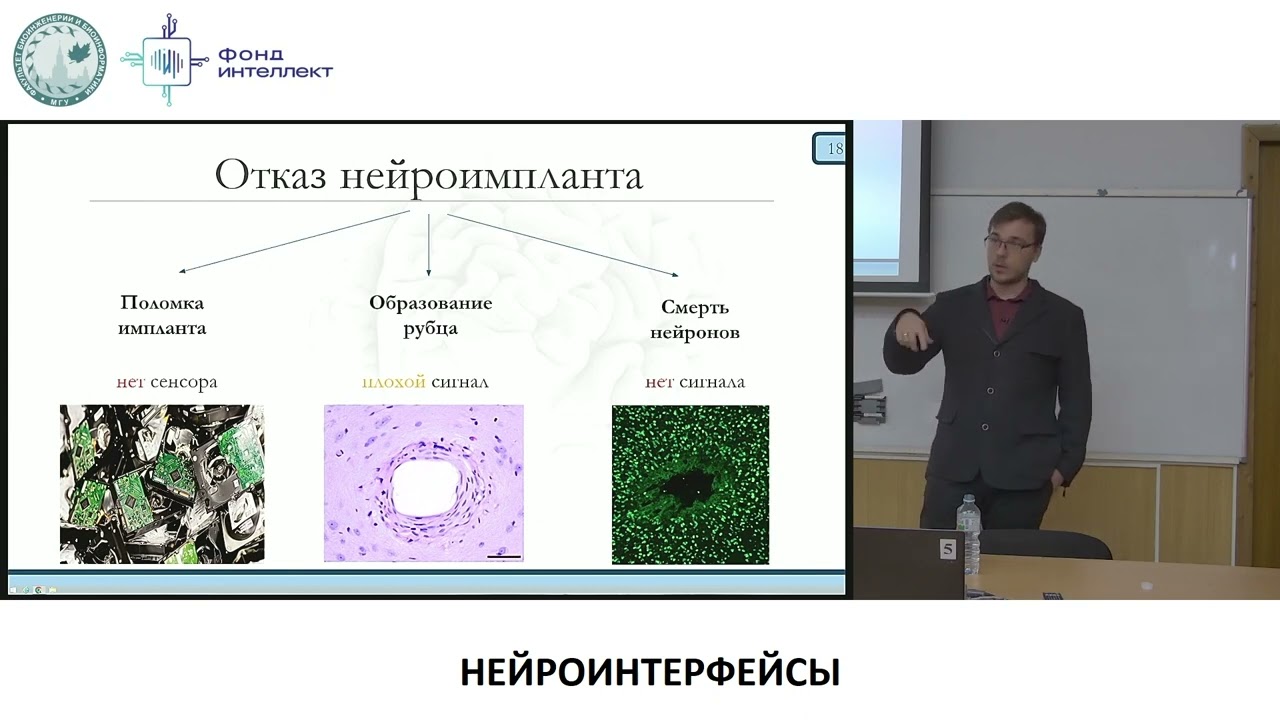
Попков В.А. - Нейроинтерфейсы от биологии до анализа данных - 4.Биосовместимость инв. нейроимплантов
648
The brain contains approximately 10^11 neurons and glial cells. Oligodendrocytes insulate axons for electrical conductivity. Microglia serve as immune cells performing pathogen recognition and synaptic pruning. Astrocytes regulate neurotransmitters, maintain blood-brain barriers, and provide metabolic support. Modern neuroscience recognizes the brain as an integrated system where all cell types participate in cognitive function. Invasive neurointerfaces offer superior spatial and temporal resolution for recording individual neuron activity but face significant biocompatibility challenges. Research shows only 25% of implants maintain function at 150 days post-implantation. Three main failure causes exist: mechanical device failure, neuronal death around the implant, and scar tissue formation. Neuroinflammation involves acute and chronic phases with microglia attempting destruction and astrocytes forming protective barriers. Management strategies include anti-inflammatories like dexamethasone, systemic interventions (low-calorie diets, hypoxic training), and local drug delivery systems.
Click to watch →

Bob Kennedy Part 3: Derivatization to the Rescue
169
In vivo neurochemical monitoring involves placing probes in the brain to measure neurotransmitters. Two main approaches exist: sampling probes that collect fractions for later analysis, and microelectrodes that can be inserted into different brain regions to detect specific neurotransmitters in real-time. The motivation for developing better methods stems from the complexity of brain function, which controls everything from sleep to eating decisions to drug-seeking behavior, across trillions of neurons in multiple brain regions. Better analytical techniques with improved temporal and spatial resolution enable more comprehensive coverage of neurotransmitter release patterns.
Click to watch →
Deep Dive
The integration of advanced nanomaterials, such as graphene-field effect transistors (GFETs), to enhance biosensor sensitivity and limits of detection.

How Graphene is Changing the World … Right Now
1.3M
Graphene Field Effect Transistors (GFETs) are advanced sensors made by growing graphene on a sapphire base with contacts and gate electrodes. They offer several advantages over traditional sensors: they are cheaper, tougher, and last longer; their high electrical conductivity enables higher efficiency with less heat loss; they can be easily tuned for specific optical characteristics; and their single-atom thickness allows easy miniaturization. GFETs can detect various substances including potassium ions, heavy metals, gases, and pH levels by adding different receptor molecules to the sensing surface.
Click to watch →

Fast detection of COVID-19 virus, the discovery of a biosensor for SARS CoV-2
24.3K
A graphene field effect transistor (GFET) biosensor has been developed for rapid detection of SARS-CoV-2 by detecting the virus's spike protein; the device uses graphene's exceptional electronic properties combined with antibody-based recognition, achieving high sensitivity (detecting spike protein down to 1 femtogram per milliliter and whole virus at 16 plaque-forming units per milliliter) and specificity, allowing direct detection from patient swab samples without lengthy laboratory preparation.
Click to watch →

BMEH | Graphene-Based Field-Effect Transistors for Biosensing: Where Is the Field Heading To?
102
Graphene-based field-effect transistors (gFETs) represent a promising next-generation biosensing technology that offers high sensitivity, fast response times, and the potential for miniaturization. These sensors work by detecting changes in the electrical properties of the graphene channel when target biomolecules bind to surface-immobilized receptors, causing charge modulation. Key challenges include operating in high-ionic-strength biological environments like blood, where the Debye screening length limits detection to approximately 1 nanometer from the graphene surface. Solutions involve using anti-fouling agents such as polyethylene glycol molecules and employing smaller synthetic receptors like nanobodies (approximately 4 nm) to stay within the sensing range. gFET biosensors have demonstrated applications in detecting cardiac biomarkers (troponin I) with detection limits around 1-4 picomolar and monitoring wound healing by detecting matrix metalloproteinase-9 (MMP9) levels, showing competitive performance compared to traditional ELISA methods while providing much faster results.
Click to watch →
25 january 2024 «Thursday´s NanoSpin-off Talks» GRAPHENEA
154
Graphene field effect transistors for biosensing operate with electrolyte gates instead of traditional back gates, forming electric double layers at the graphene-solution interface that bring ions extremely close to the channel. This capacitive coupling enables detection of biomolecular binding events with exceptional sensitivity. Key performance advantages include: smaller device dimensions enhancing sensitivity; high carrier mobility enabling rapid response; high interfacial capacitance improving signal transduction; and low operation voltages (10-100 mV) enabling low-power portable applications. Graphene demonstrates significantly better mobility and interfacial capacitance than silicon or other materials, translating to higher transconductance essential for sensitive detection. The transduction mechanism involves shifts in the Dirac point caused by electron donation/withdrawal from bound molecules or conformational changes in bio-receptors.
Click to watch →

Electronic biosensors using Field-effect transistor as the transducer - part 2
1.9K
Graphene, a single-layer honeycomb carbon lattice with SP2 hybridization, offers exceptional electronic properties including zero band gap and ambipolar conduction. CVD growth on copper foil followed by PMMA transfer enables practical device fabrication. Electrolyte-gated GFETs differ from classical MOSFETs by placing the semiconductor directly in electrolyte, where the electric double layer serves as dielectric. Non-covalent functionalization using pyrene chemistry preserves electronic properties while enabling surface modification. Transfer curves show on/off ratios exceeding 10^4, with leakage currents at least 100x lower than signal currents. However, GFETs require 20-30 minute stabilization periods and face interference from redox-active species in complex biological samples.
Click to watch →
Deep Dive
Clinical diagnostics and therapeutic applications of aptamers, comparing their efficacy and stability to traditional antibody-based assays.

Application of Aptamers in the Clinical Laboratory - Geoffrey Baird, MD, PhD
10K
Aptamers offer significant advantages over antibodies: production uses automated robots instead of animals; they are 5-10 times smaller (~150 kDa vs. antibody size); DNA chemistry is simpler and more accessible than protein biochemistry; specific derivatization is easier with DNA; all aptamers work identically in the same buffer conditions enabling straightforward multiplex assays. However, aptamers face challenges including lower affinity for small molecules (typically mid-micromolar vs. nanomolar for proteins) and limited clinical applications (only Macugen approved, others in trials).
Click to watch →
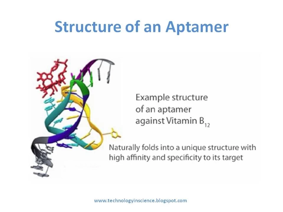
Aptamers in Therapeutics and Diagnostics | Oligonucleotide Applications
37.8K
Multiple SELEX variants enable targeted aptamer discovery: nitrocellulose membrane filtration-based methods require target-bead coupling but lack flexibility; capillary electrophoresis-based SELEX achieves success in 2-4 rounds through mobility shifts; microfluidic-based approaches enhance efficiency, as demonstrated by obtaining neurotoxin-specific aptamers in single rounds; Cell-SELEX targets whole cells and surface structures rather than purified proteins. Compared to antibodies, aptamers offer significant advantages: chemical synthesis enables reproducible, scalable production independent of cell cultures; greater specificity and affinity; easy chemical modification for customization; smaller size improves transport and tissue penetration; ambient stability eliminates cold chain requirements. However, limitations exist including potentially lower affinities, inability to bind certain targets, and costly identification processes.
Click to watch →
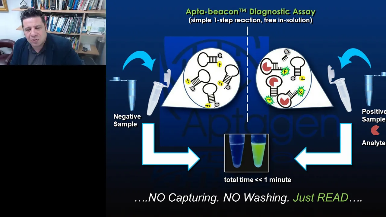
Synthetic Antibodies - The Emerging Field of "Aptamers" in Diagnostics and Drug Discovery
5.6K
Aptamers are synthetic nucleic acid ligands (DNA or RNA) that bind to targets with high affinity and specificity, offering advantages over traditional antibodies including smaller size (one-fifth of IgG antibodies), comparable or better specificity, lower manufacturing costs, and greater thermal stability; they are developed using directed molecular evolution and can be engineered into diagnostic tools like aptamer beacons for one-pot detection assays or into drug delivery vehicles called aptabodies that combine nucleic acid scaffolds with amino acids, fatty acids, and other functional groups for targeted therapy applications.
Click to watch →

AIBBC 2015: Aptamers for Therapeutic and Diagnostic Applications
10.5K
Aptamers are single-stranded DNA or RNA molecules that function as 'chemical antibodies' with unique advantages over traditional antibodies including smaller size for better tissue penetration, easier in vitro production without animal hosts, and longer shelf life at room temperature; however, they face challenges with rapid nuclease degradation requiring chemical modifications like locked nucleic acids to extend half-life, and they bind to targets through 3D structural pockets rather than simple hybridization, making them valuable tools for targeted drug delivery, cancer therapy, and multiplexed diagnostics.
Click to watch →
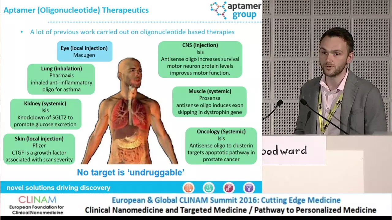
Modified Aptamers as Replacement for Antibodies in Diagnostics and Therapeutics
1.6K
Modified aptamers, which are single-stranded nucleic acid molecules (DNA or RNA) that fold into specific 3D shapes to bind targets, offer significant advantages over traditional antibodies including smaller size (5-6 kDa vs. 150 kDa), superior cell permeability, non-immunogenicity, and flexible structure; these synthetic affinity reagents can be manufactured through chemical synthesis with consistent quality and can be designed to target toxic or non-immunogenic molecules, making them valuable tools for both diagnostic applications (such as lateral flow devices and cancer staging) and therapeutic uses (including drug delivery and viral entry inhibition).
Click to watch →
Aptamer Basics
Structure Switching
Charge Dynamics
FET Transduction
Signal Readout
Response Patterns
Aptamer Basics
0:00- 1
Aptamers are DNA sequences engineered to change shape.
- 2
Unlike antibodies, they undergo large 3D conformational shifts.
Debye Screening and Stability Limits: The Case for Antibodies and MIPs
While DNA aptamer-based field-effect transistor (FET) biosensors are highly sensitive, critics highlight significant limitations when translating this technology to real-world physiological environments. A primary challenge is the 'Debye screening effect': in high-ionic-strength fluids like blood or cerebrospinal fluid, mobile ions shield the target's charge, severely hindering FET detection unless samples are heavily diluted. Furthermore, unmodified DNA aptamers are prone to rapid degradation by nucleases in vivo. Consequently, many bioengineers advocate for alternative approaches, such as traditional antibodies or synthetic Molecularly Imprinted Polymers (MIPs). These alternatives often provide superior chemical stability, easier scaling, and avoid the electrostatic shielding limitations inherent to FET-based electrical sensing.
Basic DNA structure and biochemistry, particularly how single-stranded nucleic acids fold into complex three-dimensional conformations.
The physiological role of neuropeptides as signaling molecules and chemical messengers in the nervous system.
Fundamental operating principles of Field-Effect Transistors (FETs), specifically how electrical conductivity changes in response to surface charge.
Core biosensor architecture, including the distinct roles of a biorecognition element (receptor) and a transducer.
The SELEX (Systematic Evolution of Ligands by Exponential Enrichment) methodology used to screen and engineer custom aptamers.
In vivo neurochemical monitoring and the biocompatibility challenges of implanting FET-based biosensors in living brain tissue.
The integration of advanced nanomaterials, such as graphene-field effect transistors (GFETs), to enhance biosensor sensitivity and limits of detection.
Clinical diagnostics and therapeutic applications of aptamers, comparing their efficacy and stability to traditional antibody-based assays.
779 views38likes1:54@neurosciencebeyondOriginal Release: 2024-09-02
So you have this kind of molecules, aptamers.
Can you please elaborate on what they are and how do you measure actually the, whatever you're measuring, for example, the neuropeptides, you know, this is my favorite part to explain because I think it's pretty cool.
But you basically have these DNA sequences that, in our case, are designed to structure switch.
So it really changes its morphology in 3D.
So not all aptamers are structure switching.
A lot of them have these lock and key type binding where you just have an aptamer something clicks to it like antibodies.
But in our case they were designed in vitro to undergo a large conformational change.
So DNA is very negatively charged because of its phosphodiester backbone.
So if you have 40 bases in your DNA sequence, you have 40 negative charges.
So now what we basically have is a DNA that has a certain structure.
A molecule comes in, it changes its morphology to really capture this analyte, which rearranges charge.
So now as soon as you rearrange charge, you can start to transduce that at the surface of different materials.
For example, semiconductor at the surface of field effect transistors, where now you can have two electrodes a source and drain electrode, you have a semiconducting channel and you have if it's an N-type semiconductor or you have electrons that carry the charge from the source to the drain electrode.
So when the aptamer moves its backbone, it changes how much electrons can actually pass through the semiconductor by changing the charge density at the surface.
And so we can really record that as a change in current is the current increasing is it decreasing.
And we now really know that depending on how the aptamer structure switches, whether it compresses or it elongates or something in between, we get a very different signal transduction in terms of current increasing versus decreasing, for example.
Up Next
13:18
Electrochemical Biosensors Explained: Principles and Applications
@HMCH-bt2ue
20.8K views•2023-10-30
14:12
Quantitative Real-Time PCR (qPCR): Principle, Method & Data Analysis
@animatedbiologywitharpan
129.2K views•2023-09-06

23:33
Microbial Degradation of Plastics: Biodegradation Pathways & Sustainability
@majeedhammad
2.9K views•2021-04-11
16:04
CRISPR and Genetic Engineering: How Gene Editing Works and Why It Matters
@kurzgesagt
30.5M views•2016-08-10
Related Study Plans & Knowledge Roadmaps
Structured learning paths in Biotechnology
biotechnology
AAV Production: Transfection, Prep & qPCR
01Introduction to Gene Therapy and AAV Biology
02Mammalian Cell Culture: Suspension HEK293 Cells
03Upstream Bioprocessing: Triple-Transfection Optimization
+ 2 more learning modules
18h5 modules
Explore Roadmap
biotechnology
Building Gut-on-a-Chip: PDMS, Cells & TEER
01Foundations of Microfluidics and Gut Anatomy
02Cleanroom Fabrication & PDMS Soft Lithography
03Epithelial Cell Culture and On-Chip Seeding
+ 2 more learning modules
26h5 modules
Explore Roadmap
biotechnology
Optimizing CFPS: E. coli Extract & Yield
01Fundamentals of Molecular Biology and Translation
02Introduction to Cell-Free Protein Synthesis (CFPS)
03Preparing E. coli Crude Lysate (The Extract)
+ 2 more learning modules
19h5 modules
Explore Roadmap
biotechnology
Haematococcus PBR: Design, Stress & Astaxanthin
01Biology and Life Cycle of Haematococcus pluvialis
02Bench-Scale Photobioreactor (PBR) Design and Engineering
03Culture Media Formulation and Inoculation
+ 2 more learning modules
15h5 modules
Explore Roadmap