








![★★초음파 유도하 정맥관 삽입술 Axillary v, femoral v, PICC/ MC 길은미 [2020 SECCI 온라인연수강좌]](https://i.ytimg.com/vi_webp/ukcyhZgUAcE/maxresdefault.webp)


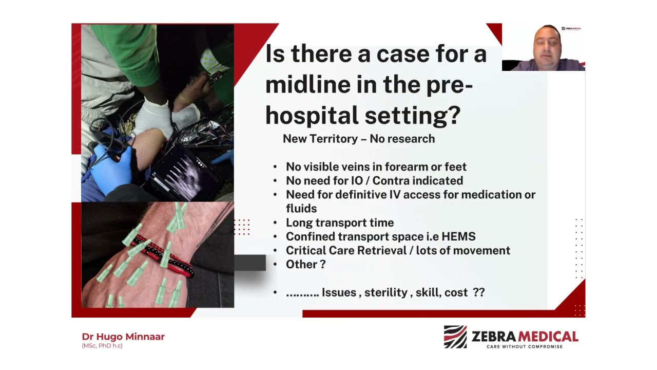




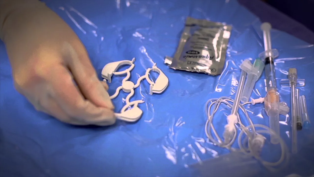
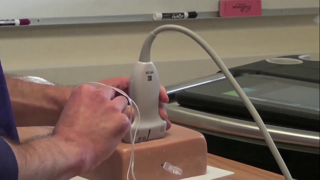


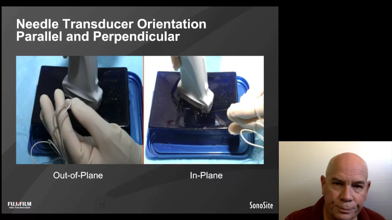
This video demonstrates a combined out-of-plane to in-plane ultrasound-guided technique for peripheral venous catheter placement, involving patient positioning with arm abduction and external rotation, vessel identification using ultrasound (such as the basilic vein approximately 6mm below skin), local anesthesia administration, and catheter advancement while maintaining ultrasound visualization to confirm successful cannulation and secure the catheter.
Ultrasound-Guided Peripheral IV Catheter Placement Technique
Added:Standard peripheral intravenous (IV) catheter insertion techniques and sterile field preservation.
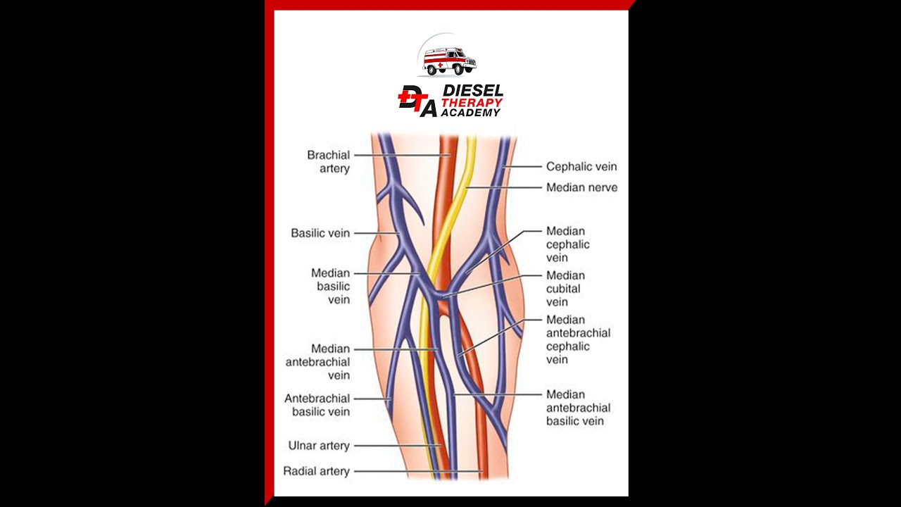
Upper extremity vascular anatomy, specifically distinguishing between veins, arteries, and nerves.
Basic ultrasound physics and knobology, including adjusting depth, gain, and choosing a linear transducer.
Sonographic appearance of tissues (hyperechoic vs. anechoic) and methods to differentiate veins from arteries (e.g., compressibility and Doppler flow).
Advanced vascular access techniques, such as ultrasound-guided midline or PICC (Peripherally Inserted Central Catheter) lines.
Clinical algorithms for patients with Difficult Intravenous Access (DIVA) and catheter length selection strategies.
Management and troubleshooting of complications unique to ultrasound-guided IVs, such as deeper vessel extravasation and thrombosis.
Applying needle-guidance motor skills to other ultrasound-guided procedures, such as arterial line placement or regional nerve blocks.
123.2K views1.3Klikes8:44Original Release: 2023-08-21
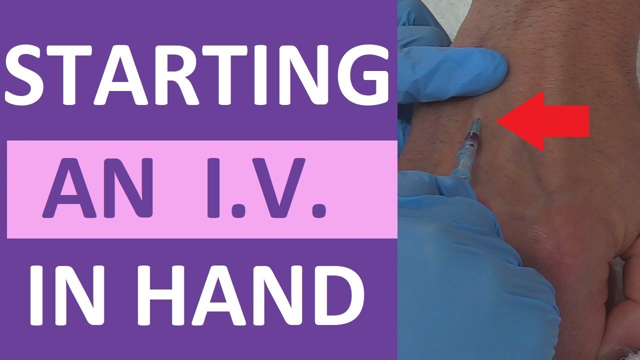
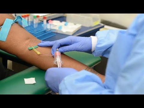
hi I'm Tom Cook I'm an Emergency Physician I practice in South Carolina with Prisma Health in the University of South Carolina and today we're going to demonstrate how to place a peripheral intravenous catheter using ultrasound there are basically three different techniques to do this one of those is to use an out of plane approach the other one is to use an in-plane approach but today we're going to demonstrate a combination of those two approaches where we'll start out of plane then readjust the transducer to go in plane before we eventually Advance the catheter into the vessel and secure it to begin with we'll go ahead and place the patient's arm so that it's abducted and externally rotated this exposes this side of the arm which has the vessels that we're interested in cannulating and of course we'll apply a tourniquet next we're going to clean the skin this particular skin cleaner has a die in it and it helps me understand what part of the skin is clean and what's not clean then I'll take the transducer and clean the end of the transducer and set it into position so I can't contaminate the tip Some people prefer to put a sterile probe cover on that I've found that this works well for me there's really no difference in terms of the ability to perform the exam next thing we'll go ahead and take some sterile gel place it on the area of the skin that we're going to examine the next thing we're going to do is make sure that we're stabilized so you'll notice I've got my hand and my arm stabilized on the bed and I'm going to stabilize my other hand on whatever sort of is convenient at the time it could be my leg it could be some other structure but it's very important to stabilize your hands because you're moving in very very precise ways to to perform the procedure so we have several different targets here these are the brachial vessels here on the right side of the screen right here uh we can see if we can compress the artery pulsates down the center of the screen and there are a couple of brachial veins there as well that we could cannulate but in this patient we're going to use this blood vessel here this is the basilic vein you can see it's pretty large in size you'll also notice that the superficial wall is somewhere around six millimeters below the surface of the skin if we move the transducer up and down the skin we can try and figure out the path of that vessel by just making sure that it stays more or less in the center of the viewing field as we move up and down the skin if we look at it in the longitudinal aspect we can see the vessel there and that will become important as we get near the end of the procedure when we're getting ready to place the catheter the next thing that I do with most of my patients but not everyone does but I do is go ahead and anesthetize the skin where I'm going to place the catheter I find that the patients appreciate it they also is the benefit that if the first attempt doesn't succeed and you have to try again the patient usually doesn't have to experience any discomfort with a second attempt so I'll go ahead and center the Target in the screen I'm just going to increase the magnification of that a little by decreasing the depth and you'll notice on the transducer there's a center Mark and that Center Mark corresponds with the center of the screen so I'm going to use that right now and I'm going to go ahead and stick the needle in you can see the needle tip there in the soft tissue I'm going to adjust inject a little bit of anesthetic and right there that should be numb in a minute or two the next thing we'll do is get the catheter the catheter again it's a longer catheter it's a two and a half inch catheter it um it's a catheter over needle setup some people make a point of placing the bevel up most the time it doesn't make too much of a difference but I'll make sure that I do that in this situation now if you go back and look at the patient you can see there's a nice little spot of blood where I anesthetized him so this makes a nice little Target for me to place the catheter needle or break the skin and if you look on the screen again that little collection of Lidocaine injected is pretty much dissipated so you got a nice clean view there now you'll notice I'm stabilizing my right hand I'm going to go ahead and introduce the catheter into the skin and immediately you can kind of see the tip right there above the vessel and I can advance it a little bit more and go ahead and cannulate this vein but for this situation I'm going to take my transducer I'm going to turn it and get a longitudinal View of the vessel now if you look on the left side of the screen you can see that needle and if I move the tip of the needle back and forth it comes in and out of plane and the importance of understanding this is if you can't find the needle just by moving it back and forth it will fall into the signal and be picked up on screen the other thing you want to keep in mind is you'll notice my left hand never moves I always keep the ultrasound signal on the target I don't move the ultrasound signal to find the needle I've moved the needle to find the signal and that's a very important distinction when you're performing this procedure in the long Axis or The in-plane View so I'll go ahead and Advance the needle making sure that I try and keep that tip in plane and now if you look you can see that the needle is plainly in the Lumen of the vessel you can also see there's some blood coming into the Hub of the needle itself I'm going to continue to advance until I just get down to about the back wall of that vessel now you notice right here it looks like it's sort of pointed down towards the deep wall of the vessel so I'm going to change that trajectory just a little bit lay it down just a little bit more and now I'm going to go ahead set my transducer down and Advance the catheter it should slide in nice and easy and when I remove the needle blood comes out of the end of the catheter hub now I'm going to attach my flush I'll go ahead and flush the catheter pull back just a little bit to make sure that I can get blood from the catheter if I need to do any laboratory studies it also helps confirm placement and then go ahead and flush the rest of the catheter with sailing now I've got these little two by twos I'm going to go ahead and clean this area up and again because I've used a long catheter this has gone a pretty decent way up into the vein and it gives me the luxury of knowing that that catheter is not going to come out even though the vein is relatively deep under the skin compared to what you would do with most peripheral catheters not placed with ultrasound then I'll go ahead and place my Tegaderm and now I have a fantastic catheter in place I can get blood for laboratory studies you can put all kinds of different solutions and medications through that and most of the time that catheter can last for three or four days or the patient's entire admission if they're admitted to the hospital
Up Next
71:20
TESS Launch: SpaceX Falcon 9 Deploys NASA Exoplanet Hunter
@SpaceX
1.1M views•2018-04-20

12:54
Triumph of Orthodoxy Icon: Byzantine Art & History Explained
@BenCallan
2.1K views•2024-08-06

9:49
FastAPI vs Flask vs Django: Choosing the Right Python Web Framework
@TechWithTim
302.5K views•2024-05-26

1:42
Game of Thrones Opening Credits: A Cinematic Analysis
@gameofthrones
46.3M views•2011-04-18
Related Study Plans & Knowledge Roadmaps
Structured learning paths in General & Interdisciplinary Studies
general-studies
Elevator Engineering: History, Physics & Cities
01From Hoists to Safety Brakes: The Dawn of Vertical Lift
02Hydraulic vs. Traction: The Engineering Physics of Elevators
03Modern Speed, Safety, and Algorithms
+ 2 more learning modules
12h5 modules
Explore Roadmap
general-studies
Rubber: Material Science, History & Geopolitics
01Origins of Natural Rubber & Mesoamerican History
02The Chemistry of Natural Rubber & Vulcanization
03The Geopolitics of the Rubber Boom & Colonization
+ 2 more learning modules
12h5 modules
Explore Roadmap
general-studies
Containerization: Evolution, Logistics & Trade
01Ancient Trade & Bulk Cargo: Amphorae to Barrels
02The Industrial Revolution & Cardboard Packaging
03The Containerization Revolution: Malcom McLean
+ 2 more learning modules
12h5 modules
Explore Roadmap
general-studies
Textile Tech: Evolution, Labor & Global Impact
01Pre-Industrial Textiles: Handloom & Cottage Industry
02Mechanization: The Spinning Jenny to the Power Loom
03The Factory System & Global Labor Transformation
+ 2 more learning modules
10h5 modules
Explore Roadmap