![Pleural Space [HD]](https://i.ytimg.com/vi/x-625AkBwDc/maxresdefault.jpg)


![Pleural Space: Part 1 of 3 [HD]](https://i.ytimg.com/vi/mD9goLXYhYU/maxresdefault.jpg)














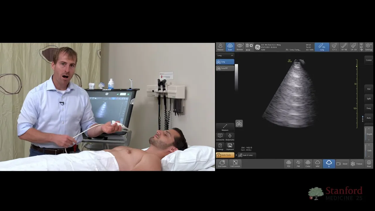

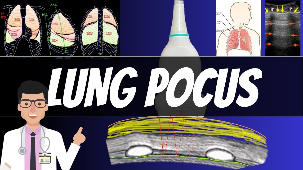


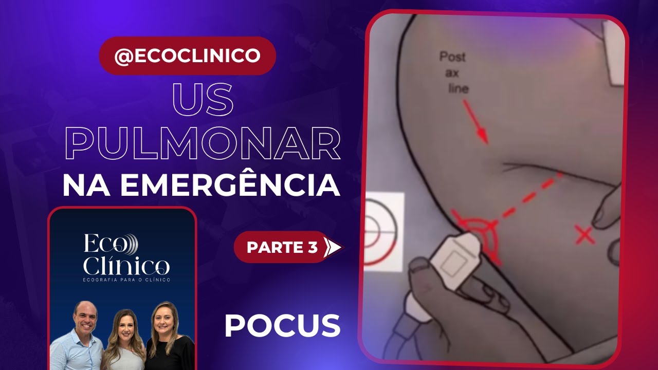
The BLUE protocol is an emergency bedside lung ultrasound technique developed by Dr. Daniel Lichtenstein that uses three key anatomical points (upper blue, lower blue, and plats points) to rapidly assess patients with breathing difficulties. The protocol classifies findings into profiles: A profile indicates absent lung sliding suggesting pneumothorax or exudative pleural disease, while B profile indicates present lung sliding with comet-tail artifacts (B lines) indicating interstitial syndrome such as pulmonary edema or pneumonia. The technique distinguishes between normal lung (newt profile), pneumonia (sea profile), and pneumothorax (lung point identification), offering accuracy comparable to or exceeding chest X-rays and physical examination for conditions like pulmonary edema, pneumonia, and pneumothorax.
BLUE Protocol Bedside Lung Ultrasound in Emergency: A Practical Guide
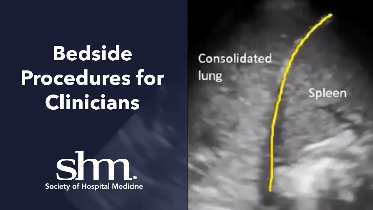
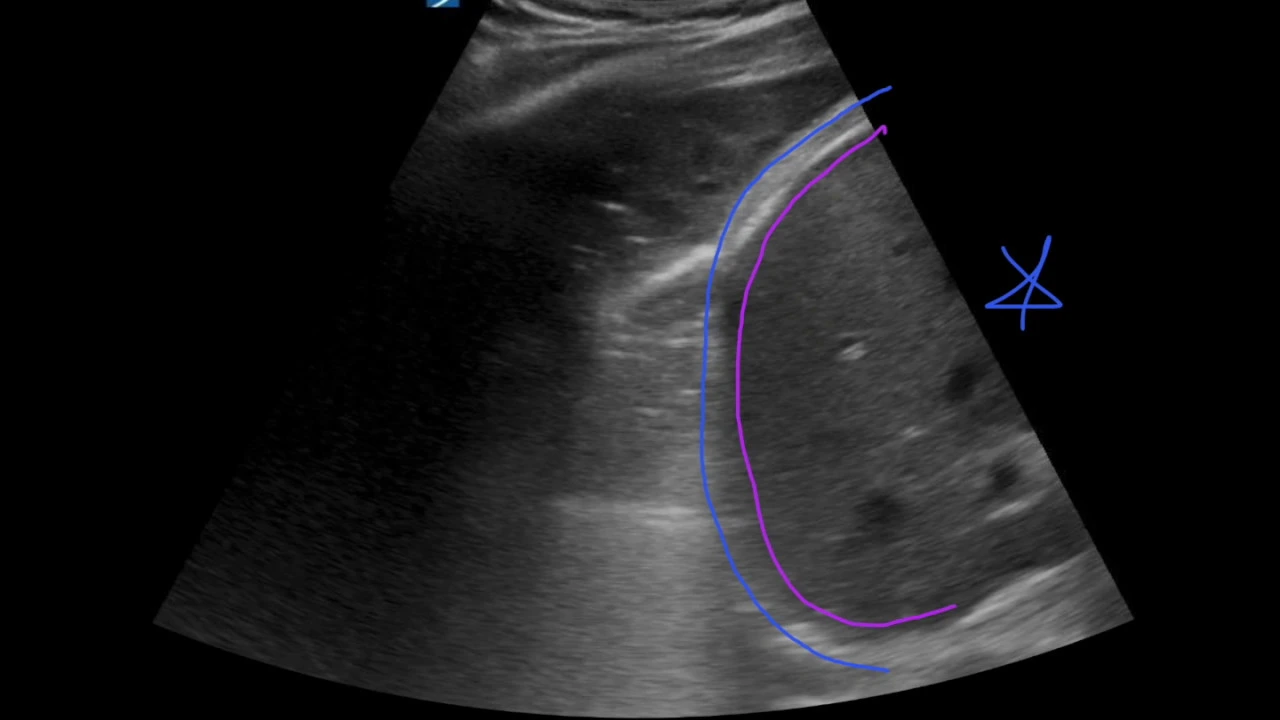
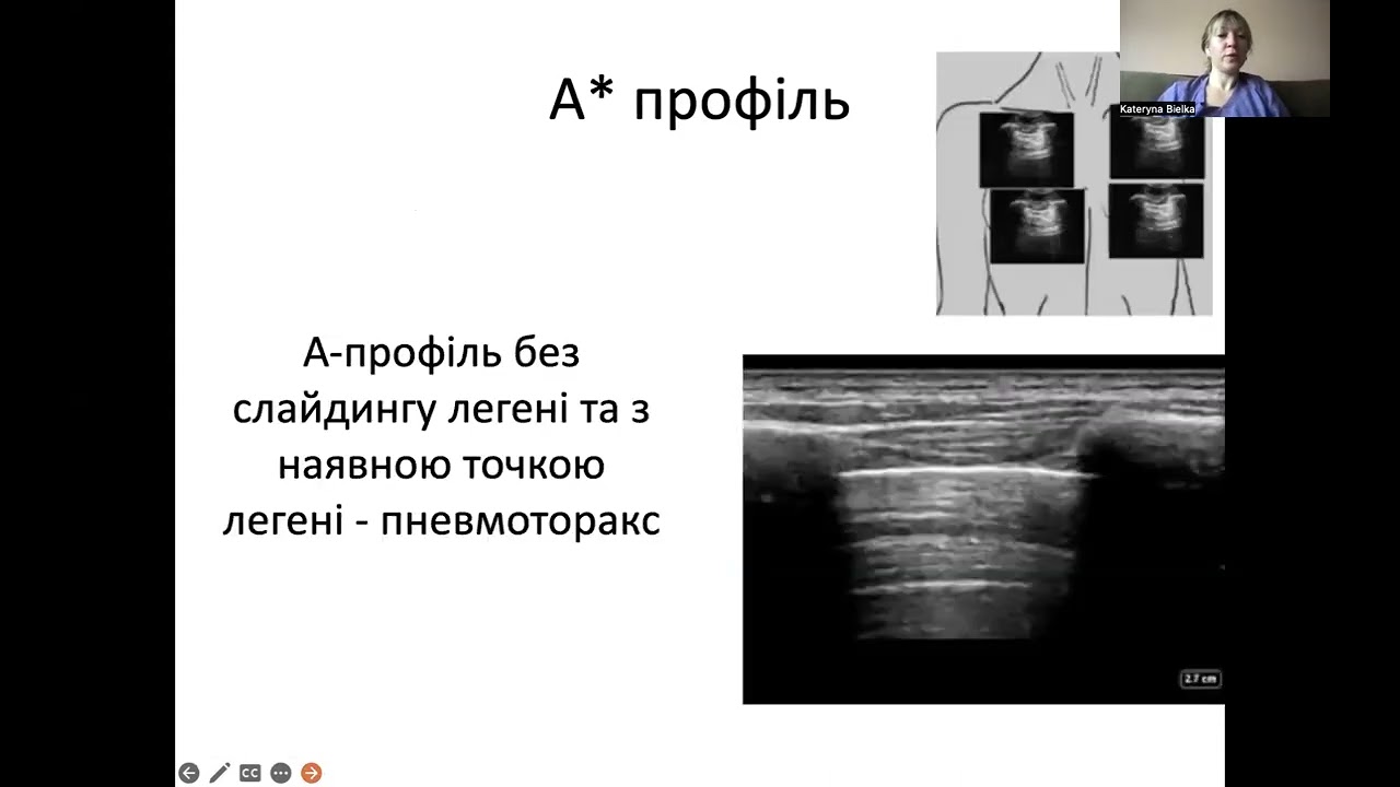
Added:our speaker today is Robert Cole painter the senior cardiac surgery physician assistant at aspire wasu Hospital in Wausau Wisconsin he is a RDMs certified along with critical care ultrasound certified by the American College of Chest Physicians she is a recognized leader in endoscopic vessel harvest and bedside ultrasound procedures Robert is also a member of the point-of-care ultrasound certification Academy committee Robert enjoys teaching and in his spare time playing and teaching guitar Robert thank you for being with us today my honor and I'm honored to be asked if it's okay I'm gonna go ahead and get started what me sure this is something I use every day this is something I'm very passionate about and I believe in this statement that came out of the cardiology literature I use this on almost every patient that goes through our service what I'd like to do here today is kind of inspire you to kind of think about this and maybe little learn a little bit more about it it's a it can be kind of an overwhelming conversation because it is such a long ultrasound that's such a big topic so I hope you guys will move forward with this this is really kind of an introduction to pleural or a lung ultrasound and most people especially physicians in the room who do ultrasound are kind of shocked by that and the reason is is if you look back to Harrisons it is pretty much on everybody's bookshelf from med school back in 2015 they're still saying that ultrasound is essentially useless for a lung alt result even up until this year Harrisons is still saying that it mean they're coming around they're saying that we can use it for pneumothorax pleural effusion but to do things like a value wait for increased lung water there they're just not there yet but it is a rapidly evolving conversation and it's because of this gentleman dr. Daniel Lichtenstein he's a intensivist out of Paris and he started doing this back in the early 80s and he has a brilliant book out there and it's I think I'm reading it for third time it's very academic and you can really get into the weeds on in this subject but what he likes to do is he likes to talk about something called ultrasound semiology and what semiology is it's the study of an object or something that tells you something else is there and this is what we do in medicine all the time we do medical semiology so if i see that i have a big sea wave on my my jugular venous pulsation I can kind of infer that I probably have some tricuspid regurge and this is what we do every day in medicine what he did was he asked the question how about if we just look in if we if we look at the image on the Left we see horizontal lines if we see the image on the right we see kind of more vertical lines that move with respiration now it's so easy that they've said that compared to a kakari ography this is kind of it's it's it's the kindergarten of of what we do with ultrasound historically what we do is we look at chest x-rays but what I can tell you that is that that x-rays infamous for not being very accurate on that same patient on the right side I was not able to find a pleural effusion yet I took five hundred and fifty milliliters off of it so back to the blue protocol people who have problems with breathing lose their airway scares me I don't like it and I was thrilled when dr. Lichtenstein introduced the blue protocol it's uh it's it's emergency ultrasound it's it's not critical care it's a little bit different and it's it's meant to be something that's utilized in the moment fast to give you direction on where to go and this is the the process this is the blue protocol first time I thought of this it really kind of scared me and I thought I was never going to be able to use it but once I kind of grasp the process I got it a little bit more nice thing about it is it's comparatively to most other modalities it's surprising surprisingly accurate and regarding accuracy if you look at the detailed performance of the blue proton call it really does a nice job much better than chest x-rays or even physical exam and stethoscope if you look at it for pneumonia because and you'll will go over all this beep this profile stuff but if you look and you see an abnormality that represents one of these profiles it's again very accurate for pneumonia so to start with the blue protocol they look at the blue points it's a series of three points the upper and lower blue the oppor is essentially at the midclavicular line at the angle of Louie it pretty much tells you what's going on with the upper lobes if you go down to that the the anterior axillary line at the level just above the armpit you have the lure lower blue point my understanding is that initially it was as was was developed to get away from the heart on the left side but if they found that it correlated to the to the middle lobes in the lingula on the left if you drop straight down and that's what they're trying to represent here and you point up towards the sternum you have what's called the plats point the posterior lateral and alveolar or plural syndromes basically what that is is it's telling you do you have a pleural effusion or do you have something going on in the lung which would represent either atelectasis or pneumonia there's two other things that came out of this description of where to look that I use all the time at the bottom of the hand if you drop straight back down this is the phrenic line if you go to the mid-axillary the posterior axillary line you're at the phrenic point I use that for sniff testing up in the office so let's start out by talking about normal lung ultrasound so you put it in the with the indicator up towards the head in the anterior the anterior chest the upper blue point and this is what you're going to look at this is the starting point for any lung ultrasound and it's called the batwing sign apparently dr. Lichtenstein must have thought it looked like a bat and what you have is you have two ribs and then you have this motion below of the musculature and subcutaneous tissues that's called the lungs slide already we've learned that if you see lung slide you've 100% ruled out pneumothorax so and that's the first question asked in the blue protocol is there a lung sliding so if lung sliding is present you have pretty much a transudative pleural space transudative mean that that pleural fluid is made and put into the pleural space and it's me to allow the the visceral pleura to rub up against each other it's a very small amount of fluid but that's what you're seeing right here a transitive process tells you essentially either you have a normal lung you have pulmonary edema you you have maybe have a pulmonary embolism or COPD asthma all it tells you is that is normal lung if it's absent you can see so batwing here pleura here there's no motion it could be representing either an exudative process or absent absent being pneumothorax if you're going to pull the pleura away from the chest wall you're not going to be able to see it and they'll become more clear a little bit later in the talk what happens pneumonia with pneumonia is pneumonia is usually a more peripheral process so when the infection abuts the lung it's very inflammatory and it gets inflammatory and gums up the pleural space so you'll lose a lot of the lung sliding so really the idea behind lung sliding is it puts things into two boxes based on their pleural presentation the second question asked is do you have an a profile or a brief profile well what's an April file in just step back that little prime up there that you see with absent is just telling you that that puts it on the right side of the column that there's no lung sliding so we're back to our batwing sign except I've increased the depth on this image a little bit and if you'll remember that that slide I had with Harrison's internal message our internal medicine it said that air is dissipated once it hits the pleural line well to me that that says that their philosophy is once you hit air all that is is really all that information is not useless and I disagree they actually call that Merlyn space not because of Phantasia it's actually somebody's name that was assigning this but this is truly where all the good stuff all the magic happens things like these horizontal lines that come in that we need to kind of find out what do they mean so this is what happens so we have a transducer and then put it up against the chest wall what's going to happen is the ultrasound beam is gonna come and it's gonna meet two reflectors it's going to hit air at the lung so there's my inner lobular fissure here and this is the transducer fissure or this is the transducer face the ultrasound beam is going to come out and it's going to hit the transducer and it's going to paint the pleura right where it's supposed to be and then it's gonna reverberate because there's such high specular reflectors it's gonna bounce back and forth and every time it does that it's going to paint a horizontal line at the same depth deeper with lower intensity and this is what it looks like you can see it's equal distant down lower intensity as it goes and that's all that tells you is that it's air you can do this with just a plain transducer if you have a linear int or a transducer go ahead and put some ultrasound gel on there once the beam comes out it goes through the jelly it hits air it'll bounce back and forth and you'll see some form of reverberation artifact the absent so it's only found in normal lungs or in pneumothorax the absence of a lines means that something else is taking them over and we'll get into that shortly so what's a beat profile be profiles a little bit different and I like to kind of start out by talking about curly be lines because everybody talks to them and they're just these little horizontal lines that are sometimes found x-ray mostly with people who have pulmonary oedema but it's found in all kinds of other things what we can also this is what it looks like in ultrasound it's the same place that it happens but it looks much a difference in to me it represents a lot more information it happens at the secondary lab you'll the secondary lobule is just the endpoint of our pulmonary system it's a bunch of Asuna at the end of the bronchial tree where the gas exchange takes place and it's it's it's it's put into little boxes of interstitial tissue or septic called interlock the other septum and that's right here this is where all the things most of the initial changes from eight lines to be lines happen with lung ultrasound what we know is as the PAMP pump fails we get as a heart fails you end up getting pulmonary edema and uh when the pressure get the back pressure gets to be enough a lines are now replaced by B lines it's instant on instant off it's the first thing that you see it's a very early indicator and it's going to be a part of echo protocols within the next five to seven years there's some amazing stuff coming out of the Italian literature right now on this and this is how they're formed here we have a new interface or transducers on the chest wall we have a new interface so the ultrasound beam is going to come and it's going to get into the inner lobular fissure because it's now filled with fluid and it's going to reverberate back and forth between the air here and the pleura now to put this into perspective I think what Ben Stein did a really good job describing this you have an ultrasound beam moving at 1540 meters per second going into a structure about seven hundred microliters that's about I think it's three one hundredths of an inch so it's very small so you're putting a huge energy into a very small space and this big explosion kind of happened and every time you the beam comes down it bounces back and forth and you end up with this this vertical artifact that's generated from the inner lobular fissure and that's a really important point because B lines are generated only from the visceral pleura and again this is what they look like you can see how they move with respiration when you see B lines that you essentially in the anterior chest you essentially have an interstitial syndrome interstitial syndrome meaning you have pulmonary edema either cardiogenic or non cardiogenic pneumonia or pulmonary fibrosis these are the criteria for using them when they say that that one of the criteria is a common tale artifact that doesn't mean that every common tale artifact is is a beeline a beeline is a type of common tale artifact we all if you look at echocardiogram parasternal long you look at the pericardium you have what looks like comet tail artifacts those are actually called are lines they arise from the pleural line 100% of the time and they move with lungs sliding now it's really important I think one of the mistakes that's done with teaching lung ultrasound is depth they see several things online where you're at four or five centimeters and you see these horizontal are these linear artery artifacts and the it's you can't really tell whether there are B lines or something else look Lichtenstein's book was very academic and he defines a whole alphabet soup of artifacts based off of ultrasound and if you're not past seven or eight centimeters there are some lines that mimic B lines that are very short that only go to three or four centimeters they're called the high lines is they don't represent it to anything there's another artifact called Z lines they're the same thing three four but three or four centimeters but they don't move with respiration and again there are they're parasites they're just artifice that don't mean anything so let's do a little bit of quiz to get through the rest of this so here we've got two pictures okay I've got right and left both upper blue I've got a rib here rib here pleura here and I'm seeing B lines so the first question is is do we see lung sliding and the answer is yes we do do we have a B profile yes we do so what's our diagnosis going to be pulmonary edema so if you see B profile diffusely across the anterior chest in the upper and lower blue chances very good chances that your R that you're dealing with some type of a pulmonary edema whether it's cardiogenic or none cardiogenic so in this image do you see lung sliding we do we have an eighth profile I don't see B lines I see reverberation artifacts coming from the pleura that are equidistant and fading with time so what do we do with this we should really look at their legs is the recommendation and there's a another really good podcast on her or webinar that kind of details this but I'll touch it in brief most deep venous thrombosis are going to found be found either in the popliteal space or in the the femoral vein approximately and we use a two-point technique we're taught with critical carriers where we look at four views per side we look at the common femoral vein the junction of the greater saphenous vein the take off of the poor Fonda and the popliteal and it's not that hard either you have compression or you don't if you don't have compression as you see here you have a DVT you can throw some color on there and make sure that it doesn't fill up so if you go down the road or the the pathway and you see a thrombosed vein that they call that 1/8 so you have an a profile DVT so you have a pulmonary embolism you have to be careful with this and I it works but it's it's not perfect 30% of all people who have a pulmonary embolism will have no residual DBT so you really should look at your per criteria or whatever you whatever risk stratification you want to use for pulmonary embolism so if you go down this pathway and you see free veins you want to look at flaps point which is again straight down from the lower blue and you may see something like this so I've got liver here on the right diaphragm here but I've got this which looks like it's liver above the diaphragm and I've got this which is called shred sign I'm not going to get into a lot of this because I really you can really get into the weeds on this but this is a consolidation if you see something like this now I'm on the left side you've got spleen here diaphragm here you've got a moderately sized pleural effusion with some consolidated lung bottom line is if you see anything that looks like fluid or it looks like liver above the diaphragm you've got a Platts point so a meaning you've got an a profile your veins are clean but you've got a positive plaques be positive platypus point that represents a pneumonia then people come in from the ER they really shouldn't have something like this if your plaques is clean you have something called a newt profile what I mean by that is you have a normal pleura you have free veins and you have nothing hanging out in your Platts point that's essentially a normal lung if I have one use for a stethoscope it's wheezing I cannot I cannot use ultrasound for that and and you can just assume that it's most likely COPD or asthma if you have any lung sliding but you have something called an a B or C profile a B meaning you have on the right side B lines on the left side you have a lines or the C profile which means I'm up the I'm up the upper blue point here and I something that comes in and out that looks like liver and it's gotta be lines surrounding it or comet tail artifacts distally that's called a sea profile or anterior consolidation that represents all of these represented pneumonia so we've looked at a little bit more than half the protocol what's nice about this is this is where in my mind the moneymaker as in emergency room well greater if there's a pretty good study out there that came out this year that shows that the majority are all going to be found on this side of the aisle with pulmonary edema and a nude profile or you're dealing with COPD or asthma being the most common things found to complete the the other side of the graft if you have lung sliding which is absence you have an a prime profile so batwing here and here's my pleura I do not see anything here as far as movement of the pleura you have to look at something called lung point we talked about lung slide and if you see it that 100% rules out pneumothorax but if you don't see that that does not tell you you have a pneumothorax it suggests it because again pneumonias are usually peripheral and they can really gum up the pleural space and and really preclude long slide so what you have to do is you you really should look for something called lung point if I place the the transducer on a patient on a patient who has a pneumothorax anteriorly here I'll see batwing sign but I will not see plural motion because the pleura is pulled away from the chest wall what I really have to do is move my transducer over to entrain find a spot where the the lung meets the chest wall and you'll get something that looks like this where the the lung comes in and out of view so here there's no pleura moving here here there's pleura moving okay you can see that point right there it's called lung point and that's what you're seeing here is it as they breathe the lung pulls away in Riya proximate to the chest wall and you're just seeing it represented on your ultrasound screen here's a better image of what this looks like you can see coming in right here in and out that tells me 100% this patient has a pneumothorax and I can treat it so back to this if I have an absent if I have no lung slide in an a prime profile I have a pneumothorax if I can't find a lung point the recommendation is is you you base it off the clinical picture if your patient is in extremis then you put in a tube if your patient is comfortable and you have time to look for for another modality whether it's chest x-ray CT you should do it and the reason that you might not see lung point it's probably my understanding is that it's a 60% of patients you won't see it it has to do with the fact that most pneumothorax at Earth the majority of new authorities can be complete in the emergency room so the entire lung would be pulled away from the chest wall there's something that I haven't seen yet and that is if you have no lung slide but you have a B Pro V Prime profile but I'm assuming it would look like this and the literature it's very rare but it would represent a pneumonia and that's the the blue protocol so just for example this is a patient that I did a couple of weeks ago we'll just call or miss some patient she had an op cab I left on Friday and she was on three liters of oxygen she's a big smoker so she's got some pulmonary issues I'm sure I come in on Sunday she's on 70% fio2 in really kind of limping along so our usual move in in in medicine is to obviously listen to him which is fairly ineffective but also to get an exam and this is her chest x-ray this one is her preoperative and this one is her post-operative on post-op day for when she's struggling and I see maybe a little plural fusion here little pleural effusion here it doesn't look like she's in CHF to me I see no pneumothorax so really for somebody who's on that much oxygen I don't find it very helpful the the radiologists that I respect agreed with me that this was kind of a small bilateral pleural effusions but pretty unremarkable so I went down my pathway I went to the upper blue the lower blue diffused B lines everywhere she's in CHF probably probably residual fluid from surgery she's just mobilizing him I want to step further and looked at collapse point so I'm sorry she's got she's obviously beat she's got lung sliding which is present even just that the suggestion of B lines tells me that she's got lung sliding because they're generated from the visceral pleura and I've got B profiles so she's got pulmonary edema went one step further and looked at her plat at this point and on the right side liver diaphragm moderately-sized pleural effusion probably compressive atelectasis here and on the left side she's got spleen here heart here this is all an elect attic lung this is this is her whole left lower lobe is down because you can see all the way through the lung to the heart you shouldn't do that when you're looking at the lung so that's all I have and I really believe this this is going to be a real part of our existence in the next several years and I hope you'd enjoyed this thank you Robert we really appreciate that that was phenomenal and incredibly educational and I for those of you on the on the webinar that don't know me if you haven't attended one of our webinars before I am non-clinical and so I loved listening to your talk Robert I think that that I learned a lot and I hope everyone in attendance did as well we actually had a couple of questions come in while you were talking and so one of them is actually going to drive you back to the slide with Mickey Mouse from Fantasia one of the audience members asked if you could review Merlin space again they weren't quite sure what Merlin space was if you could talk a little bit about that sure so back to batwing sign if i can go back a little bit in time err if you'll bear with me so batwing sign you find one get in there I promise here we go so rib here rip here pleura theoretically because you're hitting err none of this is real so the space from the the pleura here all the way down is is artifact so that's called Merlin space the space above the pleura from from the skin down to the pleura there's actually a name for that it's called key space thank you I hope that that answered the question that was asked another question that came in is for those of us learning lung ultrasound and just starting out do you have a suggestion or multiple suggestions of how to see examples of pathology or cases that they can utilize their learning absolutely I think probably one of the best places to look is on the line there's some great emergency sites for foam med foam that is free online access medical education one of my personal favorites is something called five minutes oh no it's an ear physician who has been very generous with his his time and his skills who has put a lot of videos online about educating and with pathology but if you look up fo med point-of-care ultrasound you'll get a listing also the Society of point-of-care ultrasound has an area that you can go to that lists out an enormous amount of places that have pathology thank you I would also throw a plug in for the pocus certification Academy we do a monthly Taste challenge and so you can visit our website there's a whole education section and we have had a number of lung cases recently so and I know we have a few coming up so take a look there as well one last question I think we have time for one more and the question is are you concerned but nonsan ographers will begin being called experts um no I won't I don't you know it's it's much different point of care ultrasound is much different than the comprehensive exam it's done at the bedside to make real-time decisions in patient care there are some things they can't be done in radiology like lung ultrasound I've not met radiology department where I can say or I can put in an order for a lung altra sound and there's there's I think one of the beauties of of point of care ultrasound are taking it out of the department to people who are where the clinician it's in the clinicians hands is using protocols like this to better better get a grip on on what's wrong with a patient it just doesn't have to be lung ultrasound I mean the rush protocol uses heart-lung IVC fast exam it uses a whole whole series of images to stratify what's what's going on with a patient with hypertension there are several protocols out there so I think they're know on the flip side of that Congress I am an advocate of education I do agree with every position paper out there from the American Medical Association and the American College of Radiology work I think training is paramount and there's there's some really well defined ways of getting that training which is coupled with mentorship thank you all right I know I said that last one was the last one but this one is truly the last question and Robert I think that you had really good points there and I think for anyone who is concerned about kind of the growth of focus and taking ultrasounds from a purely sonographer realm into the hands of other healthcare providers I think that anyone on our pocus certification Academy team or committee members would be happy to have conversations with you so please do not hesitate to email us focus at octa org and we would be happy to connect with you but Robert last question any any effectiveness comparison between the blue protocol and then more traditional Methodist methods of evaluating lung pathology and it seems that there may be some opportunity for false positives and so just a little bit of wondering there your thoughts on that I'm sorry can you repeat the question sure it looks like they're asking for comparison between blue protocol and traditional methods of evaluating lung pathology sure tradition traditional ways of evaluating pathology have been great you know we were given very few tools and are tooled back to look at this from a physical exam point really is the stethoscope and the stethoscope is is really poor not only in evaluating the lungs but also the heart and the studies on us are very clear they released a paper in August of this year evaluating how physicians did with lung ultrasound and it was a 50/50 chance whether they could identify at normal lung or not with a stethoscope and it's because we just stopped learning about it the chest x-ray is is marginal at best the studies on that compared to ultrasound are are really not even close ultrasound returned the pathway of a CT scan but one CT scan which i think is just an incredibly overused especially out of the in the emergency setting is I mean that's that's four or five hundred chest x-rays at one time so the question that's coming out of some of the the the opinions are what are we doing to patients by exposing them to that much radiation my opinion ultrasound is a great starting place compared to other technologies thank you Robert and thank you to everyone who was able to attend today Robert we really appreciate you spending your time with us and sharing this incredible educational webinar thank you for being here
Up Next
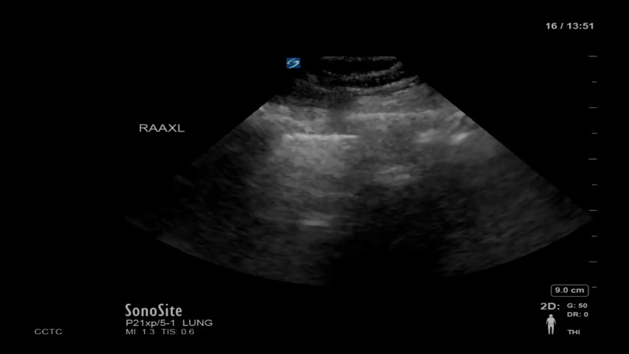
Lung Ultrasound Interpretation: A Systematic Guide
@westernsono
67.2K views•2018-03-24

Graphic Medicine: Comics for Collaborative Healthcare Communication
@nationalpatientadvocate
189 views•2023-12-04

Neuroanatomy: Central and Peripheral Nervous System Divisions Explained
@AKLECTURES
136.2K views•2014-09-20

Stages of Labor and Vaginal Birth | Childbirth Animation
@nucleusmedicalmedia
52.1M views•2017-08-18
Related Study Plans & Knowledge Roadmaps
Structured learning paths in Medicine