
























Lung ultrasound interpretation follows the BLUE protocol, which uses specific sonographic signs to diagnose respiratory conditions: A-lines indicate air in the pleural space (pneumothorax or emphysema); lung sliding and pulse confirm pleural contact and rule out pneumothorax; the lung point (transition between sliding and non-sliding pleura) is diagnostic of pneumothorax; B-lines (linear artifacts from fluid/fibrosis) indicate interstitial pathology and rule out pneumothorax; lung consolidation appears as tissue-density gray structures with characteristic signs like the spine sign; and pleural effusions are detected using the quad sign, sinusoid sign, and jellyfish sign, with qualitative assessment based on size and echogenicity.
Lung Ultrasound Interpretation: A Systematic Guide
Added:Basic anatomy of the respiratory system, including the lung lobes, pleural layers, and chest wall structures.
Fundamental physics of ultrasound, particularly how ultrasound waves interact with different media (air, fluid, bone) and the concept of acoustic impedance.
Basic ultrasound machine operation, including probe selection (linear, curvilinear, and phased array) and primary controls like depth and gain.
Pathophysiology of acute respiratory distress, including the clinical presentation of pneumothorax, pulmonary edema, pneumonia, and pulmonary embolism.
Integration of lung ultrasound with Point-of-Care Echocardiography (FoCUS) for comprehensive cardiopulmonary assessment in undifferentiated shock.
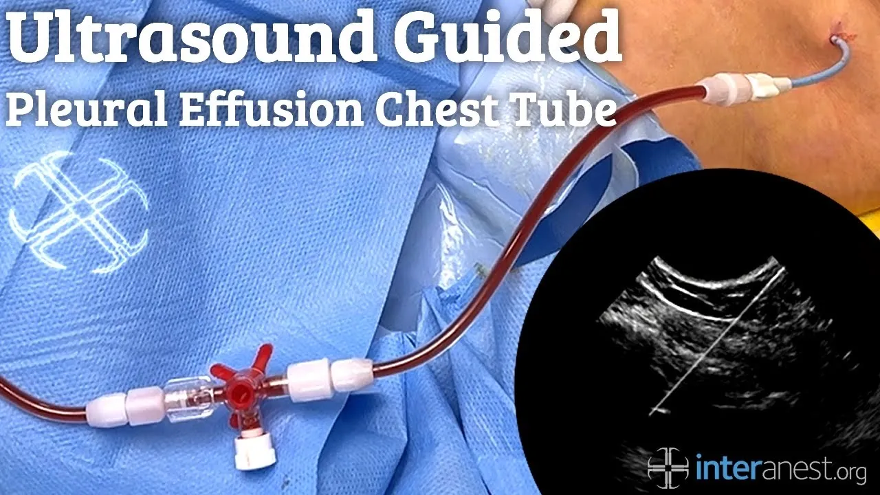
Clinical application of ultrasound-guided thoracic procedures, such as safe thoracentesis and chest tube placement.
Utilization of quantitative lung ultrasound protocols, such as calculating the Lung Ultrasound Score (LUS) to monitor ARDS progression and recruitment.
Exploration of specialized pediatric and neonatal lung ultrasound protocols, focusing on transient tachypnea of the newborn and respiratory distress syndrome.
67.2K views953likes28:39Original Release: 2018-03-24
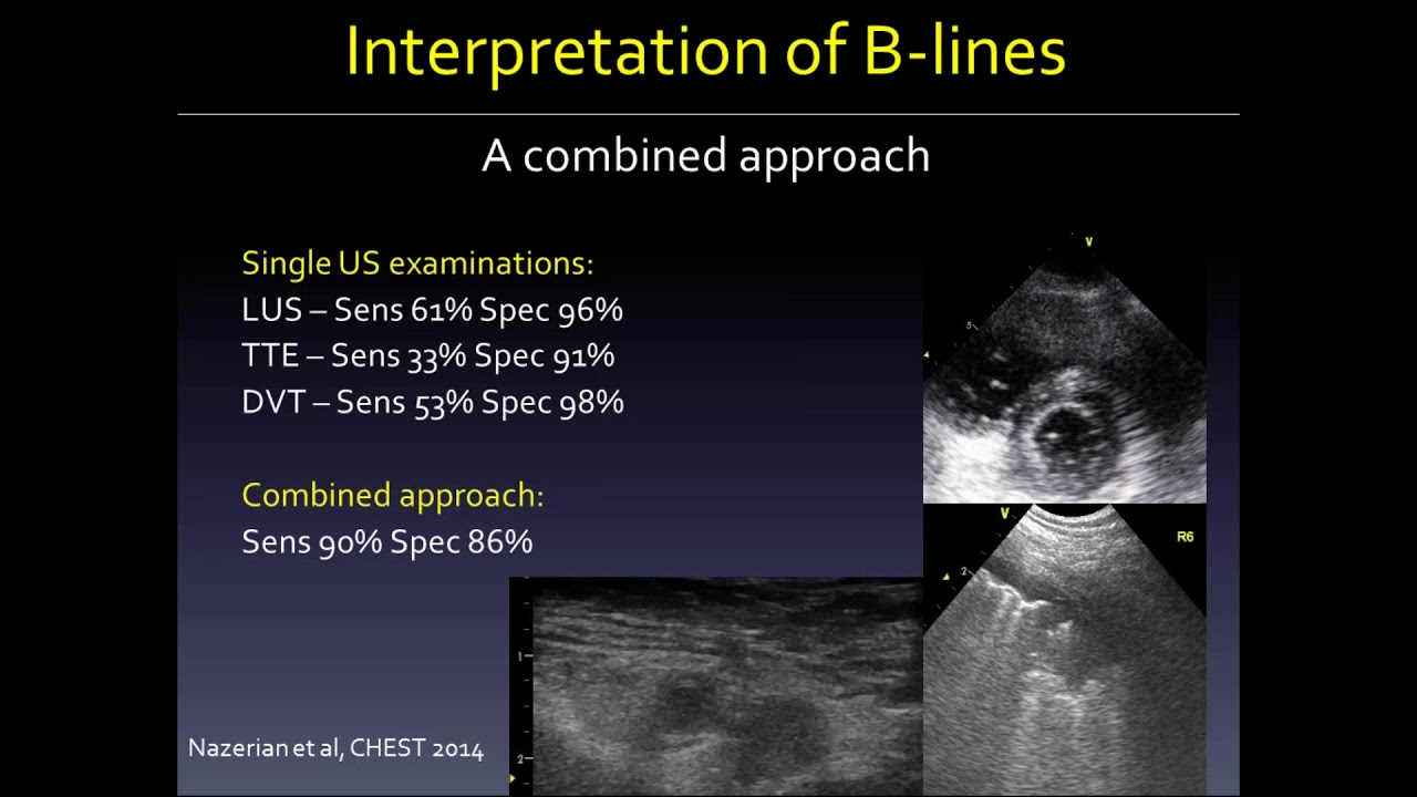
welcome back hopefully you've taken a little bit of a break and are ready to learn about lung ultrasound image interpretation so without further ado let's get started this is probably the most widely cited of the long ultrasound interpretation protocols it's named the blue protocol and was created by Liechtenstein in Maisie air in the journal chest in 2008 it allowed them to arrive at a diagnosis in patients presenting with acute respiratory failure in an ICU setting with a 90 point five percent accuracy at first glance it can seem complicated but we can break it down into a few basic patterns to recognize in different steps the first step is to assess whether there is presence of lung sliding once its presence has been established we need to us to be able to assess for the presence of a lines B lines consolidation or lung point or the point where non-sliding long begins to slide let's go over these findings individually as well as the other lung ultrasound finding of pleural effusion this is the basic long ultrasound view we initially see subcutaneous tissues a little deeper we have different white lines there are these two mildly higher lines that represent the ribs we can know they are the ribs by the characteristic shadowing deep to them the white line between them and just deep to them is the pleura the culmination of all of this is termed the bat sign if there is air below the pleura either from healthy lung earth pneumothorax we will be able to see reverberation artifact as repetitions of the pleural line have multiples of the distance between the transducer and the pleura this is the a-line pattern and indicates that there is air just below the pleura either in the lung in emphysema blebs or between the visceral and parietal pleura so a pneumothorax it's important to see both ribs and the bat sign to not mistake another artifact as a lines as you can see here it would be easiest say that this clip represents a lines however by sliding the probe over a little we can find the true pleura and see that what we were seeing was in fact reverberation artifact from the rib itself remember any strong reflector can create reverberation artifact and produce an artifact that looks like a lines this is why it's important to see bat sign because the pleura will be the first bright white line deep to the bright white lines caused by the ribs once you've found the pleural line the next question is whether or not the parietal and visceral pleura iearned contact to ensure there is no pneumothorax when the parietal and visceral pleura are touching the rubbing that happens between them during ventilation produces a kind of shimmering that's been likened to ants crawling and is termed plural sliding when patients aren't breathing or ventilation areas minimal as can happen with low tidal volume ventilation or in Regent's over descended by high peep or obstructive lung disease the only indication of the contact between the two plurals may be the effects of cardiac oscillations making them vibrate together this is termed long pulse and like lungs lighting indicates pleural contact and rules out pneumothorax in that area on occasion the pleural sliding or pulse can be difficult to see decreasing the gain even down as far as it will go well often enhance the contrast so that the pleura and it's movement can be seen more clearly other useful tricks are adjusting the depth so the pleura is in the middle of the screen switching to a linear probe to enhance resolution and using M mode as we will discuss shortly another useful trick for optimizing the image and the appearance of artifacts is ensuring the probe is perpendicular with the pleura this is achieved by slowly tilting the probe until our artifacts are optimized as is happening here we're progressively clearer and more numerous a lines appearing as we get more perpendicular with the pleura on occasion you will notice that on some of the following Clips we do not see the bat sign this is generated because we have found the bat sign I've confirmed which line is the pleural line but once this is done it may be necessary to not have boat ribs in view in order to have the best view possible of the structures we are trying to look at on your image however you should still always start by finding your boundaries and this will mean finding the bat sign to make sure the line you are looking at is the pleural line here is an image of loss of lung sliding we can still see that there is some motion happening but this is not the shimmering we saw previously and is simply due to motion of the chest wall loss of lung sliding and pulse alone do not indicate that there's definitely a pneumothorax present rather presence of lung sliding or pulse excluded in the thorax at that point here we can clearly see there's lung sliding on the right of the screen while there isn't any on the left here is an example of lung point or the intersection of sliding and not sliding along this is diagnostic of a pneumothorax this is the point where the pleura go from being in contact to not being in contact as we can see here on this chest CT the location of this point changes somewhat with inspiration and expiration which is why we see intermittent sliding and loss of sliding at the same location on the pleura it is also possible to demonstrate lung sliding the absence of lung sliding and the lung point on M mode since the subcutaneous tissues aren't moving they appear as straight lines above the pleura if the pleura is also not moving we will also see that that the artifacts below it are not moving as we see here this has been termed barcode sign given the prison presence of all straight lines on the screen if the plura are rubbing the pleural line and the artifact below it have a grainy appearance given the movement or friction this has been likened to sand and so this is called a seashore sign where above the pleura are the linear waves from the static subcutaneous tissues that look like still waters and at the level of the pleura and below are the grainy sandy shores if we see both of these signs at a given point then we have the transition from sliding to not sliding and vice versa that happens with breathing and so we have a long point which is diagnostic of a pneumothorax the reason a lung point is necessary to diagnose a pneumothorax is because other phenomena can stop the pleura from rubbing against each other such as emphysema emphysema blabs therapeutic pleurodesis pleurodesis from pneumonia etc unfortunately if there is no place where the to Clara come back into contact like in this tension pneumothorax there will be no long point and so it will not be possible to conclusively diagnose a pneumothorax all you will see is loss of lung sliding this is why it's important to integrate the clinical context into your interpretation just like with auscultation or any diagnostic aid another important thing to know is that other things that intermittently rub against the pleura can resemble a long point or create a pseudo long point this clip shows a suit a long point in a region on the left and clear anterior chest where the pericardium intermittently touches the pleura resembling a lung point as you can see what we are seeing isn't quite long sliding and we are able to see a structure the pericardial space deeper your eye will become trained to see the difference between the much like your ears become training to ear the different sounds of long auscultation that closes the chapter on a lines next let's move on to the other important artifact you will be seeing which are B lines these are linear artifacts created by fluid fibrosis or other phenomenon in the lung interstitial as we discussed in the artifact segment they need the two layers of the pleura to be in contact to be produced and so their presence rules out a pneumothorax in that lung location just like long sliding or long Falls does in order to be called B lines these lines have to meet certain criteria they have to be straight well-defined hyper echoic lines that are in the axis of the being they must arise from the pleura they must move with plural sliding they have to go all the way to the back of the screen or the far field and they should obliterate a lines along their path this doesn't mean that a lines and B lines can't coexist in one window screen we see if here that they can rather if a B line goes through an a line we lose the a line at the spot where they meet when fluid in the interstitial is causing B lines the pleura will appear regular as it does here here are the same B lines with a regular pleura but seen this time with a linear probe for better resolution when you see this it is generally caused by fluid and so non inflammatory conditions such as cardiogenic pulmonary edema should be high on your differential diagnosis when the B lines are from an inflammatory or v grossing process the pleura will tend to fear more jagged and having a regular shape as it does here you again here are B lines with an irregular jagged pleura in a patient with a RDS this time image with a linear probe if the pleura looks like this inflammatory conditions such as a RDS pneumonia or inflammatory interstitial lung disease should be higher on your differential diagnosis here they are side by side with the regular thin non inflammatory pleura on the left and the irregular jagged inflammatory pleura appearing on the right a few B lines - or less especially independent regions can be normal much like faint and inspiratory crackles independent regions however if you see them in multiple ultrasound windows in the non-dependent lung regions if they are very thick if they are numerous so three or more signaling a b-line dominant lung profile and if they arise in someone you suspect of having a respiratory pathology they are more likely to reflect significant lung disease when B lines become very numerous they can become confluent as they are here and look almost as if flashlights were shining from the pleura to the far field we know these are concluded B lines and not normal long given the absence of a lines our next long ultrasound finding is lung consolidation when this occurs the lung becomes deflated or filled with fluid and takes on tissue density it bus looks gray on ultrasound and is sometimes called the patters ation to reflect the fact that consolidated long looks like liver in the case of Thor and Slovak consolidations the lack of significant lung aeration also allows ultrasounds to be transmitted deeper allowing us to see deeper structures this gives rise to the spine sign or the visualization of the spine through the lung which we have here this helps us know that we are truly seeing consolidated lung and not a mirror imaged artifact when the consolidation is not trans lover we will see a fractal border caused by ring down artifact which are sometimes mistaken for B lines this is known as the Shred sign and signifies a non trans lower consolidation also interesting to note here is the decrease in size of consolidation on inspiration suggesting some component of atelectasis as you can see here consolidated long generally has a density similar to the livers or the spleen and so if you aren't careful you can mistake a normal abdominal organ for an abnormal lung the best way to avoid making this mistake is to find the diaphragm which you should always do in the lower lung zones anyway as you may expect an infra diaphragmatic tissue density for tissue density below the diaphragm will not be consolidated long at times there can also be tissue density above the diaphragm but not in the long as we can see here from top to bottom or left right on the screen we have long a complicated paraná monika fusion or empyema a miss shape and diaphragm and finally the liver this can make things quite complicated again finding the diaphragm will be a huge help if the tissue density is above the diaphragm but you're not sure if it's longer effusion here's another trick consolidated lung will have blood flow whereas complex effusion walked by placing the color Doppler box over the tissue density you'll be able to see whether there is blood flow in the consolidation or the tissue density or not if there is blood flow you're highly likely to be in the presence of consolidated long rather than a complex effusion another feature of consolidated lung is that there will often be some residual air remaining in the bronchi this is visible as I've ripped go xbox and lines within the constant consolidated long these are called air Branca grams just like on chest radiograph there are two types of air brach Gramps novo the more common are static air Bronk Gramps these represent air that is trapped within the bronchi and so when the lung moves it moves but it doesn't have the features of flow that we will see with our next type of air brocco gram static air bracha grams can be found with any cause of consolidation but don't really help us know what the etiology is dynamic air programs happen when there is residual air flow in the consolidate as long causing the air in the bronchi to move with inspiration and expiration they have a flowing quality sometimes late something like rail cars moving on a train track usually if there is air going into the lung it should be inflated so dynamic air Branca Graham suggests that something other than just loss of aeration or atelectasis is causing the lung to be consolidated this is almost always because there is infection in the lung tissue or pneumonia consequently the specificity of dynamic air broker grants for pneumonia is 95% unfortunately the sensitivity is much lower one important minute of air bracha grams to know is air within the stomach but again this will be under the diaphragm and devoid of blood flow around the air it is also much more likely to be on the left side mirror image artifact of the liver or spleen can mimic lung consolidation this tends to happen in the dependent region of healthy lungs where the interface of the diaphragm and aerated lung serves as a mirror and projection liver or the spleen the absence of B lines and the disappearance of the mirror image artifact with respiration or small probe movements are good clues to the presence of this normal mirror artifact you also won't likely see any of the other features consolidations that there's a spine sign another phenomenon you may observe especially with non-consolidated lungs as the curtain sign this happens at the interface between the lung and abdominal viscera at the level of the diaphragm the abdominal viscera are visible during exhalation and with inspiration the lungs inflate lower and a long artifact appears and obscures the abdominal contests on screen signifying that the first thing the ultrasound beam is encountering after the pleura is aerated lung and not a fusion or consolidation the long artifact may either be in the form of a lines like here or D lines the last long ultrasound finding will discuss is pleural effusion most pleural effusions will appear as anechoic or hypoechoic and be located in the dependent regions between pleural layers or as we can see on ultrasound between the parietal pleura on the diaphragm or chest wall and the visceral pleura on the log this gives rise to two signs that will help us detect pleural effusions the quad sign represents the formation of a four-sided anechoic or hypoechoic density the four sides are formed by the chest wall the long line and the rib shadows on each side the sinusoid sign occurs when the long line to Ward the parietal pleura on the chest wall during inspiration this means the lung is able to move reasonably well within the effusion and therefore the effusion is unlikely to be loculated the effusion is unlikely to be loculated another important sign of motion of the lung in the effusion is the jellyfish sign or lung flapping we can see as a lung floats in the effusion it is important to make sure you see the diaphragm and subdiaphragmatic abdominal viscera when looking at an infusion to make sure it is indeed within the pleura it is also important to make sure you see the lung in chest wall again to make sure the effusion you are seeing is within the pleura here it would be easy to be fooled into thinking that this anechoic space is pleural effusion however once we note the absence of long within it and the fact that it is above the parietal pleura it becomes clear that it is in the chest wall itself here's another example where determining the boundaries on diffusion is important on first glance it might be tempting to say this effusion is entirely inside the pleura however on closer inspection part of the effusion is between two diseased pericardial layers and will require much different management different formulas exist to attempt to predict how much fluid is contained inside a pleural effusion and generally perform best for moderate the fusions however for most clinical circumstances qualitative assessment dividing effusions into small or about 0 to 2.5 centimeters between the lung and the chest wall moderate or about 2.5 to 5 centimeters between the lung and the chest wall and large volume or more than 5 centimeters between the lung and chest wall will be sufficient some degree of AD Alexis is expected whenever there is a pleural effusion if the effusion is roughly larger than the consolidated long this generally represents passive atelectasis due to the effusion itself otherwise there is either more atelectasis than it's expected for the size of the effusion which may be due to recumbent C mucus plugging etc or there is a coexisting cause of lung consolidation such as pneumonia here the consolidation is mildly larger than expected given the effusion and so something else is likely contributing to the consolidation on occasion there won't be any consolidation and you'll be able to appreciate a lines or B lines where the effusion meets the lung these are termed sub a lines and sub B lines respectively apart from size and location it is also important to note the echogenicity of the fluid most of fusions will be trans dates and will appear anechoic or black exudative effusions will generally be hypoechoic or less commonly i silico ik or i / ik ik here we can see that they are de breeze floating around in the effusion this is termed plankton sign and suggest a complex or exudative effusion often when we see the plankton sign there will only be one or a few small complexities floating around in the fluid so it'll be important for you to keep a watchful eye here's an example of a mildly hypoechoic and heterogeneous complex pleural effusion with the lung above it and the liver below it again identifying all the local Anatomy and the use of color Doppler to confirm the absence of blood flow in the collection can be useful to confirm it is a pleural effusion and not a consolidated long or abdominal organ other times it would possible to cease thicker fibrin strands in the effusion again this suggests a complex or exudative effusion more often an empyema and is likely to warrant training we can also appreciate here that sometimes with necrotizing pneumonia the border between the lung and the effusion can be difficult to determine one structure you may see that can be confused for fiber and/or adhesions is the inferior pulmonary ligament that ties the base of the lung to the diaphragm seeing this one big strand in otherwise anechoic appearing effusion will help you know that is in fact the inferior pulmonary ligament that you are observing and not a complex of fusion again the clinical context is likely to help you in these kinds of situations it is also important to distinguish between fibrin or complexity in an effusion and being thickness artifact as we discussed in the artifact module this is caused by compression of its three-dimensional structure into two dimensions and represents a partial display of the lung fanning through the structure will reveal that it is in fact merely partially seen long that's it for the respiratory ultrasound modules naturally there's a lot more content out there and much more you can continue to learn about respiratory ultra sonography but hopefully this will help you begin to use ultrasound with a solid base don't forget to check out Western Sun or calm for more information tutorials and further lectures
Up Next
14:43
Basic Echocardiography Tutorial: Cardiac Ultrasound Views
@mghultrasound8334
258.2K views•2014-02-07

48:47
Graphic Medicine: Comics for Collaborative Healthcare Communication
@nationalpatientadvocate
189 views•2023-12-04

10:28
Neuroanatomy: Central and Peripheral Nervous System Divisions Explained
@AKLECTURES
136.2K views•2014-09-20

3:05
Stages of Labor and Vaginal Birth | Childbirth Animation
@nucleusmedicalmedia
52.1M views•2017-08-18
Related Study Plans & Knowledge Roadmaps
Structured learning paths in Medicine
medicine
Abnormal LFTs: Diagnostic Workup & Pathology
01Liver Anatomy and Basic Physiology
02Introduction to Liver Function Tests (LFTs)
03Hepatocellular vs. Cholestatic Patterns
+ 3 more learning modules
12h6 modules
Explore Roadmap
medicine
Analyzing Blood Smears: RBC, WBC & Platelets
01Introduction to Blood Components & Smear Preparation
02Normal Morphology of Blood Cells
03Erythrocyte Pathologies & Morphological Variants
+ 2 more learning modules
12h5 modules
Explore Roadmap
medicine
Mechanical Ventilation: Waveforms & Alarms
01Anatomy and Physiology of Breathing
02Introduction to Mechanical Ventilation & Core Modes
03Ventilator Settings and Waveform Interpretation
+ 2 more learning modules
12h5 modules
Explore Roadmap
medicine
Neuro Exam: Localization & Stroke Syndromes
01Foundations of Neuroanatomy and Vascular Supply
02The Neurological Examination: Step-by-Step
03Principles of Neurological Localization
+ 3 more learning modules
18h6 modules
Explore Roadmap