The central dogma of biology describes how genetic information flows from DNA to RNA to protein: DNA contains genes with instructions for making proteins; during transcription, RNA polymerase reads the DNA code and synthesizes messenger RNA (mRNA); mRNA then exits the nucleus and enters the cytoplasm where ribosomes read the mRNA codons (groups of three bases) and assemble amino acids into polypeptide chains; finally, these chains fold into functional proteins that perform cellular jobs.
Protein Synthesis From DNA to Protein — 3D Molecular Animation
Added:Deep Dive
Understanding the basic structure of DNA, including its double-helix shape, nucleotide subunits (Adenine, Thymine, Cytosine, Guanine), and complementary base-pairing rules.

شرح DNA بطريقة رائعة-الدرس الثانى-بيولوجى محمد عياد
75K
DNA has a double helix structure consisting of two antiparallel strands. The strands are held together by hydrogen bonds between complementary nitrogenous bases: Adenine (A) always pairs with Thymine (T) through 2 hydrogen bonds, while Guanine (G) always pairs with Cytosine (C) through 3 hydrogen bonds. Each complete helical turn contains 10 nucleotides (20 total). The sugar-phosphate backbone is on the exterior while nitrogenous bases are on the interior.
Click to watch →

वंशागति का आणविक आधार ONE SHOT | Molecular basis of Inheritance ONE SHOT | Class 12th Biology Ch 6
337.4K
DNA consists of two polynucleotide chains that run in opposite directions (antiparallel). One strand runs 5' to 3' while the other runs 3' to 5'. The two strands are held together by hydrogen bonds between complementary nitrogenous bases: adenine always pairs with thymine (double bond), and guanine always pairs with cytosine (triple bond). This complementary base pairing ensures accurate genetic information transfer. The double helix structure was proposed by James Watson and Francis Crick in 1953, for which they received the Nobel Prize. Chargaff's rules state that adenine equals thymine (A=T) and guanine equals cytosine (G=C). Adenine-thymine pairs have two hydrogen bonds, while guanine-cytosine pairs have three hydrogen bonds, making G-C bonds stronger and DNA more stable.
Click to watch →

تأسيس الحصة الثالثة - احياء توجيهي - الاستاذ حسام عياش - المناهج الجديدة 2007
24.2K
DNA (Deoxyribonucleic Acid) is the genetic material composed of two chains of nucleotides. Each nucleotide contains three components: a pentose sugar called deoxyribose (which lacks oxygen), a phosphate group, and a nitrogenous base. The four nitrogenous bases are Adenine (A), Guanine (G), Cytosine (C), and Thymine (T). DNA forms a double helix structure where bases pair complementarily: Adenine always pairs with Thymine through 2 hydrogen bonds, while Guanine always pairs with Cytosine through 3 hydrogen bonds. Chargaff's rules state that in DNA, the amount of Adenine equals Thymine (A=T), and Guanine equals Cytosine (G=C). This complementary base pairing is fundamental to DNA structure and function.
Click to watch →

DNA Structure
920
DNA is composed of nucleotides containing deoxyribose sugar, phosphate groups, and four nitrogenous bases: adenine, thymine, guanine, and cytosine. Purines (adenine, guanine) have two nitrogen rings, while pyrimidines (thymine, cytosine) have one. DNA forms a double helix with two sugar-phosphate backbones on the outside and bases on the inside. Complementary base pairing occurs through hydrogen bonds: adenine always pairs with thymine, and cytosine always pairs with guanine. Chargaff's rules state that adenine equals thymine and cytosine equals guanine in any DNA sample.
Click to watch →

DNA Structure 4- DNA structure
538
DNA is composed of nucleotides, each containing a phosphate group, pentose sugar (deoxyribose), and nitrogen base. There are four nitrogen bases: guanine, cytosine, adenine, and thymine. Chargaff's rules state that guanine equals cytosine and adenine equals thymine in number. Bases are classified as purines (adenine, guanine - larger, double-ring) or pyrimidines (cytosine, thymine - smaller, single-ring). A purine always binds to a pyrimidine: adenine with thymine, guanine with cytosine. This complementary base pairing forms the rungs of the DNA double helix ladder.
Click to watch →
Deep Dive
Familiarity with the fundamental differences between DNA and RNA, such as ribose vs. deoxyribose sugar and Uracil replacing Thymine.

DNA & RNA | Difference Between DNA and RNA | Full Concept | Class 12 Biology
133.9K
DNA (Deoxyribonucleic Acid) and RNA (Ribonucleic Acid) are nucleic acids composed of nucleotide monomers, each containing a pentose sugar, a nitrogenous base, and a phosphate group. DNA contains deoxyribose sugar and the nitrogenous bases adenine, guanine, cytosine, and thymine, while RNA contains ribose sugar and the bases adenine, guanine, cytosine, and uracil. The key structural difference lies in the sugar component: DNA has deoxyribose (lacking an oxygen atom at the 2' carbon position), whereas RNA has ribose (with a hydroxyl group at the 2' carbon position). This sugar difference, along with the presence of thymine in DNA and uracil in RNA, distinguishes these two fundamental genetic molecules.
Click to watch →
Molecular Basis of Inheritance | In One Shot | NCERT line to line | Dr. Rakshita Singh
564.6K
DNA (Deoxyribonucleic Acid) and RNA (Ribonucleic Acid) are the two types of nucleic acids that store and transmit genetic information. DNA contains deoxyribose sugar and thymine base, while RNA contains ribose sugar and uracil base. The deoxyribose sugar in DNA lacks an oxygen atom at the 2' carbon position, making DNA more stable than RNA. This structural difference explains why DNA serves as the primary genetic material for long-term storage, while RNA is involved in protein synthesis and gene regulation.
Click to watch →

Biomolecules | NEET Class 12 Chemistry | NCERT 3D Animation
234.3K
Nucleic acids transmit genetic information and are carriers of heredity. Basic components include pentose sugar, phosphate group, and nitrogenous bases. DNA has beta-D-2-deoxyribose sugar and bases adenine, guanine, cytosine, thymine. RNA has beta-D-ribose sugar and bases adenine, guanine, cytosine, uracil (replaces thymine). Bases are categorized as purines (adenine, guanine) or pyrimidines (cytosine, thymine, uracil). A nitrogenous base attached to C1' of sugar is a nucleoside. A base-sugar-phosphate unit is a nucleotide. DNA has one hydroxyl group on sugar, RNA has two.
Click to watch →
PLUS TWO PUBLIC EXAM 2026 CHEMISTRY MS PREDICTION 42+ MARK SURE | MS SOLUTIONS
74.4K
DNA and RNA differences are a guaranteed question type in the Plus Two Public Exam Chemistry paper. Students will be asked to differentiate between DNA and RNA. Key differences include: DNA contains deoxyribose sugar while RNA contains ribose sugar, DNA contains thymine while RNA contains uracil, DNA is double-stranded while RNA is single-stranded, and DNA is more stable than RNA. Students should understand these fundamental differences.
Click to watch →

CUET 2024 Chemistry Top 150 Most Expected Questions 🔥
146.5K
RNA contains ribose sugar, while DNA contains deoxyribose sugar. RNA contains uracil instead of thymine. The ribose sugar in RNA has a hydroxyl group at the 2' position, while deoxyribose in DNA has only a hydrogen.
Click to watch →
Deep Dive
Basic knowledge of eukaryotic cell anatomy, specifically the location and roles of the nucleus, cytoplasm, and ribosomes.
Eukaryotic Cell Structure & Organelles | A-level Biology | OCR, AQA, Edexcel
255.1K
Eukaryotic cells contain membrane-bound organelles including the nucleus (the largest organelle containing DNA packed with histone proteins as chromatin, surrounded by a double nuclear envelope with nuclear pores that allow mRNA and ribosomes to pass through), the nucleolus (a dark-staining region involved in ribosome production), and ribosomes (organelles made of ribosomal RNA and protein that translate genetic material into proteins, found either attached to the endoplasmic reticulum or floating freely in the cytoplasm).
Click to watch →

Fundamental Unit of Life Class 9 Oneshot || Scence class 9 || CBSE NCERT new syllabus class 9
109.6K
The nucleus is the control center of the cell (headquarters) that directs and controls all cellular activities. In eukaryotic cells, a well-defined nucleus is present, while in prokaryotic cells, no well-defined nucleus is present. The nucleus contains chromosomes, DNA, and proteins, and is responsible for inheritance of traits from parents to offspring. Cells are classified into two types: (1) Prokaryotic cells - small in size, no well-defined nucleus, contain a single chromosome (examples: bacteria). (2) Eukaryotic cells - larger in size, have a well-defined nucleus, contain more than one chromosome (examples: amoeba, human cells). Cytoplasm is the liquid part of the cell located between the cell membrane and the nucleus, containing cell organelles that work to keep the cell alive. Cytoplasm consists of cytosol (aqueous medium) and cell organelles (specialized structures performing specific functions).
Click to watch →
Eukaryotic Cell Structure | Dr Najma Nissa
1.3K
Eukaryotic cells consist of three main components: (1) Plasma membrane - the outermost boundary present in all cells, (2) Nucleus - the central structure containing genetic material, (3) Cytoplasm - the material between plasma membrane and nucleus. Cytoplasm consists of cytosol (water-like fluid) and all organelles except the nucleus. Protoplasm is the living content inside the cell membrane, consisting of cytoplasm plus the nucleus. Key relationships: Protoplasm = Cytoplasm + Nucleus; Cytoplasm = Protoplasm - Nucleus; Cytosol = Cytoplasm - Organelles. Cytosol is water-like fluid, while cytoplasm is gel-like when organelles are suspended in it. Animal cells have a round shape with plasma membrane as the outer boundary and a centrally located nucleus containing nucleolus and chromatin. Key organelles include: (1) Endoplasmic reticulum - rough type (with ribosomes for protein synthesis) and smooth type (without ribosomes for lipid synthesis), (2) Ribosomes - free in cytoplasm or attached to RER, (3) Mitochondria - powerhouses of the cell, (4) Lysosomes - digestive organelles, (5) Golgi body - involved in packaging and distribution, (6) Centrosome - involved in cell division, (7) Peroxisomes - microbodies involved in oxidation reactions.
Click to watch →
Cell biology | L-3 | Eukaryotic cell ( Animal cell and Plant cell | Useful for TGT, PGT, GIC , NEET
196
Eukaryotic cells are cells with a well-developed nucleus, derived from Greek words 'eu' (true) and 'karyon' (nucleus). The largest cell is the ostrich egg, while the longest cell is the nerve cell (neuron). The nucleus contains: nuclear membrane (double-layered), nuclear pores, nucleolus, chromatin (genetic material with DNA and RNA), and nucleoplasm (gel-like fluid). The nucleus is called the 'brain of the cell' or 'control room' because it controls all cellular activities. In prokaryotic cells, DNA is naked, circular, and lacks histone proteins, while in eukaryotic cells, DNA is covered by a nuclear membrane, linear, double-stranded, and associated with histone proteins (H1, H2A, H2B, H3, H4). Eukaryotic cells exhibit compartmentalization with membrane-bound organelles: single-membrane organelles (ER, Golgi, lysosomes, peroxisomes) and double-membrane organelles (nucleus, mitochondria, chloroplasts). Eukaryotic genes are monocistronic and called split genes, containing both coding regions (exons) and non-coding regions (introns). Ribosomes in eukaryotic cells are 80S in the cytoplasm and 70S in mitochondria and chloroplasts, supporting the endosymbiotic theory. Animal cells contain: plasma membrane, cytoplasm, nucleus, endoplasmic reticulum (rough and smooth), Golgi apparatus, mitochondria, and centrioles. The rough ER breaks down to form the Golgi apparatus, which modifies, sorts, and packages proteins. Mitochondria are the powerhouses of the cell, responsible for cellular respiration through glycolysis (cytoplasm), Krebs cycle (mitochondrial matrix), and electron transport chain (inner mitochondrial membrane). Mitochondria contain their own 70S ribosomes and circular DNA. Centrioles are cylindrical structures with a 9+2 arrangement of microtubules, involved in cell division.
Click to watch →
Nucleus Structure and Function | Video 4
146.3K
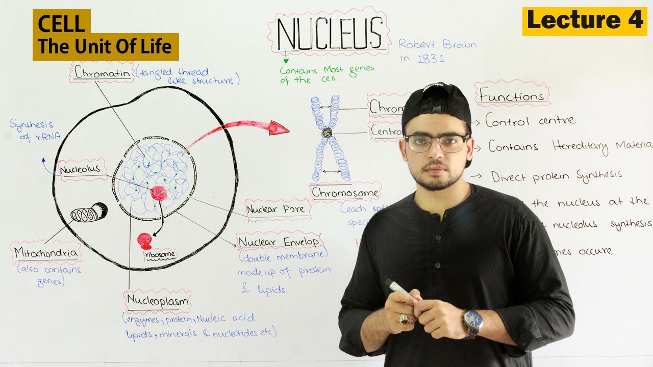
The nucleus is the control center of the eukaryotic cell, containing most of the cell's genes stored as DNA within chromatin structures; it is enclosed by a double membrane called the nuclear envelope with nuclear pores that regulate transport between nucleoplasm and cytoplasm, and contains the nucleolus where ribosomal RNA synthesis occurs, enabling the nucleus to direct protein synthesis and maintain hereditary material transfer between generations.
Click to watch →
Deep Dive
The concept of the Central Dogma of Molecular Biology (DNA -> RNA -> Protein) as a general framework for genetic information flow.

Cómo se hacen las proteínas (traducción): Crash Course Biología #35
148.2K
The central dogma describes how genetic information flows from DNA to RNA to protein. DNA stores genetic instructions in the nucleus, while messenger RNA (mRNA) acts as a courier, carrying these instructions out of the nucleus. mRNA uses the same four nucleotides as DNA (A, U, C, G) but substitutes uracil for thymine. Proteins are built from amino acids, which use a completely different chemical language than nucleotides. This fundamental communication challenge is solved through the process of translation, where ribosomes decode mRNA sequences into specific amino acid chains.
Click to watch →

Лекция 3. Транскрипция, трансляция. Окштейн И.Л., МФТИ
66.1K
The central dogma describes the flow of genetic information: DNA is transcribed into RNA, which is then translated into proteins. This unidirectional flow (DNA → RNA → protein) is fundamental to molecular biology. DNA replication creates identical copies for cell division. Transcription copies genetic information from DNA to RNA using RNA polymerase, which binds to promoters and synthesizes RNA complementary to the template strand. Eukaryotic pre-mRNA undergoes extensive processing including splicing (removing introns and joining exons using snRNPs), 5' capping, and polyadenylation. The processed mRNA is exported through nuclear pores to the cytoplasm. Translation begins when the small ribosomal subunit binds to mRNA and locates the start codon (usually AUG). The initiator tRNA carrying methionine recognizes the start codon, and the large subunit joins to form the complete ribosome. The ribosome has three binding sites: A site for incoming aminoacyl-tRNA, P site for peptidyl-tRNA, and E site for empty tRNA. Translation proceeds through elongation (aminoacyl-tRNA binding, peptide bond formation, translocation) and termination (stop codon recognition and polypeptide release).
Click to watch →
The genetic code
505.2K
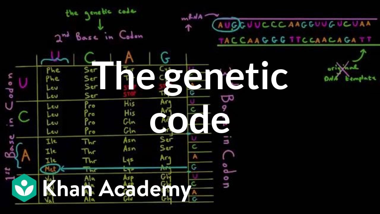
The central dogma describes how genetic information flows from DNA to RNA to protein. A gene is a DNA segment coding for one polypeptide chain. During transcription, RNA polymerase synthesizes complementary mRNA using one DNA strand as a template, with RNA containing uracil instead of thymine. The mRNA then travels to ribosomes for translation, where the ribosome reads three-nucleotide codons to assemble the corresponding amino acid chain. Only mRNA reaches the ribosome; DNA remains in the nucleus.
Click to watch →
El Dogma Central de la Biología: ADN, ARN y Proteínas.
217.4K
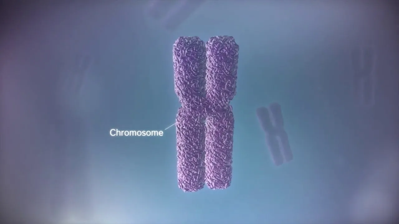
The Central Dogma of Biology describes the flow of genetic information from DNA to RNA to proteins, explaining how genetic information is stored in DNA (composed of nucleotides with phosphate groups, sugars, and nitrogenous bases adenine, guanine, cytosine, and thymine), transcribed into RNA, and translated into proteins through processes including DNA replication (semi-conservative copying), transcription (DNA to mRNA synthesis), and translation (mRNA to protein synthesis at ribosomes using codons and amino acids).
Click to watch →
Molecular Basis Of Inheritance-II 🔥🔥| Biology Crash Course | Neet 2021
175.7K
The central dogma describes the flow of genetic information: DNA → RNA → Protein. The genetic code is triplet (3 bases per amino acid), nearly universal, and degenerate (multiple codons per amino acid). Transcription synthesizes RNA from DNA template using RNA polymerase, involving initiation, elongation, and termination. Prokaryotes use single RNA polymerase with sigma and rho factors. Eukaryotes produce pre-mRNA that undergoes splicing to remove introns. Translation converts mRNA codons into proteins using ribosomes and tRNA molecules, building polypeptide chains until stop codons are reached.
Click to watch →
Prerequisite Knowledge
- Concept 01Understanding the basic structure of DNA, including its double-helix shape, nucleotide subunits (Adenine, Thymine, Cytosine, Guanine), and complementary base-pairing rules.
- Concept 02Familiarity with the fundamental differences between DNA and RNA, such as ribose vs. deoxyribose sugar and Uracil replacing Thymine.
- Concept 03Basic knowledge of eukaryotic cell anatomy, specifically the location and roles of the nucleus, cytoplasm, and ribosomes.
- Concept 04The concept of the Central Dogma of Molecular Biology (DNA -> RNA -> Protein) as a general framework for genetic information flow.
Subsequent Learning
- Step 01Exploring post-translational modifications (e.g., phosphorylation, glycosylation) and how proteins fold into their functional 3D tertiary structures.
- Step 02Studying gene regulation mechanisms, such as operons in prokaryotes and transcription factors in eukaryotes, which control when and how much protein is produced.
- Step 03Investigating the consequences of genetic mutations (like point mutations or frameshifts) on protein synthesis and their link to genetic diseases like sickle cell anemia.
- Step 04Applying this knowledge to modern biotechnology and medicine, such as the mechanism of action behind mRNA vaccines and recombinant protein production (e.g., insulin manufacturing).
Gene Expression
Protein Synthesis
Gene Expression
0:06Playing Section
- 1
Explains DNA packaging and the role of genes in protein creation.
- 2
Covers transcription, where RNA polymerase produces messenger RNA from DNA.
- 3
Describes initial RNA processing and its movement to the cytoplasm.
Exceptions to the Central Dogma of Molecular Biology
While standard 3D animations depict a linear, unidirectional flow of genetic information from DNA to RNA to protein, this classical "Central Dogma" is incomplete. Modern molecular biology recognizes significant exceptions that challenge this simplistic pathway. For instance, retroviruses utilize reverse transcription to copy RNA back into DNA, reversing the expected flow. Additionally, RNA editing can alter mRNA sequences after transcription, meaning the final protein sequence is not always a direct reflection of the genomic DNA. Furthermore, the vast majority of transcribed RNA consists of non-coding RNAs (ncRNAs) that are never translated into proteins, yet perform crucial cellular functions. Finally, prions demonstrate that proteins can transmit biological information by altering the conformation of other proteins without any nucleic acid intermediary. Introducing these phenomena helps students understand that genetic expression is a complex, multi-directional network rather than a rigid, one-way assembly line.
Deep Dive
Exploring post-translational modifications (e.g., phosphorylation, glycosylation) and how proteins fold into their functional 3D tertiary structures.

Post Translational Modification | Protein Folding & Others | B.Sc. Zoology 3rd Semester |
20.4K
Post-translational modification refers to chemical changes that occur to proteins after they are synthesized, which are essential for activating proteins and enabling them to perform their biological functions. These modifications include proteolytic cleavage (removing non-functional sections like signal peptides), protein folding (transforming linear polypeptide chains into functional 3D structures through primary, secondary, tertiary, and quaternary levels), and covalent attachments such as phosphorylation, glycosylation, sulfation, methylation, and hydroxylation. Chaperones assist in proper protein folding and can unfold misfolded proteins. The specific arrangement of amino acids and the bonds formed (peptide, hydrogen, disulfide, ionic, and hydrophobic) determine the protein's final structure and function.
Click to watch →

Post Translational Modifications
136.8K
Post translational modifications are chemical alterations that polypeptide chains undergo after translation to become functional proteins, including phosphorylation (adding phosphate groups to serine, threonine, or tyrosine via protein kinases), methylation (adding methyl groups via methyl transferase), glycosylation (adding sugar components affecting protein folding), proteolysis (enzymatic cutting of inactive precursor proteins), acetylation (adding acetyl groups, especially to histones for gene regulation), and lipidation (adding lipid components for membrane integration).
Click to watch →
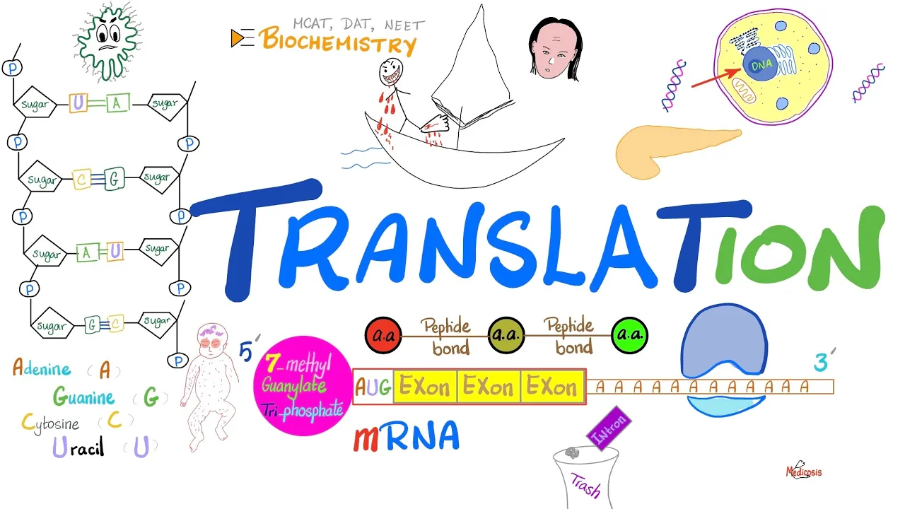
Translation (mRNA to Proteins) & Ribosomes (rER) | Post-translational Modification 🧬
64.8K
Proteins achieve functional forms through post-translational modifications after translation. Key modifications include: lipidation anchors proteins to membranes, phosphorylation adds phosphate groups using kinases, carboxylation adds carboxyl groups (vitamin K-dependent), glycosylation adds oligosaccharides, and hydroxylation adds hydroxyl groups. Collagen requires hydroxylation for triple helix stability; vitamin C deficiency causes scurvy with weak bones and bleeding. Blood clotting proteins require gamma-carboxylation (vitamin K-dependent) for calcium binding. These modifications enable diverse cellular functions from a single polypeptide sequence, demonstrating how protein functionality emerges through precise enzymatic processing.
Click to watch →

MOLECULAR BASIS OF INHERITANCE in One Shot Part 2 - All Theory, Tricks & PYQs | Class 12 | NEET
2.8M
After translation, proteins undergo various modifications including: (1) cleavage of signal sequences; (2) addition of chemical groups (phosphorylation, glycosylation, etc.); (3) formation of disulfide bonds; (4) folding into three-dimensional structures. These modifications are essential for protein function and localization.
Click to watch →
Биохимия. Лекция 37. Матричные биосинтезы. Трансляция (синтез белка).
19.8K
After translation, newly synthesized polypeptides undergo various modifications to become functional proteins. Key modifications include: N-terminal methionine removal by specific peptidases; disulfide bond formation between cysteine residues to stabilize structure; glycosylation—addition of carbohydrate groups in the ER and Golgi; assembly into multi-subunit complexes like hemoglobin (4 subunits) and collagen (triple helix); ubiquitin ligation marking proteins for degradation; phosphorylation and acetylation regulating protein activity; methylation affecting protein-DNA interactions. Protein folding is the process by which polypeptide chains acquire their functional three-dimensional structure. Chaperone proteins assist proper folding by preventing misfolding and aggregation, isolating hydrophobic regions, and facilitating correct conformational changes. Heat shock proteins (Hsp70, Hsp60) are major chaperones whose expression increases under cellular stress. Misfolded proteins can aggregate into amyloid fibrils with cross-beta sheet structure, causing diseases like Alzheimer's and amyloidosis.
Click to watch →
Deep Dive
Studying gene regulation mechanisms, such as operons in prokaryotes and transcription factors in eukaryotes, which control when and how much protein is produced.

Genregulation bei Prokaryoten und Eukaryoten in 3mins erklärt - Biologie kurz und knapp - BioIxi
5.7K
Gene regulation controls when and how much protein is produced from genetic information. Prokaryotes use operon models with substrate induction (substrate presence promotes enzyme formation for its breakdown) and end product repression (accumulated end products inhibit enzyme synthesis). Eukaryotes employ more complex mechanisms: epigenetic regulation through DNA methylation (silencing) and histone acetylation (promoting transcription); transcriptional control via promoters, transcription factors, enhancers, and silencers; RNA processing including capping, polyadenylation, and alternative splicing (producing multiple protein variants from one gene); and translation regulation through RNA interference. These multi-layered mechanisms allow precise control of gene expression in response to cellular needs.
Click to watch →

Prokaryotic and Eukaryotic Control
117
Prokaryotes regulate gene expression primarily through operons (like the lac operon) where repressor proteins bind to operator regions to block RNA polymerase from transcribing genes, while eukaryotes employ more complex multi-layered control mechanisms including chromatin packaging (DNA wrapped around histones), transcription factors binding to promoters and enhancers, alternative splicing of mRNA, RNA interference, and post-translational modifications to precisely control when and how much protein is produced.
Click to watch →

Phoenix 2.0: Biology Most Important Video for NEET 2025 | Unacademy NEET Toppers | Udaan
412.3K
Gene regulation controls when and how much of each gene product is produced. In prokaryotes, regulation occurs primarily at transcription through operons. In eukaryotes, regulation occurs at multiple levels including transcription, RNA processing, and translation.
Click to watch →

Lecture 3 - Translation & Gene Expression
269
Gene regulation controls when and how much protein is produced. In prokaryotes, the lac operon demonstrates dual regulation: CAP (catabolite activator protein) binds when glucose is absent, activating transcription; the lac repressor binds when lactose is absent, blocking transcription. Both conditions must be met for activation. Eukaryotes use more complex mechanisms: chromatin structure (DNA wrapped around histones) controls accessibility, with condensed regions inaccessible for transcription. Epigenetic mechanisms like DNA methylation silence genes. Transcription factors bind promoter sequences (TATA box, CAAT box) to help RNA polymerase bind. Enhancers contain regulatory proteins that activate transcription. mRNA lifespan also affects protein production. These mechanisms allow cells to precisely control gene expression in response to environmental and developmental signals.
Click to watch →
Gene Regulation || Regulation of mRNA forming Genes || Gene Regulation in Prokaryotes and Eukaryotes
132
Gene regulation controls when and how much protein is produced from genes, occurring at multiple levels including transcription, post-transcriptional processing, transport, translation, and post-translational modification. In prokaryotes, regulation primarily occurs at the transcriptional level through operons, which are functional units containing a promoter, operator, and structural genes. There are two main types of operons: inducible operons (like the lac operon) that are activated by the presence of a substrate, and repressible operons (like the trp operon) that are repressed by the presence of a product. Regulatory proteins act as activators or repressors to control transcription rates.
Click to watch →
Deep Dive
Investigating the consequences of genetic mutations (like point mutations or frameshifts) on protein synthesis and their link to genetic diseases like sickle cell anemia.

Molecular Basis of Inheritance | Part 8 | NCERT Line to Line | Thannambikkai Batch
114.7K
Point mutations are sudden changes in DNA base pairs. Sickle cell anemia results from a single base pair mutation in the beta globin gene, changing glutamate to valine at position 6. Frameshift mutations occur when one or two nucleotides are inserted or deleted, altering the reading frame and all subsequent amino acids. However, insertion or deletion of three or its multiple nucleotides changes one or multiple codons without altering the reading frame, as the triplet nature of the code is maintained.
Click to watch →

Molecular Basis of Inheritance Full Chapter in 1 Shot | CBSE Class 12 Biology | Stuti Mam
328
A point mutation is a change in a single nucleotide base in the DNA sequence. For example, in sickle cell anemia, a single base change (A to T) in the beta-globin gene causes the amino acid glutamic acid to be replaced by valine. A frameshift mutation occurs when nucleotides are inserted or deleted from the DNA sequence in numbers not divisible by three, shifting the reading frame and changing all downstream codons. Frameshift mutations usually result in non-functional proteins.
Click to watch →

نظري وراثة القسم الرابع
33.1K
Point mutations include deletion, insertion, and substitution. Deletion and insertion cause frameshift mutations that change the reading frame, affecting all downstream codons. Substitution changes one nucleotide to another, potentially changing one amino acid. Sickle cell anemia is caused by a substitution mutation where Adenine replaces Thymine in the DNA sequence. This changes the codon from GAG (glutamic acid) to GTG (valine). This single amino acid change causes hemoglobin to form abnormal shapes, resulting in sickle-shaped red blood cells. This is a classic example of how a single nucleotide change can cause a genetic disease.
Click to watch →

10-Chromosomal abnormalities and mutations-Genetics
42.2K
Point mutations involve single base substitutions with distinct effects. Nonsense mutations create premature stop codons, terminating protein synthesis early. Missense mutations change one amino acid to another; conservative missenses preserve protein function (similar amino acids), while non-conservative missenses disrupt function (different amino acids). Sickle cell anemia demonstrates this: a GAG→GTG mutation in the beta-globin gene changes glutamic acid to valine in hemoglobin. The altered hemoglobin is less soluble and polymerizes under low oxygen, causing red blood cells to sickle. These rigid cells block blood vessels, reducing oxygen delivery and causing pain crises. Sickle cell trait (AS) carriers have one normal and one mutated allele, usually asymptomatic. Sickle cell disease (SS) with two mutated alleles causes severe, often fatal complications.
Click to watch →

Translation 2
127
Point mutations change single nucleotides: silent mutations (same amino acid), missense mutations (different amino acid), and nonsense mutations (premature stop codon). Frameshift mutations from insertions/deletions shift the reading frame, altering all downstream amino acids. Sickle cell anemia exemplifies how a single point mutation (glutamic acid to valine) causes severe physiological consequences through protein misfolding.
Click to watch →
Deep Dive
Applying this knowledge to modern biotechnology and medicine, such as the mechanism of action behind mRNA vaccines and recombinant protein production (e.g., insulin manufacturing).
Science & Tech | Class - 12B | Science & Tech | Application of Biotechnology - 2 | Mr. A. Saravanan
18.3K
Recombinant vaccines use genetically engineered DNA to produce antigens that trigger immune responses without using the actual pathogen. The Hepatitis B vaccine (1997) was the first recombinant vaccine, produced by inserting the antigen gene into yeast cells. mRNA vaccines use messenger RNA to instruct cells to produce specific antigens, triggering immune responses. Examples include Pfizer-BioNTech and Moderna COVID-19 vaccines. mRNA vaccines offer rapid development and high efficacy but require cold chain storage. Both approaches represent advances in vaccine technology that provide safer alternatives to traditional vaccines.
Click to watch →
Master Cadre Biology Preparation 2026 | Applications of Biotechnology One Shot By Ramandeep Ma'am
201
Recombinant protein production involves inserting genes into host organisms to produce therapeutic proteins. Recombinant human insulin is produced in E. coli, replacing extraction from animal pancreases. Human growth hormone is produced for dwarfism treatment. TPA (clot-dissolving protein) is produced in mammalian cells for treating heart attacks and strokes. Interferon is produced in E. coli for antiviral and anticancer applications. These examples show biotechnology's role in modern medicine.
Click to watch →

Healthcare Biotechnology || insulin production || vaccine production|| Part 9 Biotechnology class 12
8.7K
Healthcare biotechnology applies biological processes to medicine, including insulin production through recombinant DNA technology where the insulin gene is inserted into E. coli bacteria to produce human insulin, and vaccine production using various approaches such as killed/attenuated pathogens, recombinant proteins, DNA vaccines, viral vector vaccines, plant-based vaccines, and mRNA vaccines to stimulate the immune system and provide protection against diseases.
Click to watch →
जैव प्रौद्योगिकी और उसके उपयोग One Shot | Chapter 12 Class 12 Biology | 10/10 पक्के 🔥 | BSEB 2026
18.7K
This section covers medical biotechnology applications. Insulin is a hormone produced by pancreatic beta cells that regulates blood sugar levels. Diabetes mellitus occurs when insulin production is insufficient. Historically, insulin was extracted from slaughtered animals, which was problematic due to religious restrictions and disease transmission risks. Modern biotechnology enables production of human insulin using recombinant DNA technology. The human insulin gene was inserted into E. coli bacteria, which produce the A and B chains separately. These chains are combined in the laboratory with disulfide bonds formed between them. Humulin, developed by Genentech in 1983, was the first commercially produced recombinant human insulin. Gene therapy is a technique for treating genetic disorders by introducing functional genes into patient cells. The first successful gene therapy was performed in 1990 on Ashanti DeSilva, a 4-year-old girl with ADA-SCID (Adenosine Deaminase Severe Combined Immunodeficiency). Her T lymphocytes were modified with a functional ADA gene and reinfused, restoring immune function.
Click to watch →
Vaccins et vaccination (Immunologie appliquée 210-6B2-HU)
475
Recombinant vaccines are produced using genetic engineering techniques. For example, the hepatitis B vaccine is produced by inserting the gene for the hepatitis B surface protein into yeast cells, which then produce the protein. mRNA vaccines (such as those developed by Pfizer and Moderna for COVID-19) contain genetic material that instructs cells to produce a specific protein from the pathogen. The mRNA is then degraded by the cell, and the protein is produced. The immune system recognizes this protein as foreign and mounts an immune response. mRNA vaccines do not contain any live pathogen and cannot cause the disease. They can be developed rapidly because they do not require growing the actual pathogen.
Click to watch →
Gene Expression
Protein Synthesis
Gene Expression
0:06- 1
Explains DNA packaging and the role of genes in protein creation.
- 2
Covers transcription, where RNA polymerase produces messenger RNA from DNA.
- 3
Describes initial RNA processing and its movement to the cytoplasm.
Exceptions to the Central Dogma of Molecular Biology
While standard 3D animations depict a linear, unidirectional flow of genetic information from DNA to RNA to protein, this classical "Central Dogma" is incomplete. Modern molecular biology recognizes significant exceptions that challenge this simplistic pathway. For instance, retroviruses utilize reverse transcription to copy RNA back into DNA, reversing the expected flow. Additionally, RNA editing can alter mRNA sequences after transcription, meaning the final protein sequence is not always a direct reflection of the genomic DNA. Furthermore, the vast majority of transcribed RNA consists of non-coding RNAs (ncRNAs) that are never translated into proteins, yet perform crucial cellular functions. Finally, prions demonstrate that proteins can transmit biological information by altering the conformation of other proteins without any nucleic acid intermediary. Introducing these phenomena helps students understand that genetic expression is a complex, multi-directional network rather than a rigid, one-way assembly line.
Understanding the basic structure of DNA, including its double-helix shape, nucleotide subunits (Adenine, Thymine, Cytosine, Guanine), and complementary base-pairing rules.
Familiarity with the fundamental differences between DNA and RNA, such as ribose vs. deoxyribose sugar and Uracil replacing Thymine.
Basic knowledge of eukaryotic cell anatomy, specifically the location and roles of the nucleus, cytoplasm, and ribosomes.
The concept of the Central Dogma of Molecular Biology (DNA -> RNA -> Protein) as a general framework for genetic information flow.
Exploring post-translational modifications (e.g., phosphorylation, glycosylation) and how proteins fold into their functional 3D tertiary structures.
Studying gene regulation mechanisms, such as operons in prokaryotes and transcription factors in eukaryotes, which control when and how much protein is produced.
Investigating the consequences of genetic mutations (like point mutations or frameshifts) on protein synthesis and their link to genetic diseases like sickle cell anemia.
Applying this knowledge to modern biotechnology and medicine, such as the mechanism of action behind mRNA vaccines and recombinant protein production (e.g., insulin manufacturing).
22.8M views412.2Klikes2:42@yourgenomeOriginal Release: 2015-01-07
here is a cell the basic unit of all living tissue in most human cells there is a structure called a nucleus the nucleus contains the genome in humans The genome is split between 23 pairs of chromosomes each chromosome contains a long strand of DNA tightly packaged around proteins called histones within the DNA are sections called genes these genes contain the instructions for making proteins when a gene is switched on an enzyme called RNA polymerase attaches to the start of the gene it moves along the DNA making a strand of messenger RNA out of free bases in the nucleus the DNA code determines the order in which the free bases are added to the messenger RNA this process is called transcription before the messenger RNA can be used as a template for the production of proteins it needs to be processed this involves removing and adding sections of RNA the messenger RNA then moves out of the nucleus into the cytoplasm protein factories in the cytoplasm called ribosomes bind to the messenger RNA the ribosome reads the code in the messenger RNA to produce a chain made up of amino acids there are 20 different types of amino acid transfer r na molecules carry the amino acids to the ribosome the messenger RNA is read three bases at a time as each triplet is readed a transfer RNA delivers the corresponding amino acid this is added to a growing chain of amino acids once the last amino acid has been added the chain Falls s into a complex 3D shape to form the protein
Up Next

8:28
Eukaryotic mRNA Processing: Exons, Introns & Splicing
@AKLECTURES
94.3K views•2015-02-17

50:12
Bacterial Communication: Quorum Sensing and Biofilm Formation Explained
@JHUAAP
12.6K views•2011-06-28

5:35
Enteric Nervous System Explained: The Gut's Brain | Neurobiology Lecture
@alumniu6029
438 views•2018-09-12

7:08
Bacteriophages: Earth's Deadliest Killers and Future Antibiotics
@kurzgesagt
34.6M views•2018-05-13
Related Study Plans & Knowledge Roadmaps
Structured learning paths in Biology
biology
RNAi: Biogenesis, RISC & Gene Silencing
01Molecular Biology Foundations: DNA to Protein
02Introduction to RNA Interference (RNAi)
03Molecular Biogenesis of miRNA and siRNA
+ 2 more learning modules
10h5 modules
Explore Roadmap
biology
Mycorrhizae: Signaling, Biophysics & Ecology
01Foundations of Mycorrhizal Symbiosis
02Molecular Signaling and Root Colonization
03Biophysics and Bioenergetics of Nutrient Exchange
+ 1 more learning modules
11h4 modules
Explore Roadmap
biology
Circadian Clocks: TTFL, SCN & Periphery
01Foundations of Molecular Biology & Gene Expression
02Introduction to Chronobiology and Actograms
03The Core TTFL: Molecular Architecture of the Clock
+ 3 more learning modules
14h6 modules
Explore Roadmap
biology
Population Genetics: Math, Drift & Selection
01Genetic & Probability Foundations
02The Hardy-Weinberg Principle and Baseline Models
03Mathematical Modeling of Natural Selection
+ 3 more learning modules
18h6 modules
Explore Roadmap