Spasticity and rigidity are differentiated by three key features: (1) Spasticity causes asymmetric resistance (much more resistance in one direction than the other, e.g., high extension resistance with low flexion resistance), while rigidity shows equal resistance in all directions; (2) Spasticity is velocity-dependent (faster movements feel more resistant), whereas rigidity is not velocity-dependent; (3) Spasticity typically causes weak extremities and characteristic postures, while rigidity does not cause weakness or characteristic postures. The cogwheel phenomenon in Parkinson's disease is simply rigidity plus tremor, with the cogwheeling occurring at the same frequency as the tremor.
Distinguishing Spasticity vs Rigidity: Stanford Bedside Exam
Added:Deep Dive
Basic neuroanatomy of the motor system, specifically distinguishing between the pyramidal (corticospinal) and extrapyramidal pathways.

Анатомия ЦНС: проводящих пути. Пирамидальные и экстрапирамидальные пути.
89.5K
Motor pathways control voluntary and involuntary movements. Pyramidal pathways control voluntary movements: the corticospinal tract begins in the fifth layer of the postcentral gyrus (Betz cells) and terminates in the anterior horn cells of the spinal cord. It divides into: (1) Lateral corticospinal tract (85%) that crosses at the pyramids of the medulla and controls distal muscles, and (2) Anterior corticospinal tract (15%) that crosses at the anterior white commissure and controls axial muscles. The corticobulbar tract controls cranial nerve motor nuclei and terminates in the brainstem. Extrapyramidal pathways control involuntary movements, muscle tone, and posture: (1) Red nucleus-spinal tract (controls proximal muscles, crosses at ventral tegmentum), (2) Tectospinal tract (controls head and eye movements, crosses at ventral tegmentum), (3) Vestibulospinal tract (controls balance and posture, does not cross), (4) Reticulospinal tract (controls muscle tone and posture, crosses at anterior white commissure), and (5) Olivospinal tract (controls muscle tone and posture, crosses at anterior white commissure). The spinocerebellar pathways carry unconscious proprioception to the cerebellum: the Gowers pathway (dorsal spinocerebellar) carries unconscious proprioception from the lower limb and lower trunk, passing through the gracile nucleus of the medulla, traveling up through the brainstem, and entering the cerebellum through the inferior cerebellar peduncle. The Flexig pathway (ventral spinocerebellar) carries unconscious proprioception from the upper limb and upper trunk, passing through the cuneate nucleus, crossing twice, and entering the cerebellum through the inferior cerebellar peduncle. Both pathways terminate in the Purkinje cells of the cerebellar cortex.
Click to watch →
Chapitre 5 - 1ère Partie : Organisation du système nerveux et du neurone - Cours de Biologie
656K
Motor pathways divide into direct and indirect routes: (1) Pyramidal (direct) pathways - provide fast, precise control of individual muscles, primarily from primary motor cortex to spinal motoneurons, responsible for skilled movements like playing piano; (2) Extrapyramidal (indirect) pathways - provide slower, more diffuse control of posture and muscle tone through multiple brainstem relays, working with premotor cortex. Together these systems enable both precise voluntary movements and automatic postural adjustments.
Click to watch →

Via piramidal y extrapiramidal.
34.6K
The pyramidal pathway (corticospinal tract) is a descending motor pathway that carries voluntary motor commands from the cerebral cortex to spinal motor neurons, with 90% of fibers crossing at the pyramidal decussation to form the lateral corticospinal tract and 10% remaining ipsilateral as the anterior corticospinal tract; in contrast, the extrapyramidal pathways (including rubrospinal, tectospinal, reticulospinal, and olivospinal tracts) regulate involuntary movements such as posture, muscle tone, and reflexes, originating from brainstem structures rather than the cerebral cortex.
Click to watch →

Neuroanatomie 12 : Voies motrices (neuroanatomie systématisation)
51.8K
The nervous system contains two major motor pathways: the pyramidal system (voluntary motor control) and the extrapyramidal system (automatic/involuntary motor control). The pyramidal system originates from the precentral gyrus of the frontal lobe, using a bineuronal pathway with cortical origin, decussation, and termination in spinal motoneurons. The extrapyramidal system involves central gray nuclei (thalamus, striatum, subthalamic nucleus, substantia nigra) and brainstem nuclei (red nucleus, vestibular nuclei, reticular formation, olive) that modulate movement through multiple descending tracts. The cortex is organized into six layers, with large pyramidal cells (Betz cells) in layer V serving as protoneurons. The homunculus maps motor cortex representation: facial areas inferiorly, hand representation prominently in the middle, and lower limb representation medially on the medial surface of the precentral gyrus.
Click to watch →
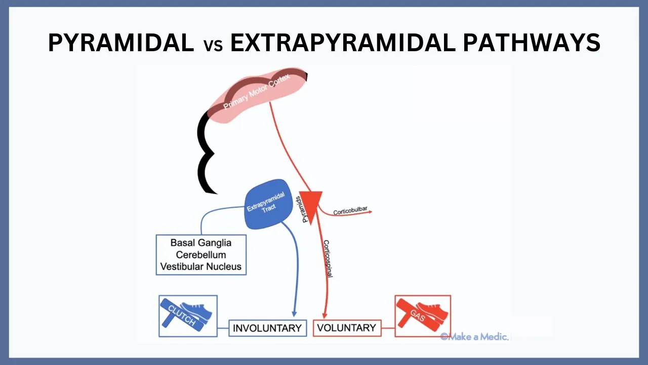
Pyramidal vs Extrapyramidal Pathways Explained
22.4K
The pyramidal pathway originates from the primary motor cortex and controls voluntary movements through the corticospinal and cortical bulbar tracts, while the extrapyramidal system (comprising the basal ganglia, cerebellum, and vestibular nucleus) modulates these signals to make movements smoother and more fluent; stroke affects the pyramidal pathway, whereas Parkinson's disease affects the extrapyramidal tracts due to degeneration of dopaminergic neurons in the substantia nigra.
Click to watch →
Deep Dive
The physiology of the muscle stretch reflex arc and how muscle tone is regulated by the central nervous system.

PHYSIOLOGY RR CLASS BY DR ASHISH ( PART 3)
18.4K
The stretch reflex (also called deep tendon reflex) is a monosynaptic reflex arc: stretching a muscle activates muscle spindle receptors → Ia afferent fibers → spinal cord → alpha motor neuron → muscle contraction. This negative feedback mechanism maintains muscle length. Gamma motor neurons continuously activate intrafusal muscle fibers, maintaining muscle spindle sensitivity and contributing to muscle tone. Without gamma motor neuron activity, muscle spindles would become less sensitive to stretch, reducing the stretch reflex response.
Click to watch →

Physiology | Muscle Spindle | INI-CET 2021 | Dr Reena Kaur
2.6K
The stretch reflex is a monosynaptic reflex arc where tendon tapping stretches extrafusal fibers, which stretches intrafusal fibers and activates Ia afferents directly on alpha motor neurons, causing contraction that returns the spindle to original length. However, gamma motor neurons continuously discharge to prevent spindle slack, maintaining constant afferent discharge for continuous muscle tone monitoring. Spindle loading occurs through extrafusal stretching (exogenous) or gamma motor neuron discharge (endogenous). Spindle unloading occurs when extrafusal fibers contract. This continuous loading ensures the brain constantly monitors muscle length and tension for posture and movement control.
Click to watch →
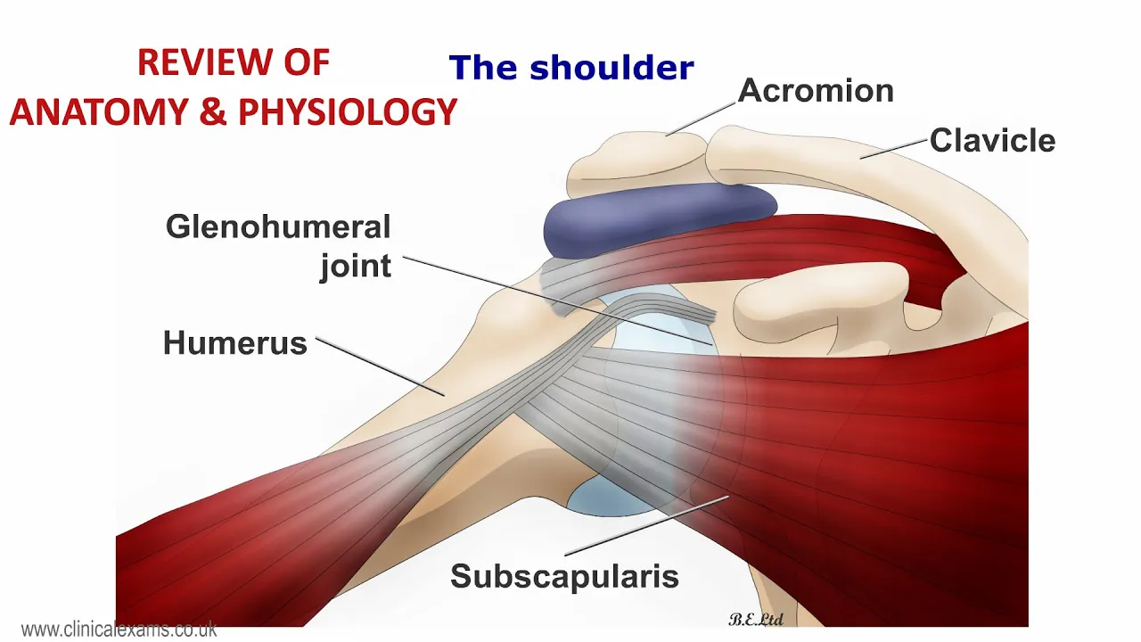
MUSCULOSKELETAL EXAM - REVIEW OF ANATOMY & PHYSIOLOGY
150
The myotatic stretch reflex is a monosynaptic reflex arc that maintains muscle tone. Muscle spindles (intrafusal fibers arranged in capsules) detect muscle stretch and send feedback via Ia and II afferent fibers to the spinal cord. These afferents synapse directly with alpha motor neurons in the ventral horn, which then cause the stretched muscle to contract. Gamma motor neurons modulate spindle sensitivity by adjusting intrafusal fiber tension, allowing central nervous system control over reflex sensitivity.
Click to watch →
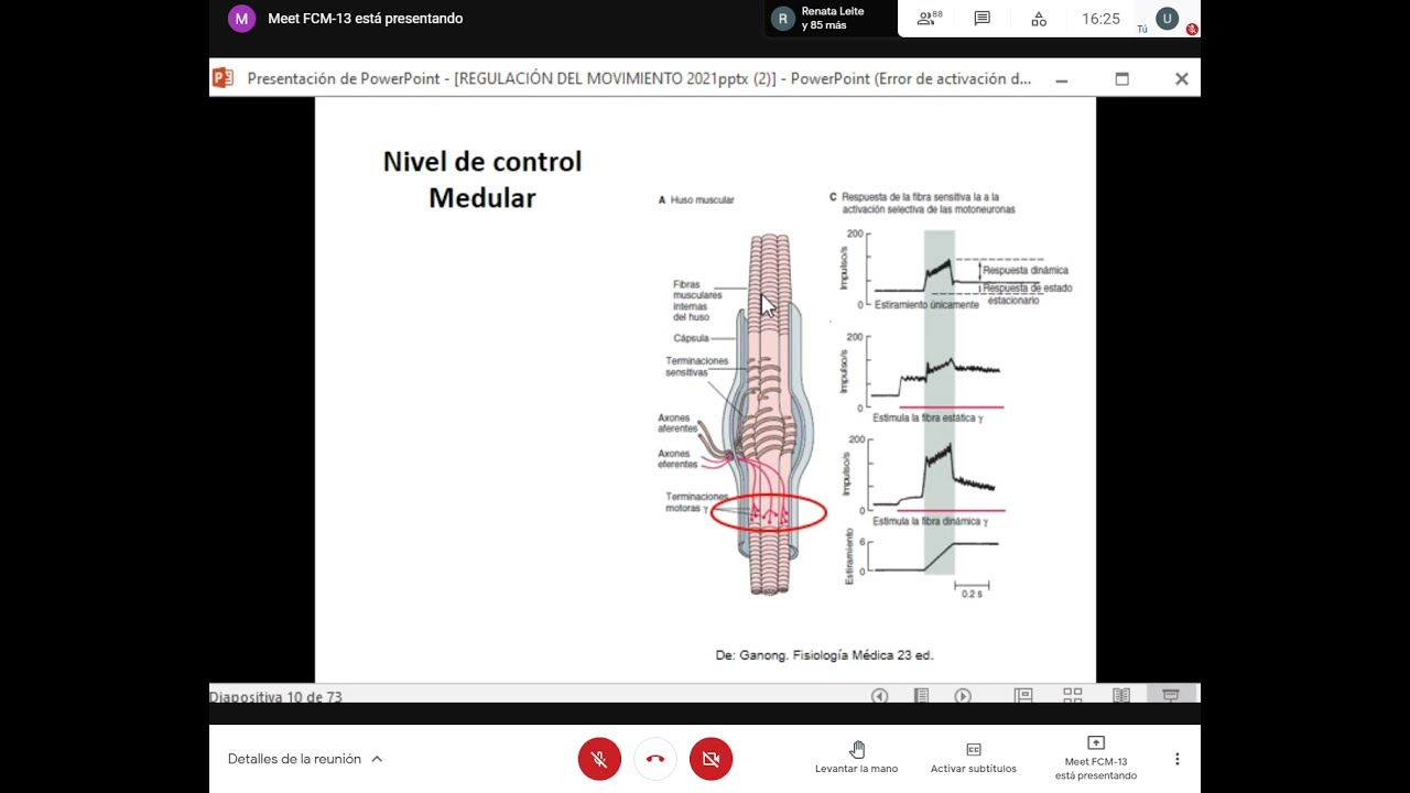
TyTl: Teórico de fisiología: Sistema equilibrio motor
5.1K
The stretch reflex is a fundamental reflex arc that regulates muscle length. It consists of: (1) a receptor (muscle spindle) that detects muscle stretch, (2) sensory fibers (type Ia or II) that transmit information to the spinal cord, (3) a sensory neuron that synapses directly with a motor neuron, and (4) an effector (skeletal muscle) that contracts in response. When a muscle is stretched, the reflex causes contraction, which reduces the muscle length. This reflex operates continuously to maintain muscle tone and posture, as muscles are constantly being stretched during daily activities like standing or walking.
Click to watch →

Clase de Repaso TYTL - Regulación del movimiento - Prof Juan Jailara
5.3K
The stretch reflex is a fundamental reflex arc that regulates muscle length through a specific pathway: muscle spindle receptors detect stretch, afferent Ia fibers transmit this information to the spinal cord, motor neurons cause the stretched muscle to contract, and gamma motor neurons adjust spindle sensitivity. Gamma motor neurons innervate intrafusal fibers within muscle spindles, allowing the nervous system to continuously adjust muscle tone based on desired muscle length. This gamma loop enables the body to maintain appropriate muscle tension for different postures and movements, from standing to sitting, by modulating the sensitivity of the stretch reflex. Muscle tone is defined as the resistance to stretch that skeletal muscle presents even when voluntarily relaxed, and is fundamental for maintaining posture and performing movements like standing and walking.
Click to watch →
Deep Dive
Fundamentals of the clinical neurological examination, including how to perform and assess passive range of motion in a patient's limbs.

Peripheral Nervous System Examination - Complete Procedure
728
Passive movement assessment involves evaluating range of motion for shoulders (flexion, extension, abduction, adduction, rotation), elbows (flexion, extension, forearm rotation), wrists, hips, knees, and ankles while encouraging patient relaxation. Lift each arm/leg and allow it to fall freely to assess muscle tone, noting rigidity types: clasp-knife (upper motor neuron), lead-pipe/plastic rigidity (Parkinson's), and cog-wheel rigidity (Parkinson's with tremor). Active movement assessment requires the patient to move limbs in all normal planes of movement, noting range achieved and control. Demonstrate movements rather than issuing complex instructions. This comprehensive assessment helps identify limitations due to neurological or musculoskeletal pathology.
Click to watch →

Neurological Examination of the Limbs - Explanation
3.6M
The neurological examination of the limbs involves a systematic assessment of muscle tone, power, coordination, and sensation in both arms and legs, following a standardized sequence that begins with gait observation and Romberg's test, followed by inspection for muscle wasting and posture abnormalities, then testing tone through passive movements, assessing power through resisted movements, evaluating coordination with finger-nose tests and heel-shin tests, examining reflexes using a tendon hammer, and finally testing four sensory modalities (pin prick, light touch, proprioception, and vibration) across dermatomal patterns to identify neurological deficits.
Click to watch →

Semiología del Sistema Nervioso Clase Practica 1 Examen Neurológico
7.9K
Passive range of motion examination involves moving each joint through its full range of motion, typically 10-20 repetitions per joint, proceeding systematically through the body. Muscle tone assessment involves evaluating resistance to passive movement: in hypotonia, the limb moves easily with minimal resistance; in hypertonia, resistance is increased. The neck tone can be assessed by supporting the chin with fingers and gently pushing downward. These examination techniques are essential for neurological assessment.
Click to watch →
Orthopedic Examination (Palpation, Sensory & Motor Testing, Camera Moving) 🩺 ASMR Medical RP
11.9K
Active range of movement testing includes cervical extension and flexion, rotation, and lateral side bending. Shoulder testing includes flexion, abduction, medial and lateral rotation, and elbow flexion and extension. Passive range of movement testing is performed by the examiner supporting and moving the patient's head through extension, flexion, rotation, and lateral side bending. The examiner observes for restriction or pain during both active and passive movements.
Click to watch →

EXAMEN FISICO NEUROLOGICO (CONTINUACION DEL TEMA PARES CRANEALES)
1K
The neurological motor examination includes assessment of active voluntary movements, passive movements, and muscle tone. Active movements are performed by the patient at command, testing voluntary motor function. Passive movements are performed by the examiner to assess muscle tone. The examination evaluates head movements (flexion, extension, rotation), upper limb movements (flexion, extension, pronation, supination), and lower limb movements. This systematic approach helps identify motor deficits and localize neurological lesions.
Click to watch →
Deep Dive
Familiarity with general clinical vocabulary regarding altered muscle tone, such as hypertonia and hypotonia.
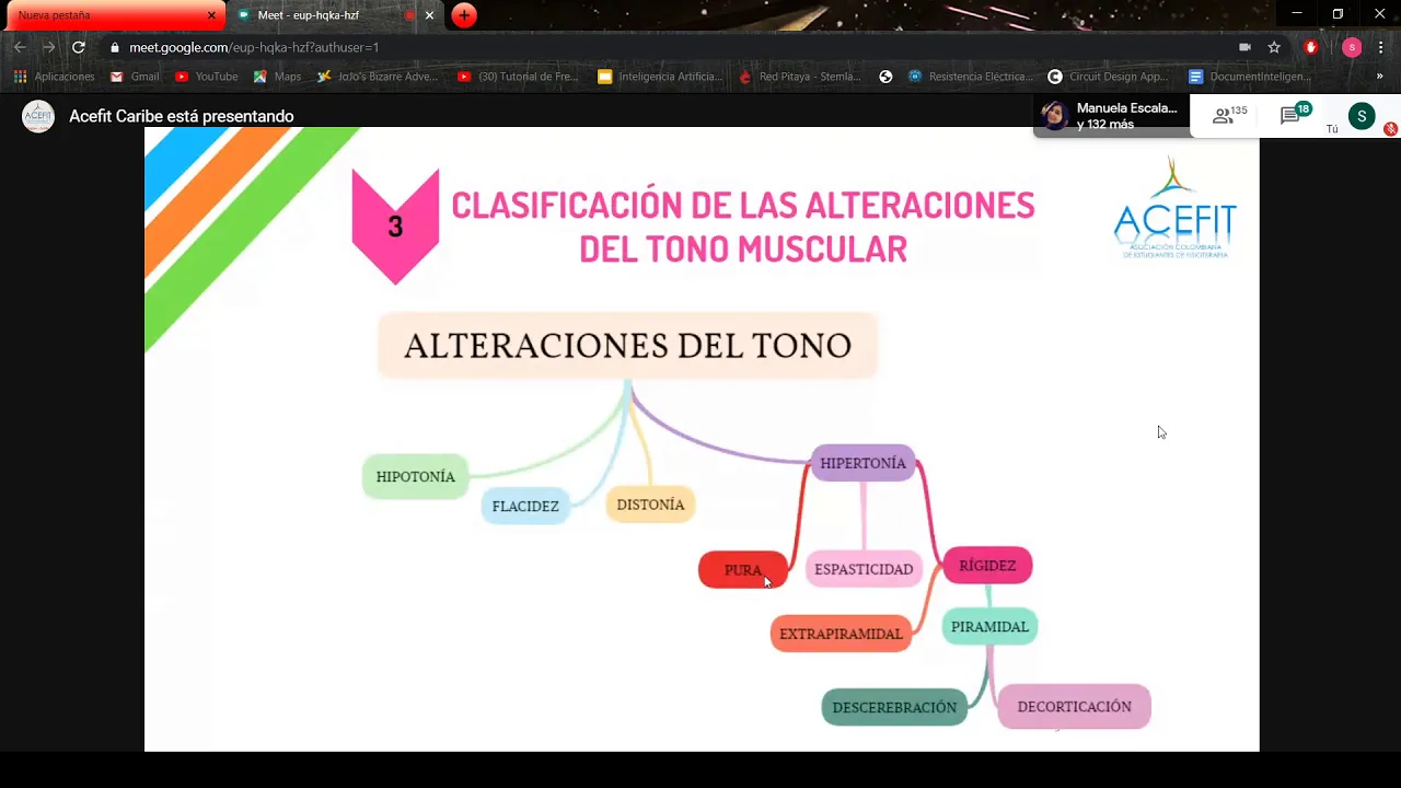
Alteraciones del tono muscular en paciente con patología neurológica
1.7K
Muscle tone alterations are classified into hypotonia and hypertonia. Hypotonia differs from flaccidity: hypotonia involves reduced excitatory influence on motor neurons with decreased stretch reflex sensitivity, while flaccidity represents complete loss of motor innervation with zero resistance. Hypertonia includes spasticity (hyperexcitability of stretch reflex with increased response magnitude and expanded stimulation area) and rigidity (increased resistance to passive movement that remains constant throughout the range). Spasticity presents with clasp-knife sign, clonus, hyperreflexia, and specific muscle group involvement. Rigidity shows uniform resistance without velocity dependence.
Click to watch →

Introduction to Clinical Neurology, Part 2 of 2
170
This section covers muscle tone disorders and their clinical significance. Muscle tone is the continuous passive partial contraction maintaining posture and readiness for movement, providing resistance to passive stretch through the stretch reflex. Hypotonia (decreased tone) results from lower motor neuron dysfunction, spinal cord lesions, or certain neurological conditions, presenting with reduced resistance to passive movement and floppy joints. Hypertonia (increased tone) results from upper motor neuron lesions and extrapyramidal disorders, presenting with increased resistance to passive movement. Hypertonia includes spasticity (velocity-dependent resistance increasing with faster movement) and rigidity (constant resistance regardless of velocity). Cogwheel rigidity shows rhythmic, ratchet-like resistance with clicking sounds, while lead pipe rigidity shows uniform resistance throughout movement.
Click to watch →

Clinical Neurology Series - Muscle Tone
3.1K
Abnormal muscle tone includes hypotonia (decreased tone) and hypertonia (increased tone). Hypotonia indicates lower motor neuron disease from gamma loop disruption, with peripheral causes (motor unit/sensory nerve damage) showing weakness, while central causes (cerebral lesions) lack weakness. Cerebral/spinal shock shows transient hypotonia after acute pyramidal injury. Hypertonia manifests as rigidity (extrapyramidal, velocity-independent, diffuse involvement) or spasticity (pyramidal, velocity-dependent, focal involvement). Rigidity affects all muscle groups equally throughout range of motion, while spasticity varies by muscle group and movement speed.
Click to watch →

9:50 Анатомо-физиологические особенности костно-мышечной системы у детей
4.1K
Abnormal muscle tone includes: hypotonia - decreased resistance to passive movements, flabby muscles; hypertonia - increased resistance, dense muscles; dystonia - alternating areas of increased and decreased tone; asymmetry - increased tone in lower extremities with decreased in upper, or vice versa. Muscle hypertonia includes spasticity (resistance only at the beginning of active movement, then decreases - scissors phenomenon) and rigidity (constant or increasing resistance with repeated movements - waxy doll phenomenon, lead pipe phenomenon, cogwheel rigidity). Spasticity results from interruption of central nervous system influence on anterior horn cells and release of segmental reflex apparatus. Muscle hypotonia features include postural abnormalities, hypotonic flabby abdomen (frog-like abdomen), scapular winging, external clubfoot, and increased joint mobility with excessive extension.
Click to watch →

Sémiologie Neurologique : EXAMEN NEUROLOGIQUE
2.6K
Muscle tone is assessed by passive movement of the patient's limbs. Normal tone provides resistance to passive movement. Hypotonia (reduced tone) is felt as flaccidity during passive movement. Hypertonia (increased tone) is felt as resistance. Hypertonia has two main types: spastic and plastic (rigid). Spastic hypertonia is velocity-dependent, meaning resistance increases with faster passive movement. It is characteristic of pyramidal tract lesions. Plastic hypertonia (rigidity) is velocity-independent, providing constant resistance regardless of movement speed. It is characteristic of extrapyramidal disorders such as Parkinson's disease. Spastic hypertonia shows increased resistance with faster passive movement and may include clonus (rhythmic oscillations) and the clasp-knife phenomenon (initial resistance followed by sudden release).
Click to watch →
Prerequisite Knowledge
- Concept 01Basic neuroanatomy of the motor system, specifically distinguishing between the pyramidal (corticospinal) and extrapyramidal pathways.
- Concept 02The physiology of the muscle stretch reflex arc and how muscle tone is regulated by the central nervous system.
- Concept 03Fundamentals of the clinical neurological examination, including how to perform and assess passive range of motion in a patient's limbs.
- Concept 04Familiarity with general clinical vocabulary regarding altered muscle tone, such as hypertonia and hypotonia.
Subsequent Learning
- Step 01Diagnostic localization of lesions, attributing spasticity to Upper Motor Neuron (UMN) lesions and rigidity to basal ganglia dysfunction.
- Step 02Differentiating specific clinical presentations of rigidity (such as lead-pipe and cogwheel rigidity) and spasticity (clasp-knife phenomenon).
- Step 03Clinical grading systems used in practice, such as the Modified Ashworth Scale for measuring spasticity.
- Step 04Pharmacological and therapeutic interventions tailored to each condition, such as baclofen or botulinum toxin for spasticity, and dopaminergic agents for rigidity.
Rigidity Exam
Rigidity Exam
0:08Playing Section
- 1
Demonstrates eliciting rigidity via passive limb movement.
- 2
Key exam instruction: patient must keep limb completely floppy.
- 3
Differentiates rigidity from spasticity by resistance patterns.
Limitations of the Pyramidal/Extrapyramidal Dichotomy and Manual Bedside Assessment
While the traditional bedside exam neatly categorizes increased muscle tone into velocity-dependent spasticity (pyramidal) and velocity-independent rigidity (extrapyramidal), modern neurophysiology and clinical studies challenge this strict dichotomy. Critics argue that this manual distinction is highly subjective, suffers from poor inter-rater reliability, and oversimplifies complex pathophysiology. In many mixed neurological disorders—such as stroke, traumatic brain injury, or cerebral palsy—patients exhibit overlapping features of both spasticity and rigidity due to co-existing damage across interconnected motor pathways. Furthermore, neurorehabilitation specialists increasingly advocate for quantitative, instrumented assessments (utilizing electromyography and biomechanical torque sensors) over subjective manual manipulation, arguing that traditional bedside exams cannot reliably isolate active neural reflex hypertonia from passive, non-neural structural muscle stiffness.
Deep Dive
Diagnostic localization of lesions, attributing spasticity to Upper Motor Neuron (UMN) lesions and rigidity to basal ganglia dysfunction.

Neurology localization part 1
141.3K
Neurological localization relies on distinguishing upper motor neuron (UMN) from lower motor neuron (LMN) lesions through characteristic clinical findings: UMN lesions cause spastic weakness with brisk reflexes, velocity-dependent resistance, and Babinski sign, while LMN lesions produce flaccid weakness with absent reflexes, profound atrophy, fasciculations, and fibrillation potentials; UMN pathways include the corticospinal tract (cortex to spinal cord) and corticobulbar tract (cortex to brainstem motor nuclei), whereas LMN pathways involve anterior horn cells, nerve roots, plexuses, and peripheral nerves.
Click to watch →

Неврологический осмотр. Вебинар от Абрамовой Е.В.
4.9K
Upper motor neuron (UMN) lesions are in the brain or spinal cord above the nerve root exit. Lower motor neuron (LMN) lesions are in the nerve roots or peripheral nerves. UMN lesions cause spastic paralysis with increased tone, hyperreflexia, and delayed muscle atrophy. LMN lesions cause flaccid paralysis with decreased tone, hyporeflexia, and early severe muscle atrophy. Lesion localization depends on which spinal cord segments are affected: C1-C5 lesions affect both forelimbs and hindlimbs with UMN signs. C6-T2 lesions affect only forelimbs with LMN signs. T3-L3 lesions affect only hindlimbs with UMN signs. L4-S3 lesions affect only hindlimbs with LMN signs.
Click to watch →

Hypertonia (Clasp-knife, Cogwheel rigidity, Ankle Clonus, Lead pipe rigidity)
22K
Hypertonia in upper motor neuron lesions includes spasticity (velocity-dependent, affects antigravity muscles, shows sudden release) caused by pyramidal tract lesions with increased alpha motor neuron activity, and rigidity (non-velocity-dependent, affects both agonist and antagonist muscles equally) caused by extrapyramidal tract lesions with increased gamma motor neuron activity; specific manifestations include clasp-knife rigidity (inverse stretch reflex), ankle clonus (gastrocnemius muscle), patellar clonus (quadriceps muscle), cogwheel rigidity (intermittent tone), and lead pipe rigidity (uniform tone), with Babinski sign being positive in pyramidal lesions and negative in extrapyramidal lesions.
Click to watch →

CNS Bedside Techniques Lecture 15 | Localization of Motor Lesion | Upper vs Lower Motor Neuron
181
Motor lesions can be localized by distinguishing between upper motor neuron (UMN) and lower motor neuron (LMN) involvement: UMN lesions cause increased tone, hyperreflexia, and positive Babinski sign, with localization to motor cortex (hemiplegia, apraxia), internal capsule (contralateral hemiplegia), brainstem (crossed hemiplegia with cranial nerve involvement), or spinal cord (contralateral hemiplegia below lesion level); LMN lesions cause muscle wasting, hypotonia, hyporeflexia, and negative Babinski sign, with localization to cranial nerve nuclei, anterior horn cells, nerve roots, or peripheral nerves. Brown-Séquard syndrome demonstrates spinal cord hemisection with ipsilateral UMN weakness and position/vibration loss plus contralateral pain/temperature loss. Muscle diseases (myopathies) show proximal wasting with normal reflexes, while neuropathies show distal involvement with sensory loss.
Click to watch →

Pathophysiology of spasticity following UMN lesions
97.1K
Spasticity, a manifestation of hypertonia following upper motor neuron lesions, occurs because the removal of descending inhibitory pathways eliminates the 'brakes' on lower motor neurons, causing them to fire more frequently and produce increased muscle tone; in the upper limb, this results in a characteristic flexed posture (flexed elbow, wrist, and fingers) because flexor muscles are generally more powerful than extensors.
Click to watch →
Deep Dive
Differentiating specific clinical presentations of rigidity (such as lead-pipe and cogwheel rigidity) and spasticity (clasp-knife phenomenon).

Rigidity and spasticity (Lead pipe , Cog wheel , Clasp knife) #mbbs #medicine #medicos #neetpg #nnp
616
Rigidity is an increase in muscle tone causing resistance to passive movement throughout the range of motion, with two main types: lead pipe rigidity (constant resistance throughout movement, seen in basal ganglia disorders) and cogwheel rigidity (intermittent resistance with free movement periods, seen in Parkinsonism); spasticity is an abnormal increase in muscle tone or stiffness seen in upper motor neuron lesions, characterized by maximum initial resistance followed by free movement (clasp knife phenomenon).
Click to watch →
Neurology | Upper Motor Neuron vs Lower Motor Neuron Lesion | UMN vs LMN Lesion
880.5K
Spasticity (upper motor neuron) is velocity-dependent resistance in one direction only, with a clasp-knife phenomenon where resistance suddenly gives way. Rigidity (e.g., Parkinson's disease) is velocity-independent with resistance in both directions and associated tremors. Spasticity lacks tremors and shows weakness, while rigidity typically preserves strength.
Click to watch →

CNS BEDSIDE TECHNIQUES Lecture 13 | Bulk, Tone & Power of Muscles + Reflexes
335
Muscle tone refers to resistance during passive movement. Increased tone (hypertonia) has two main types: spasticity and rigidity. Spasticity shows increased resistance during initial movement degrees followed by decreased resistance, described as 'clasp-knife rigidity' - like a knife opening easily after initial resistance. This occurs in upper motor neuron lesions. Rigidity shows constant resistance throughout movement, described as 'lead-pipe rigidity' - like bending a lead pipe. This occurs in basal ganglia lesions. Cogwheel rigidity, seen in Parkinson's disease, combines rigidity with jerky movements. Hysterical rigidity depends on applied force - more force produces more resistance. Decreased tone (hypotonia) occurs in lower motor neuron lesions and early stroke stages like spinal shock and cerebral dysfunction.
Click to watch →

Diagnosing Parkinson's Disease by Dr. Steve McGee (Stanford Skills Symposium)
147.3K
Rigidity is distinguished from spasticity by several key characteristics. Spasticity involves weakness and velocity-dependent resistance (more resistance with faster movements), with significantly different resistance in flexion versus extension directions, creating characteristic postures. In contrast, rigidity does not cause weakness, has equal resistance in both directions, is not velocity-dependent, and lacks characteristic postures. The cogwheel phenomenon in Parkinson's disease is simply rigidity plus tremor at the same frequency.
Click to watch →

Muscle tone and its regulation. Special reference to Spasticity and Rigidity by SB Deshpande
491
Spasticity and rigidity are distinct hypertonia types with different characteristics. Spasticity is velocity-dependent (resistance increases with rapid movement speed) and shows clasp knife phenomenon (initial resistance followed by yielding). It involves anti-gravity/extensor muscles, shows exaggerated tendon reflexes with possible clonus, and results from upper motor neuron lesions. Rigidity is velocity-independent (constant resistance regardless of movement speed) without yielding, involves flexor muscles, shows cogwheel rigidity, and results from basal ganglia disorders. Spasticity lacks tremors; rigidity may show tremors. These differences guide clinical diagnosis and treatment approaches.
Click to watch →
Deep Dive
Clinical grading systems used in practice, such as the Modified Ashworth Scale for measuring spasticity.

AOCPMR Journal Club with Dr. Elwert
117
The Modified Ashworth Scale is the most commonly used scoring system for spasticity, originally developed in the 1960s and modified in the 1980s and 2006. The scale ranges from 0 to 4: Grade 0 = no increase in muscle tone; Grade 1 = slight increase manifested by catch and release at end of range; Grade 1+ = slight increase with catch beyond half range of motion; Grade 2 = marked increase throughout most of range of motion but easily moved; Grade 3 = considerable increase in muscle tone making passive movement difficult but still possible; Grade 4 = affected joint is rigid in flexion or extension and does not move.
Click to watch →
![Modified Ashworth Scale EXPLAINED [with Demonstrations]](https://i.ytimg.com/vi/ZmJU9-Oe3vs/maxresdefault.jpg)
Modified Ashworth Scale EXPLAINED [with Demonstrations]
35K
The Modified Ashworth Scale (MAS) is a standardized clinical tool used to assess muscle spasticity by applying a quick passive stretch to a muscle at its maximally shortened position, with grades ranging from 0 (no resistance) to 4 (rigidity), where grade 1 shows minimal resistance at end-range, grade 1+ shows catch at mid-range, grade 2 shows moderate resistance at early range, grade 3 shows high resistance making movement difficult, and grade 4 indicates complete rigidity preventing movement to end-range.
Click to watch →

Residência SES DF | Fisioterapia: Revisão Final com Rhebecca Dumaresq
502
The Modified Ashworth Scale grades spasticity from 0 to 4 based on passive movement resistance: 0 = no increase in tone, 1 = slight increase at end of movement, 1+ = slight increase in first half of movement, 2 = moderate increase throughout movement, 3 = marked increase making passive movement difficult, 4 = severe increase preventing passive movement. This scale is essential for documenting spasticity severity and monitoring treatment response in neurological conditions.
Click to watch →

Escala de Ashworth modificada
7.2K
The Modified Ashworth Scale, originally created by Ashworth in 1964 and modified by Bohannon and Smith in 1989, is the most commonly used clinical instrument for assessing muscle spasticity, measuring passive resistance to joint movement on a 0-4 scale where 0 indicates no resistance and 4 indicates a clonus lasting more than 10 seconds; while it is widely regarded as a gold standard for spasticity evaluation due to its ease of use, high reliability, and applicability across all joints, it has limitations including potential speed-dependent responses and limited research validity, and is often used alongside the Modified Tardieu Scale which offers greater sensitivity by testing muscle resistance at both rapid and slow movement velocities.
Click to watch →
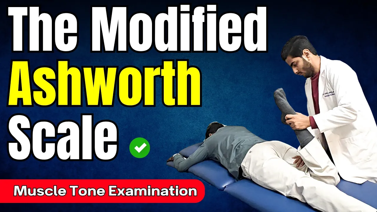
Master the Modified Ashworth Scale FAST | Spasticity Assessment Made Easy
4.1K
The Modified Ashworth Scale (MAS) is a five-point clinical tool used to assess spasticity by measuring resistance to passive movement: Grade 0 indicates normal muscle tone with no resistance; Grade 1 shows a sudden catch or resistance at the end range of motion followed by release; Grade 1+ demonstrates catch/resistance from mid-range to end range; Grade 2 indicates increased resistance throughout the full range of motion but movement remains possible; Grade 3 shows considerable resistance making passive movement difficult; Grade 4 represents complete rigidity with no movement possible even with strong force applied.
Click to watch →
Deep Dive
Pharmacological and therapeutic interventions tailored to each condition, such as baclofen or botulinum toxin for spasticity, and dopaminergic agents for rigidity.
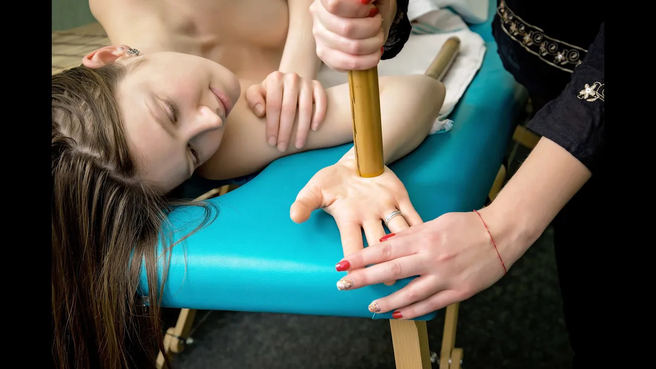
Let's SMASH Spasticity!
11.6K
Treatment selection depends on spasticity distribution: focal vs. diffuse. For focal spasticity, botulinum toxin injections weaken overactive muscles for 3-4 months with no systemic side effects. For diffuse spasticity, oral medications are preferred. Baclofen provides exogenous GABA, working around wake-up, lunch, dinner, and bedtime with doses ranging from 5-40 mg. Tizanidine (Xanx) addresses nighttime spasms and cramps but causes sedation. These medications can be combined for comprehensive symptom control.
Click to watch →
MS and Pain - Ben Thrower, M.D. - January 2016
51K
Spasticity treatment progresses to medications when conservative measures fail. Baclofen effectively reduces spasticity but causes drowsiness and floppy weakness. Tizanidine works differently but requires blood pressure monitoring. Clonazepam, the longest-acting benzodiazepine, has lowest addiction potential. Dantrium shows better efficacy in spinal cord injury than MS. Botulinum toxin injections target focal spasticity but paralyze injected muscles, limiting use to non-functional areas. Each medication has unique mechanisms, side effect profiles, and appropriate patient selection criteria.
Click to watch →

Spasticity Management
22.2K
Spasticity treatment progresses from oral medications to invasive interventions based on severity and response. Four main oral agents exist: baclofen (most common, concerns about neuro recovery inhibition), tizanidine (shorter-acting, interacts with CYP1A2 inhibitors), diazepam (highly effective but habit-forming with severe withdrawal), and dantrolene (direct muscle action with hepatotoxicity risk). Treatment follows 'low and slow' principles with gradual titration and never abrupt cessation. Botulinum toxin injections provide focal, reversible treatment by blocking acetylcholine release, with onset in 24-72 hours and reversal in 4-6 months. For refractory cases, intrathecal baclofen pumps deliver microgram-level dosing directly to cerebrospinal fluid, offering precise control with benefits including reversibility and functional independence. Major complications include overdose, catheter issues, and infection requiring careful management.
Click to watch →

Cerebral Palsy: Etiology, Pathophysiology, Complications, Treatment
271.7K
Spasticity management uses anti-spasmodic medications acting centrally or peripherally. Central-acting drugs include baclofen (enhances GABA action), diazepam (benzodiazepine enhancing GABA), and tizanidine (inhibits glutamate release). Peripheral-acting drugs include botulinum toxin injections (inhibits acetylcholine release at neuromuscular junction) and dantrolene (inhibits calcium release from sarcoplasmic reticulum). Intrathecal baclofen pump delivery may be used when oral medications are ineffective.
Click to watch →
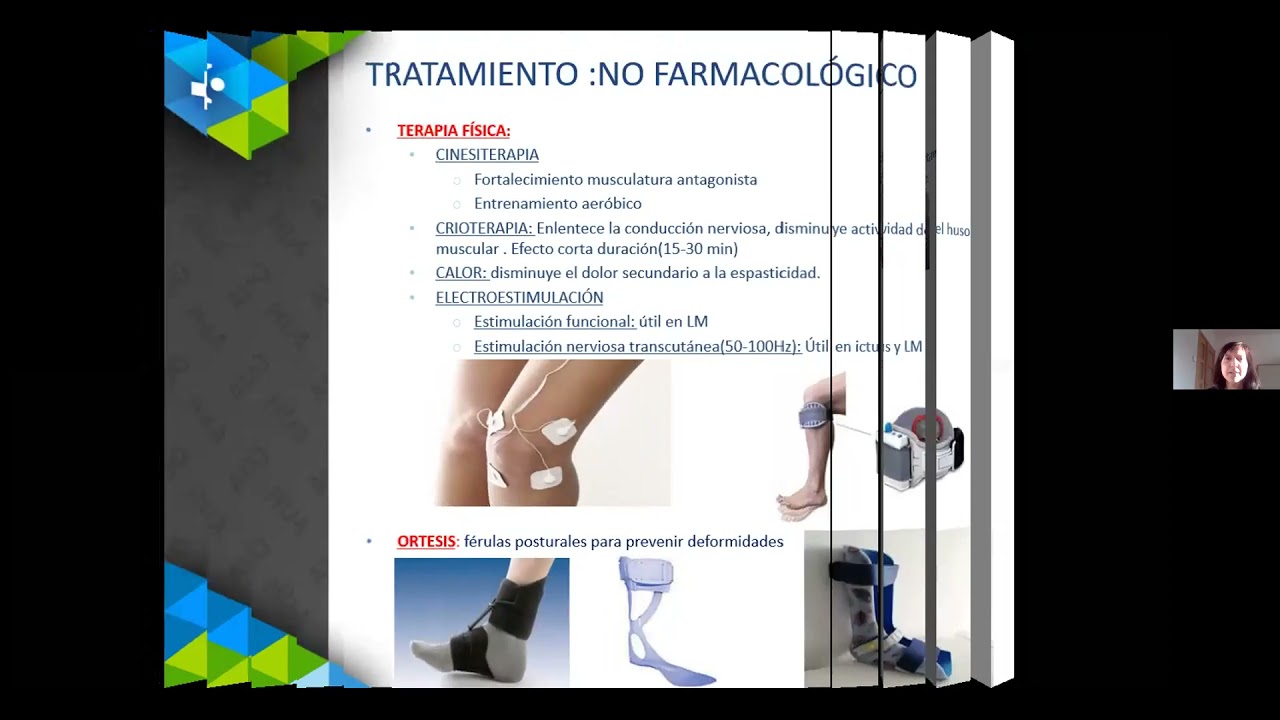
Tratamiento de la espasticidad con toxina botulínica
2.7K
Botulinum toxin type A (Botox) is the first-line treatment for focal spasticity, working by blocking acetylcholine release at the neuromuscular junction, inhibiting nociceptive neurotransmitter release, and reducing gamma motor system activity to decrease muscle tone; it is administered via intramuscular injection with effects beginning in 2-3 days, peaking at 4 weeks, and lasting 3-4 months, with dosing ranging from 25-30 units per injection site for adults and weight-based calculations for pediatric patients.
Click to watch →
Rigidity Exam
Rigidity Exam
0:08- 1
Demonstrates eliciting rigidity via passive limb movement.
- 2
Key exam instruction: patient must keep limb completely floppy.
- 3
Differentiates rigidity from spasticity by resistance patterns.
Limitations of the Pyramidal/Extrapyramidal Dichotomy and Manual Bedside Assessment
While the traditional bedside exam neatly categorizes increased muscle tone into velocity-dependent spasticity (pyramidal) and velocity-independent rigidity (extrapyramidal), modern neurophysiology and clinical studies challenge this strict dichotomy. Critics argue that this manual distinction is highly subjective, suffers from poor inter-rater reliability, and oversimplifies complex pathophysiology. In many mixed neurological disorders—such as stroke, traumatic brain injury, or cerebral palsy—patients exhibit overlapping features of both spasticity and rigidity due to co-existing damage across interconnected motor pathways. Furthermore, neurorehabilitation specialists increasingly advocate for quantitative, instrumented assessments (utilizing electromyography and biomechanical torque sensors) over subjective manual manipulation, arguing that traditional bedside exams cannot reliably isolate active neural reflex hypertonia from passive, non-neural structural muscle stiffness.
Basic neuroanatomy of the motor system, specifically distinguishing between the pyramidal (corticospinal) and extrapyramidal pathways.
The physiology of the muscle stretch reflex arc and how muscle tone is regulated by the central nervous system.
Fundamentals of the clinical neurological examination, including how to perform and assess passive range of motion in a patient's limbs.
Familiarity with general clinical vocabulary regarding altered muscle tone, such as hypertonia and hypotonia.
Diagnostic localization of lesions, attributing spasticity to Upper Motor Neuron (UMN) lesions and rigidity to basal ganglia dysfunction.
Differentiating specific clinical presentations of rigidity (such as lead-pipe and cogwheel rigidity) and spasticity (clasp-knife phenomenon).
Clinical grading systems used in practice, such as the Modified Ashworth Scale for measuring spasticity.
Pharmacological and therapeutic interventions tailored to each condition, such as baclofen or botulinum toxin for spasticity, and dopaminergic agents for rigidity.
358.6K views4.9Klikes2:26@StanfordMedicine25Original Release: 2016-04-05
we have resting tremor rigidity brady kinesia the rigidity you elicit by asking the patient to keep the limbs floppy and explain to them that you're just going to maybe flex and extend the elbow you can test it at the elbow you can test it at the wrist and one of the hardest things for patients to do is tell them to relax the minute you tell them to relax people do kind of get tense and so you want to kind of repeatedly do this and I often use the words Joslin please try and keep your limb as floppy as you can there we go what you're going to perceive with rigidity is increased resistance to movement and when you feel increased resistance to movement really there are three choices but two big choices one would be spasticity the other is rigidity and those two phenotypes differ someone with spasticity will have a weak extremity of course and with spasticity the resistance you feel in one direction is much different than the resistance you feel in the other direction so for example the resistance with flexion may be very slight a lot of resistance to extension there's a big difference between flexion and extension in spasticity and that's what creates the characteristic postures is someone with hemiparesis and then the other feature is spasticity is its velocity dependent the faster you go the more resistance you feel rigidity in contrast the extremity is not weak the resistance that you feel when the patient's trying to be floppy is the same with flexion and extension there are no characteristic postures and it is not philosophy dependent so and then sometimes you know the rigidity of Parkinson's disease has that cogwheel character but really all that is is rigidity plus tremor the cog wheeling is the same frequency as the tremor the preceding program is copyrighted by the Board of Trustees of the Leland Stanford junior University please visit us at med.stanford.edu
Up Next
3:34
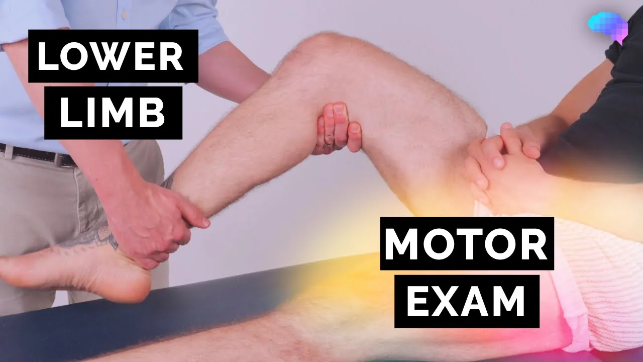
Lower Limb Motor Assessment: OSCE Procedure Guide
@geekymedics
222.6K views•2024-01-04

48:47
Graphic Medicine: Comics for Collaborative Healthcare Communication
@nationalpatientadvocate
189 views•2023-12-04

5:05
Cerebellum Examination: Stanford Medicine 25 Bedside Skills
@StanfordMedicine25
1.2M views•2014-03-18

3:05
Stages of Labor and Vaginal Birth | Childbirth Animation
@nucleusmedicalmedia
52.1M views•2017-08-18
Related Study Plans & Knowledge Roadmaps
Structured learning paths in Medicine
medicine
Abnormal LFTs: Diagnostic Workup & Pathology
01Liver Anatomy and Basic Physiology
02Introduction to Liver Function Tests (LFTs)
03Hepatocellular vs. Cholestatic Patterns
+ 3 more learning modules
12h6 modules
Explore Roadmap
medicine
Analyzing Blood Smears: RBC, WBC & Platelets
01Introduction to Blood Components & Smear Preparation
02Normal Morphology of Blood Cells
03Erythrocyte Pathologies & Morphological Variants
+ 2 more learning modules
12h5 modules
Explore Roadmap
medicine
Mechanical Ventilation: Waveforms & Alarms
01Anatomy and Physiology of Breathing
02Introduction to Mechanical Ventilation & Core Modes
03Ventilator Settings and Waveform Interpretation
+ 2 more learning modules
12h5 modules
Explore Roadmap
medicine
Neuro Exam: Localization & Stroke Syndromes
01Foundations of Neuroanatomy and Vascular Supply
02The Neurological Examination: Step-by-Step
03Principles of Neurological Localization
+ 3 more learning modules
18h6 modules
Explore Roadmap