This video demonstrates a systematic approach to assessing lower limb motor function through muscle tone evaluation (testing for spasticity or flaccidity), muscle power testing (hip flexion/extension, knee flexion/extension, ankle dorsiflexion/plantarflexion, toe movements, and foot inversion/eversion), and reflex examination (knee jerk and ankle jerk reflexes). The assessment follows a structured sequence from proximal to distal muscles, evaluating both voluntary movement strength and involuntary reflex responses to identify neurological deficits.
Lower Limb Motor Assessment: OSCE Procedure Guide
Added:Deep Dive
Basic neuroanatomy of the lumbosacral plexus and the specific nerve roots (L1-S2) supplying the lower limbs.

The Lumbosacral Plexus - Nerves - Anatomy Series
109.4K
The lumbosacral plexus, formed by the ventral rami of spinal nerves L1-S4, supplies the lower extremities through two main divisions: the lumbar plexus (producing the obturator and femoral nerves from L2-L4) and the sacral plexus (producing the sciatic nerve from L4-S3). The thigh has three compartments with distinct nerve supplies: anterior compartment (quadriceps, sartorius) by femoral nerve, medial compartment (adductors) by obturator nerve, and posterior compartment (hamstrings) by sciatic nerve. The leg has three compartments: anterior compartment (tibialis anterior, extensors) by anterior tibial nerve, lateral compartment (fibular muscles) by superficial fibular nerve, and posterior compartment (plantar flexors) by tibial nerve. Key clinical syndromes include tibial nerve injury causing plantar flexion weakness and inability to walk on tiptoes (talipes calcaneovalgus), and common fibular nerve injury causing dorsiflexion weakness and foot drop (talipes equinovarus).
Click to watch →
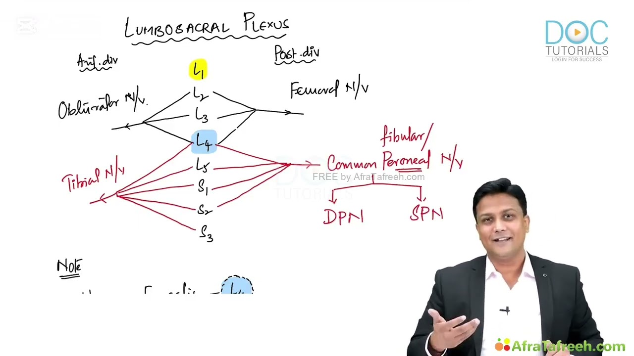
1. Lumbosacral Plexus & Major Nerves | Lower Limb Anatomy | Dr. Azam | Medical Maven
352
The lumbosacral plexus originates from lumbar (L1-L5) and sacral (S1-S3) nerve roots and divides into anterior and posterior divisions, forming five major nerves: obturator nerve (L2-L4), femoral nerve (L2-L4), tibial nerve (L4-S3), common peroneal nerve (L4-S2), and its branches deep and superficial peroneal nerves. The sciatic nerve is not a separate nerve but a combination of tibial and common peroneal nerves kept together in a sheath, originating from L4-S3. The lower limb compartments are supplied by these nerves: anterior thigh by femoral nerve, medial thigh by obturator nerve, posterior thigh and leg by tibial nerve, anterior leg by deep peroneal nerve, lateral leg by superficial peroneal nerve, and foot muscles by medial and lateral plantar nerves (branches of tibial nerve).
Click to watch →

Innervation du membre inférieur
52.1K
The lumbosacral plexus integrates the lumbar plexus (L1-L4 ventral rami) and sacral plexus (L4-L5 and S1-S4 ventral rami) to innervate the lower limbs and pelvic region. The lumbar plexus produces key nerves: iliohypogastric (L1, motor-sensory to abdomen and thigh), genitofemoral (L1-L2, sensory to groin and motor to cremaster), lateral femoral cutaneous (L2-L3, sensory to lateral thigh), femoral nerve (L2-L4, largest lumbar branch providing motor to sartorius, quadriceps, adductors and sensory to anterior thigh), and obturator nerve (L2-L4, motor-sensory to medial thigh). The sacral plexus forms the sciatic nerve (largest nerve, L4-S3) and pudendal nerve (perineal and pelvic innervation). The obturator nerve divides into anterior and posterior branches, innervating adductor muscles and hip/knee joints.
Click to watch →

Введение в анатомию для остеопатов. 1 день. Часть 1
14.1K
The lumbosacral plexus is formed by the ventral rami of spinal nerves L1-S4. This plexus innervates the lower limb and pelvic region. The sacral plexus specifically innervates the gluteal region, thigh, and lower limb. The lumbosacral trunk (L4-L5) connects the lumbar and sacral plexuses. The major nerves arising from this plexus include the femoral nerve (anterior thigh), obturator nerve (medial thigh), and sciatic nerve (posterior thigh and lower limb).
Click to watch →

Lumbosacral plexus
89.7K
The lumbo sacral plexus comprises the lumbar plexus (L1-L4) and sacral plexus (S1-S4), connected by the lumbar sacral trunk to form the sciatic nerve. Unlike the brachial plexus (C5-T1), which is prone to injury due to its mobile axillary location, the lumbo sacral plexus is deeper and less exposed. Key nerves include: ilioinguinal/iliohypogastric (L1) for groin/abdominal wall; genitofemoral (L1-L2) for genital/femoral regions; lateral femoral cutaneous (L2-L3) for lateral thigh; femoral nerve (L2-L3-L4) for anterior thigh muscles; obturator nerve (L2-L3-L4) for hip adductors; sciatic nerve (L4-S3) for posterior thigh and leg; and pudendal nerve (S2-S4) for external genitalia/perineum. Understanding these nerve origins and distributions is essential for clinical diagnosis of spinal pathology.
Click to watch →
Deep Dive
The physiological mechanism of the deep tendon reflex arc (myotatic reflex).

deep tendon reflex
1.8K
The deep tendon reflex (myotactic or stretch reflex) is a monosynaptic reflex arc where muscle spindles detect stretching forces and trigger reflexive muscle contraction through 1a and 2 afferent neurons synapsing with alpha motor neurons, with gamma motor neurons maintaining spindle tension for proper sensing; this reflex is graded 0-4 during physical examination, where hyperreflexia (clonus) indicates upper motor neuron lesions and hyporeflexia suggests peripheral nerve damage.
Click to watch →
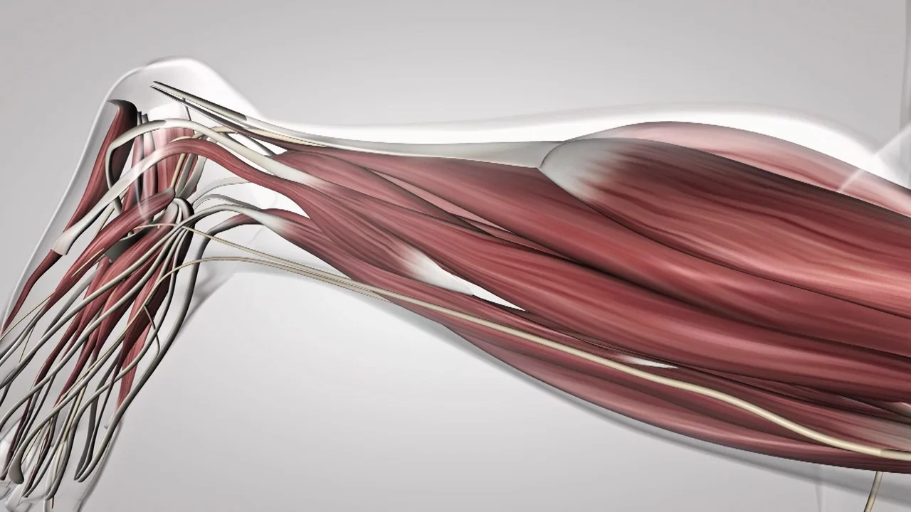
Au coeur des organes : Le réflexe myotatique
473.5K
The myotatic reflex is an involuntary muscle contraction triggered by stretching, which maintains posture through a neural circuit: muscle spindles detect stretching and send electrical nerve impulses via sensory neurons to the spinal cord, where they trigger motor neurons to release acetylcholine at the neuromuscular junction, causing muscle contraction; this reflex arc operates entirely within the spinal cord without brain involvement, enabling rapid postural adjustments.
Click to watch →

03_Le réflexe myotatique : Les éléments anatomiques du reflexe myotatique partie 02
106
The myotatic reflex is a spinal reflex arc where muscle stretch triggers contraction. The muscle spindle (fuseau neuromusculaire) acts as the mechanoreceptor detecting stretch. Sensory information travels via unipolar axons in the dorsal root ganglion to synapses in the spinal cord gray matter. The spinal cord integrates this sensory input and converts it into motor output via alpha motor neurons in the anterior horn. These motor neurons project to effector muscles, causing contraction of the stretched muscle and relaxation of its antagonist. This reflex maintains muscle tone and protects against excessive stretching.
Click to watch →

LIVE : Révisions BAC SVT 2017 : Neurone et fibre musculaire !
4.7K
The myotatic reflex is a rapid, involuntary, stereotyped response to muscle stretching. The reflex arc involves: muscle spindles detecting stretch, sensory neurons sending signals via afferent nerves to the spinal cord, synapses in the gray matter connecting sensory to motor neurons, motor neurons sending signals via efferent nerves to muscles, and finally muscle contraction at the neuromuscular junction. This reflex is rapid because it involves only two neurons, stereotyped because the response is always the same, and involuntary because it cannot be consciously controlled.
Click to watch →

Exercices - Terminale S2 - SVT: Travaux Dirigés / Réflexe Myotatique / Sujet Bac 2006 / M. Gnasse
3.8K
In spinal animals, muscle tension can be measured during progressive stretching of the Achilles tendon. Experimental curves reveal that muscle stretching triggers progressive contraction, with contraction remaining elevated as long as stretching persists. The mechanism involves: (1) mechanoreceptors (neuromuscular spindles or Golgi tendon organs) convert mechanical stretching into electrical receptor potentials, (2) these potentials generate nerve impulses transmitted via sensory fibers to the lumbosacral spinal cord, (3) monosynaptic connections transform sensory signals into motor signals, (4) motor signals travel via motor fibers to the muscle, (5) at the neuromuscular junction, signals trigger contraction through the motor end plate. This demonstrates the reflex arc pathway.
Click to watch →
Deep Dive
Understanding the Medical Research Council (MRC) scale for muscle power grading (0 to 5).
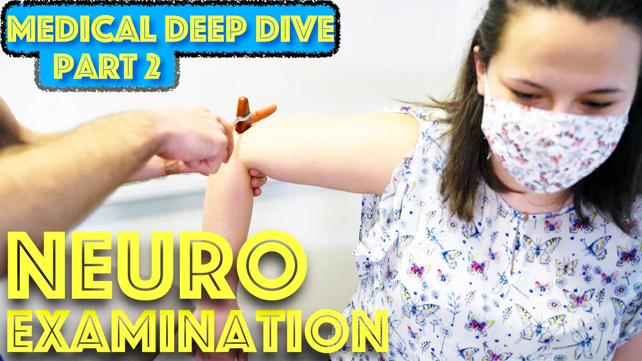
Neurological Examination - Power, Reflexes, Sensation, and Coordination - Dr Gill
177.5K
The MRC scale grades muscle power from 0 to 5: Grade 5 indicates normal power where the patient can push and pull against the examiner with reasonable strength; Grade 4 shows weakness but the patient can still move against resistance; Grade 3 allows movement against gravity without resistance; Grade 2 permits movement only when gravity is eliminated (e.g., arm can be moved side-to-side if held); Grade 1 indicates no visible movement but muscle contraction can be felt; Grade 0 represents complete absence of movement. Upper motor neuron lesions typically cause weakness in extensors of the upper limbs and flexors of the lower limbs.
Click to watch →
Neurology: Clinical Skills - Motor, Sensory, & Reflex Neurological Exam #neurology #ubcmedicine
1.6M
The MRC scale quantifies muscle power with grades: Grade 5 indicates normal strength against full resistance; Grade 4 indicates reduced strength against resistance (further broken into 4+, 4, and 4-); Grade 3 indicates movement against gravity with resistance removed; Grade 2 indicates movement only when gravity is removed; Grade 1 indicates a flicker of movement in the muscle; Grade 0 indicates no movement at all. When testing power, clinicians should isolate the muscle being tested, immobilize the proximal joint, and always compare muscle power on both sides before moving to the next muscle.
Click to watch →
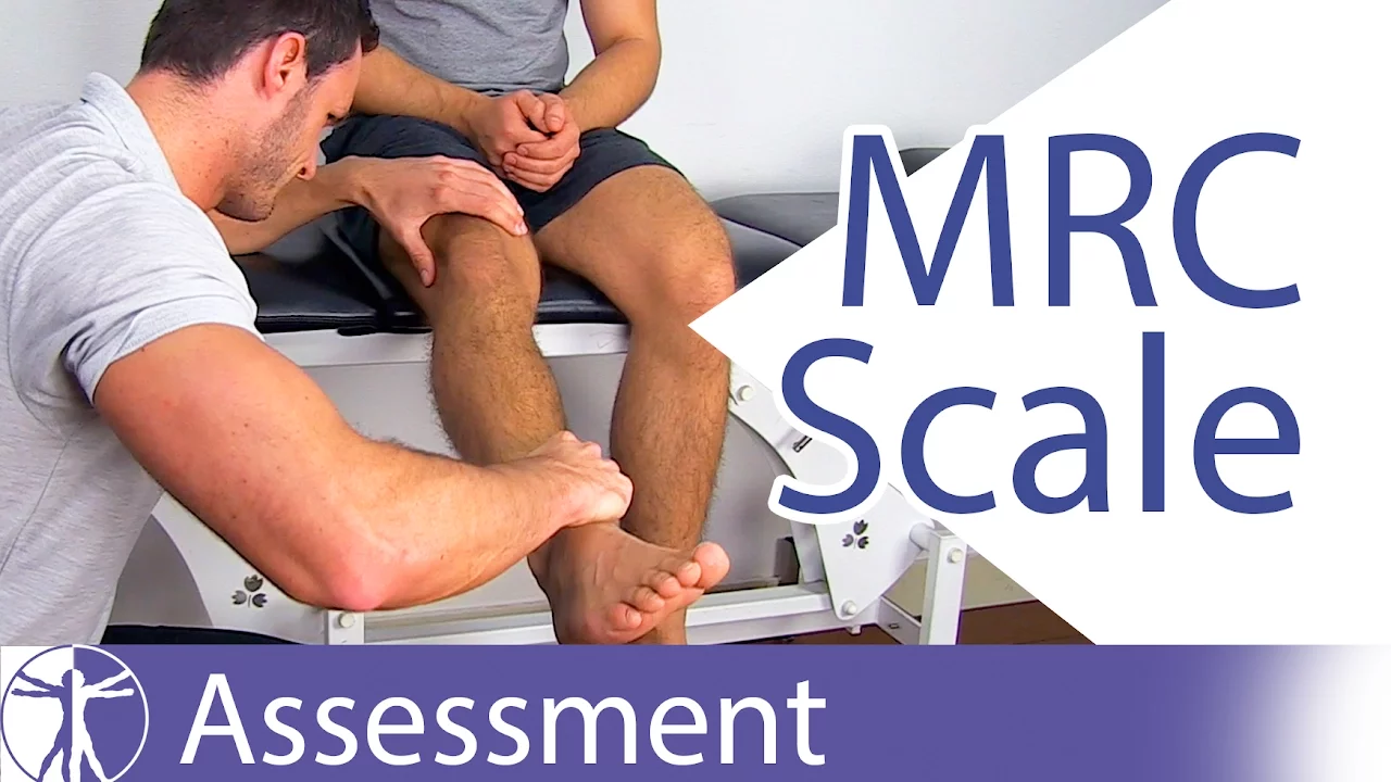
MRC Muscle Strength Grading: A Comprehensive Guide
578K
The MRC (Medical Research Council) Scale rates muscle strength on a 0-5 scale: Grade 0 indicates no muscle contraction; Grade 1 shows only a trace or flicker of movement without limb movement; Grade 2 allows full range of motion only when gravity is eliminated (tested in side-lying position); Grade 3 enables movement against gravity without resistance; Grade 4 demonstrates weakness with moderate resistance; and Grade 5 represents full strength against maximum resistance. For accurate assessment, clinicians must compare both limbs and use appropriate patient positioning for each grade.
Click to watch →

Video Atlas of the Detailed Neurologic Examination 4
19.2K
The Medical Research Council (MRC) scale grades muscle power from 0-5: 0=no movement, 1=contraction felt but no joint movement, 2=movement with gravity eliminated, 3=movement against gravity but less than normal, 4=movement against gravity and resistance but not normal, 5=normal. Accurate testing requires patient cooperation. For brachial plexus examination, test serratus anterior by having patient push arms down against resistance (scapula should hold against chest wall). Test rhomboids by having patient push back against resistance with hands on hips. Test supraspinatus and infraspinatus by having patient rotate arm outward against resistance.
Click to watch →
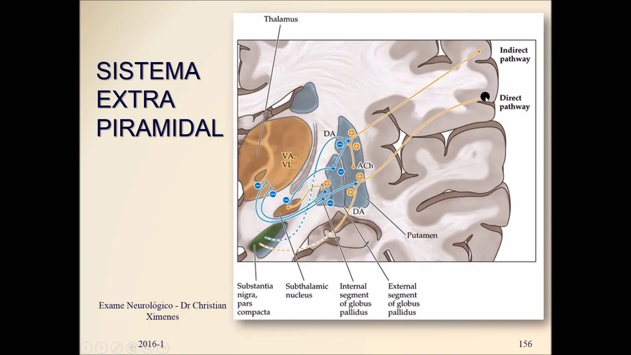
Força Muscular - Semiologia Neurológica
4K
The Medical Research Council (MRC) scale grades muscle strength from 0 to 5: Grade 0 = no muscle contraction (paralysis); Grade 1 = visible contraction but no movement; Grade 2 = movement possible but cannot overcome gravity (horizontal movement only); Grade 3 = movement against gravity but no resistance; Grade 4 = movement against some resistance but not full resistance; Grade 5 = normal strength. This standardized scale allows consistent documentation of motor deficits. The scale is essential for tracking disease progression and treatment response in neurological conditions.
Click to watch →
Deep Dive
The fundamental differences between Upper Motor Neuron (UMN) and Lower Motor Neuron (LMN) lesions and their clinical signs.
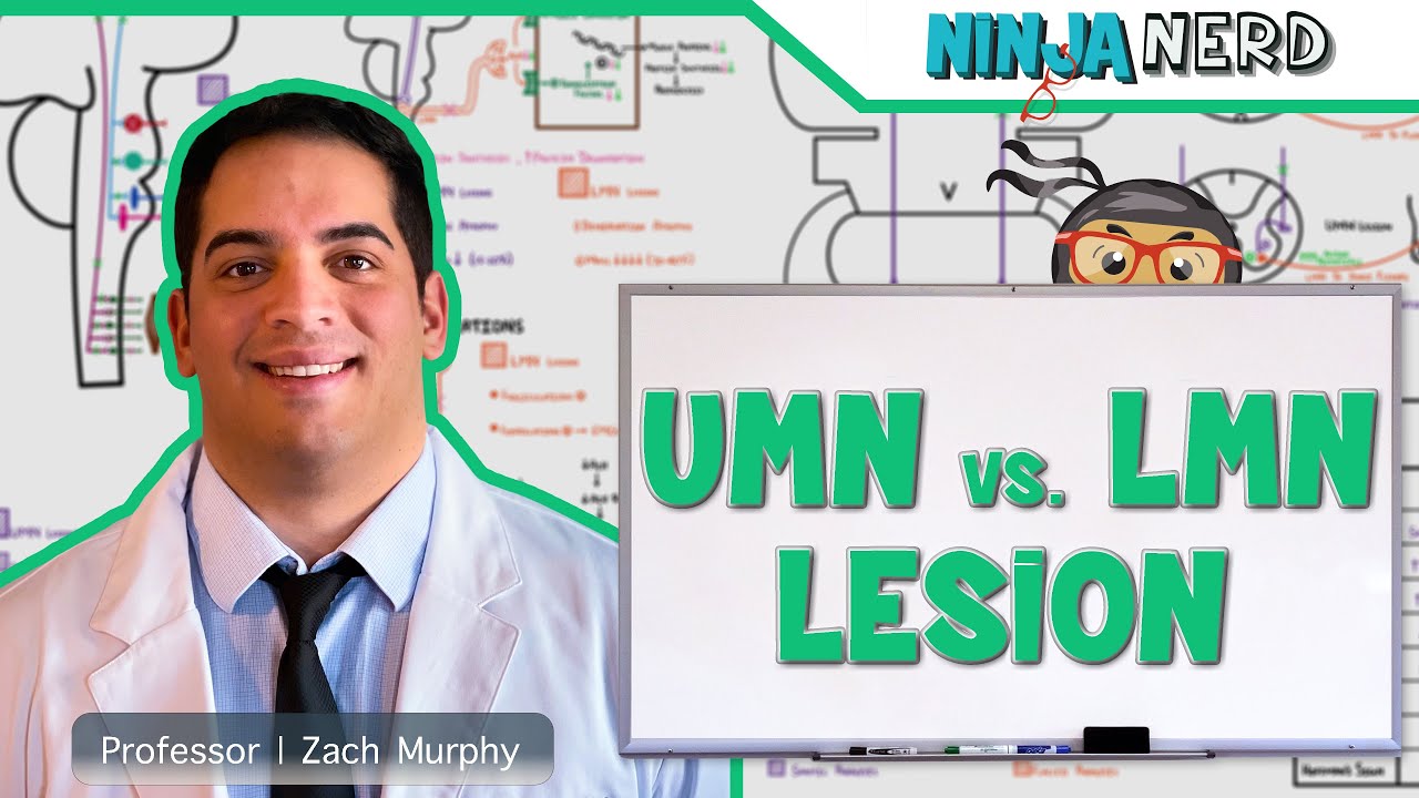
Neurology | Upper Motor Neuron vs Lower Motor Neuron Lesion | UMN vs LMN Lesion
880.5K
Upper motor neuron lesions (affecting the corticospinal/corticobulbar tracts from cortex to lower motor neurons) cause hypertonia, hyperreflexia, spastic paralysis, and minimal muscle atrophy (disuse atrophy), while lower motor neuron lesions (damaging anterior gray horn cell bodies, axons, or axon terminals) produce hypotonia, hyporeflexia, flaccid paralysis, and severe muscle atrophy (denervation atrophy); upper motor lesions show positive Babinski sign, pronator drift, and Hoffman's sign, whereas lower motor lesions exhibit fasciculations/fibrillations.
Click to watch →
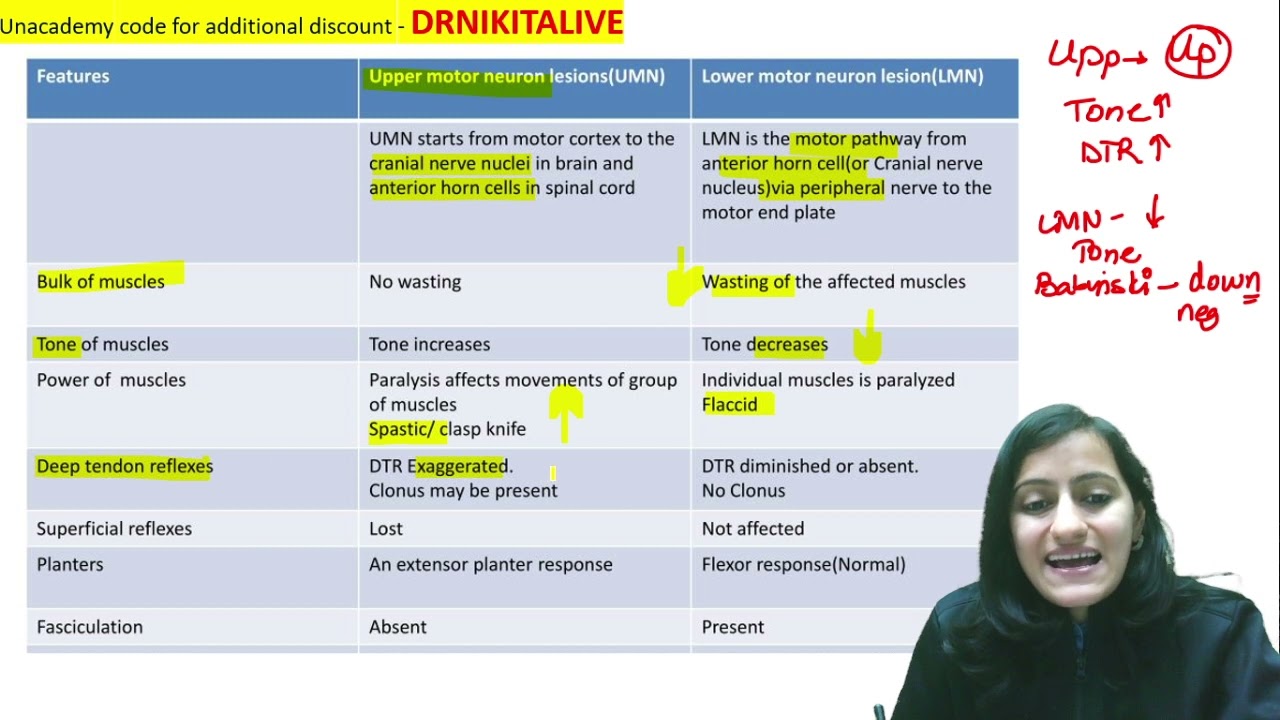
Mnemonic of the day - UMN vs LMN lesion|Medicine Physiology | Dr. Nikita Nanwani
62.4K
Upper Motor Neuron (UMN) lesions cause spastic paralysis with increased muscle tone (hypertonia), hyperreflexia, and Babinski sign, while Lower Motor Neuron (LMN) lesions cause flaccid paralysis with decreased muscle tone (hypotonia), hyporeflexia, and muscle atrophy.
Click to watch →
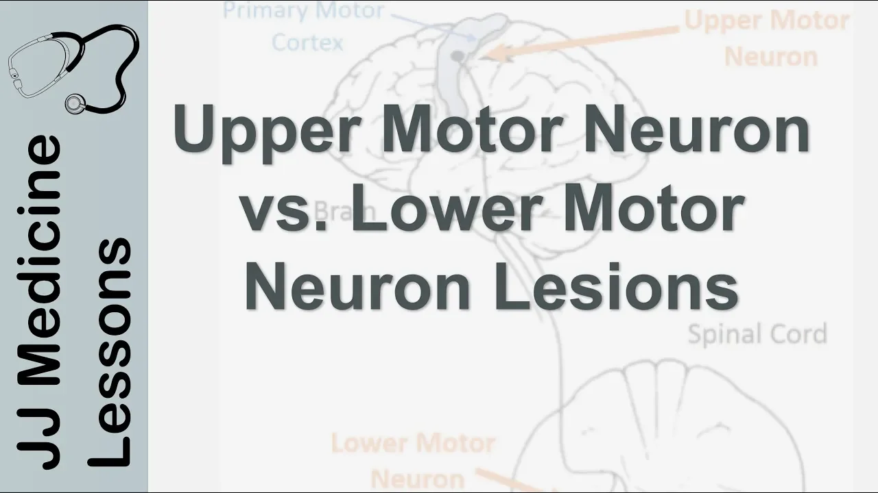
Upper and Lower Motor Neuron Lesions | UMN vs LMN, Differences and Mnemonic
258K
Upper motor neuron (UMN) lesions, originating from the brain or brainstem and synapsing on lower motor neurons in the spinal cord's ventral horn, cause hypertonicity, hyperreflexia, spasticity, disuse atrophy, and a positive Babinski sign (toes point upward), while lower motor neuron (LMN) lesions, located in the spinal cord's ventral horn and directly innervating skeletal muscles, cause hypotonia, hyporeflexia, flaccidity, denervation atrophy, and fasciculations with a negative Babinski sign (toes point downward); the mnemonic 'U = Up = Hyper' and 'L = Low = Hypo' helps remember these opposing characteristics.
Click to watch →
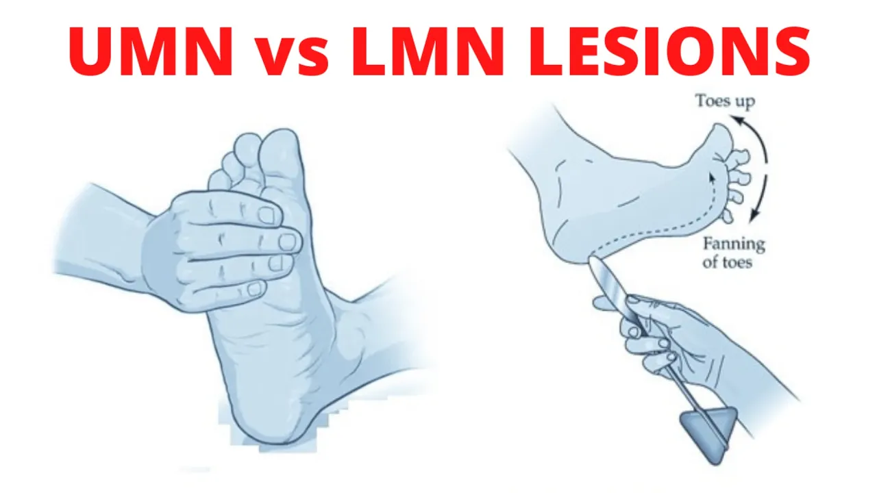
UMN vs LMN Lesion
5.2K
Upper motor neuron (UMN) lesions, which involve cortical neurons giving origin to corticospinal and corticobulbar tracts, produce spastic paralysis with increased muscle tone, hyperreflexia, clonus, and Babinski sign, while lower motor neuron (LMN) lesions, which involve anterior horn cells directly innervating skeletal muscles, produce flaccid paralysis with decreased muscle tone, areflexia, fasciculations, and muscle atrophy; UMN lesions affect large body areas below the lesion level and can be contralateral or ipsilateral, whereas LMN lesions affect small areas at the lesion level and are always ipsilateral.
Click to watch →

Internal Medicine - Dr Allam | CNS_1 | AllTebFamily.com
74.3K
The key clinical distinction between upper motor neuron (UMN) and lower motor neuron (LMN) lesions lies in their characteristic signs: UMN lesions cause hyperreflexia, spasticity, and no muscle wasting, while LMN lesions cause hyporeflexia, flaccidity, and muscle atrophy. This differentiation is essential for neurological diagnosis, as UMN lesions result from damage to the corticospinal tract (pyramidal system) above the spinal cord, whereas LMN lesions involve damage to the anterior horn cells or peripheral nerves at or below the spinal cord level.
Click to watch →
Prerequisite Knowledge
- Concept 01Basic neuroanatomy of the lumbosacral plexus and the specific nerve roots (L1-S2) supplying the lower limbs.
- Concept 02The physiological mechanism of the deep tendon reflex arc (myotatic reflex).
- Concept 03Understanding the Medical Research Council (MRC) scale for muscle power grading (0 to 5).
- Concept 04The fundamental differences between Upper Motor Neuron (UMN) and Lower Motor Neuron (LMN) lesions and their clinical signs.
Subsequent Learning
- Step 01Conducting a comprehensive Lower Limb Sensory Assessment (covering dermatomes, light touch, pinprick, vibration, and proprioception).
- Step 02Integrating motor and sensory findings to localize neurological lesions (e.g., distinguishing between radiculopathy, peripheral neuropathy, and spinal cord compression).
- Step 03Performing advanced coordination and gait assessments, including Romberg's test and the heel-to-shin test.
- Step 04Formulating a clinical differential diagnosis based on abnormal findings such as hyperreflexia, clonus, or focal muscle wasting.
Leg Exam
Reflexes
Leg Exam
0:03Playing Section
- 1
Test lower limb strength via lifting and pushing.
- 2
Assess knee flexion, extension, and ankle movement.
- 3
Evaluate toe control and foot inversion/eversion.
Limitations of Standardized OSCE Checklists and the Need for Functional and Quantitative Assessment
While traditional OSCE guides emphasize a rigid, step-by-step approach to lower limb motor exams (tone, power, reflexes, myotomes) for standardized testing, critics argue this method promotes rote memorization over clinical reasoning. In practice, traditional manual muscle testing (using the MRC 0-5 scale) suffers from poor inter-rater reliability and subjective bias, particularly in detecting subtle weakness. Opposing perspectives advocate for incorporating quantitative clinical tools, such as hand-held dynamometry, which provide objective, reproducible data. Furthermore, critics point out that isolated, segmented bedside exams lack ecological validity; they recommend prioritizing functional assessments, such as gait analysis and task-oriented mobility tests (e.g., the Timed Up and Go test), which correlate more accurately with a patient's real-world disability, daily functioning, and rehabilitation outcomes.
Deep Dive
Conducting a comprehensive Lower Limb Sensory Assessment (covering dermatomes, light touch, pinprick, vibration, and proprioception).

Lower Limb Neuro Exam
271.8K
Sensory testing evaluates five essential modalities along specific dermatomal patterns (L1-S2). Light touch assessment using a cotton swab confirms dorsal column-medial lemniscus pathway integrity; terminology includes anesthesia (loss), hypoesthesia (decreased), hyperesthesia (increased), and paresthesias (numbness/tingling). Pain sensation testing with pointed stimuli confirms lateral spinothalamic tract function, with terms analgesia (loss) and hyperalgesia (increased). Temperature testing follows the same dermatomal approach. Vibration sense testing using tuning forks proceeds distally to proximally, confirming dorsal column and peripheral nerve integrity. Proprioception testing with eyes closed evaluates position sense in three-dimensional space. Stocking-glove neuropathy refers to bilateral distal sensory loss commonly seen in diabetic patients.
Click to watch →

Real Person ASMR - In-Depth Sensation Exam (Vibration, Sharp or Dull and More)
247.1K
Lower limb evaluation mirrors upper body testing but focuses on legs and feet, including two-point discrimination on toes, vibration sense, temperature sensation, air current detection, wetness detection, and proprioception testing. Proprioception specifically involves moving toes up and down with eyes closed to assess joint position awareness. Since lower limbs are controlled by different brain regions, this comprehensive assessment helps identify potential lesions or neurological abnormalities.
Click to watch →
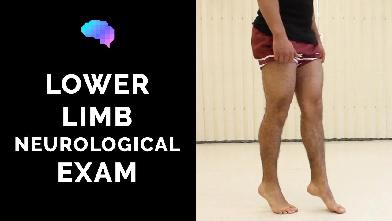
Lower Limb Neurological Examination - OSCE guide (old version) | UKMLA | CPSA | PLAB 2
2.2M
This segment covers the sensory and coordination components of lower limb neurological examination. Sensory testing evaluates multiple modalities: light touch using cotton wool, pinprick sensation using a safety pin, vibration sensation using a tuning fork, and joint position sense (proprioception) by moving the big toe up and down while the patient identifies direction with eyes closed. Coordination testing assesses cerebellar function through tasks including heel-to-shin movement, rapid alternating movements, and finger-to-nose tests. The examination concludes with documentation of findings, noting that normal results include intact gait, normal tone, power of 5/5 bilaterally, present reflexes, normal sensation across all modalities, and intact coordination. This comprehensive assessment helps identify neurological deficits affecting the peripheral nerves, spinal cord, or brainstem.
Click to watch →
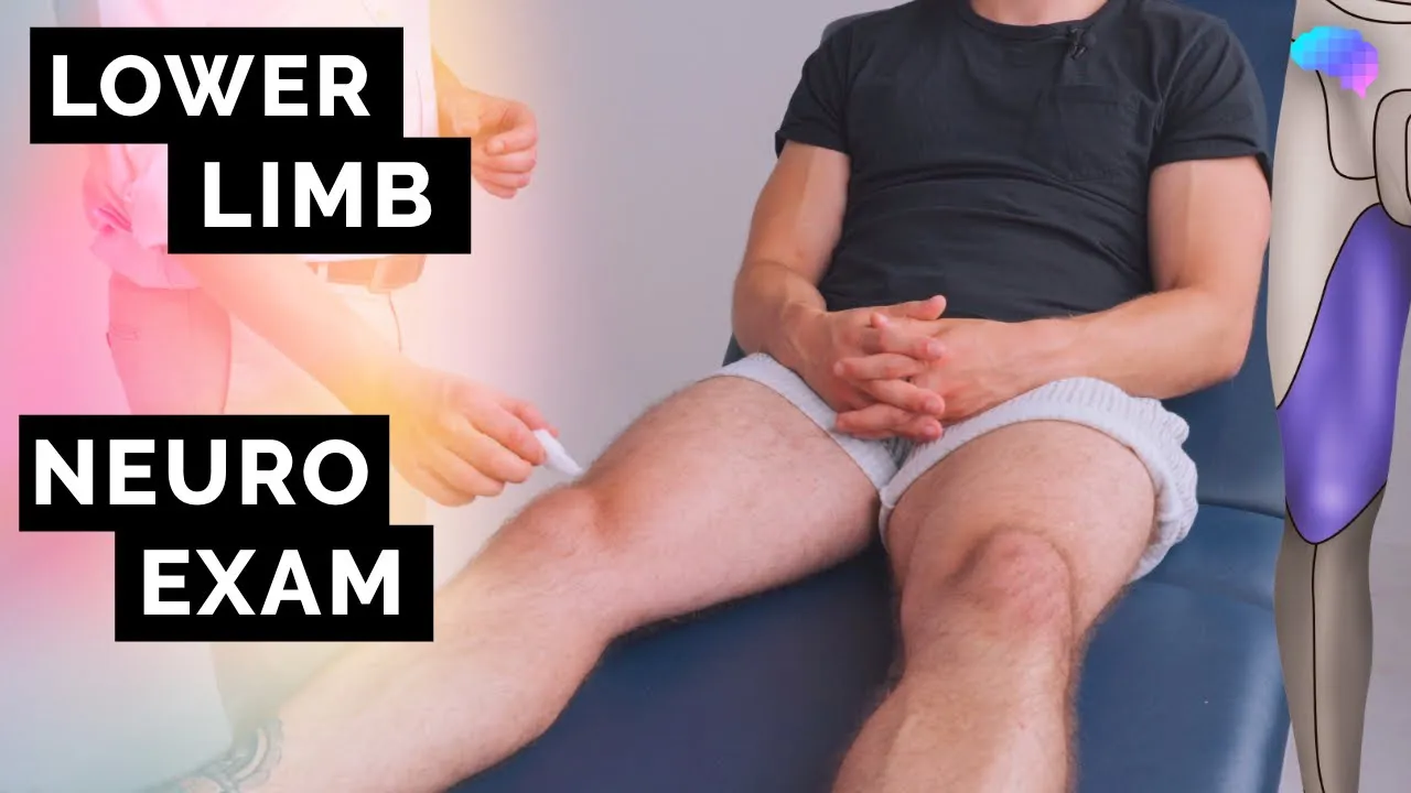
Lower Limb Neurological Examination | OSCE Guide | NEW | UKMLA | CPSA | PLAB 2
616.8K
A complete lower limb neurological examination involves assessing gait, tone, power, reflexes, sensation (light touch, pinprick, vibration), proprioception, and coordination to evaluate the function of peripheral nerves, spinal cord, and brainstem pathways.
Click to watch →
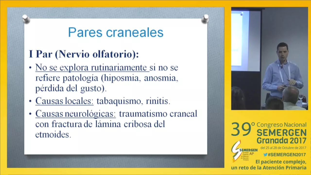
Taller. Exploración neurológica básica
54K
Sensory examination is divided into superficial (light touch, pain) and deep (proprioception, vibration) components. Superficial sensation should be examined from proximal to distal in all dermatomes. Deep sensation is examined distally first, assuming proximal sensation is normal if distal testing is intact. Superficial sensation testing uses cotton wisp for light touch and pinprick for pain. Deep sensation testing uses tuning fork for vibration at metacarpal heads, radial styloid, tibial tuberosity, and knee joint. Proprioception is tested by asking the patient to identify joint movement direction with eyes closed. Reflexes are graded from 0 to 4: 0=absent, 1=diminished, 2=normal, 3=brisk without significance, 4=hyperreflexia with clonus (pathologically significant).
Click to watch →
Deep Dive
Integrating motor and sensory findings to localize neurological lesions (e.g., distinguishing between radiculopathy, peripheral neuropathy, and spinal cord compression).

Peripheral Neuropathy with Hyperexcitability
5.7K
The sensory-motor-reflex approach localizes neurological lesions: radiculopathy shows radicular pain with precipitating/relieving factors and root-specific reflex changes; plexopathy shows pain in plexus location radiating along multiple nerves without clear precipitants; peripheral neuropathy shows symmetrical sensory-motor involvement without root-specific patterns. This systematic approach distinguishes between these entities, guiding appropriate diagnostic workup and treatment strategies.
Click to watch →

PG CLUB NEUROLOGY - Dr Sanjeev & Dr Davison
662
Spinal cord involvement produces bilateral motor and sensory symptoms. Nerve root involvement causes shooting pain on one side (radicular pain). Neuropathy causes distal weakness. The presence of both motor and sensory symptoms with bilateral involvement strongly suggests spinal cord pathology. The pattern of sensory involvement (pain and temperature crossing over) is characteristic of spinal cord lesions.
Click to watch →

Localization of Peripheral Nervous System Lesions
9.3K
This video teaches medical students how to localize peripheral nervous system lesions by understanding the motor and sensory functions of major nerves in the arm (ulnar, median, musculocutaneous, radial, axillary) and leg (femoral, obturator, sciatic with tibial and common fibular branches), distinguishing nerve injuries from radiculopathies through characteristic patterns of weakness, sensory loss, and reflex changes. For example, ulnar neuropathy causes weakness in finger adduction and thumb adduction with sensory loss in the ulnar distribution, while C8 radiculopathy presents with similar motor deficits but includes sensory loss in the medial forearm and radicular pain. Similarly, femoral neuropathy causes knee extension weakness and thigh sensation loss, whereas L4 radiculopathy may also affect foot dorsiflexion and inversion. Understanding these differences is essential for proper diagnosis and management of peripheral nerve disorders.
Click to watch →

#Davidson #Neurology 2 #Brainstem
539
Peripheral neuropathy patterns: glove and stocking distribution (most common), diabetic neuropathy (most common cause). Spinal cord lesions can be localized based on sensory level, motor level, and autonomic level. The pattern of sensory loss (dissociated sensory loss, Brown-Séquard syndrome) helps localize the lesion. The key is to identify which tracts are affected.
Click to watch →

HÉRNIA DE DISCO COLUNA LOMBAR O PASSO A PASSO da AVALIAÇÃO Clínica de Fisioterapia Dr. Robson Sitta
24.1K
Radiculopathy results from nerve root compression and presents with characteristic neurological deficits. Radicular pain typically affects regions below the knee, while somatic pain from musculoskeletal compression localizes to the gluteal or thigh regions. Clinical assessment includes myotome testing for motor function—for example, L5 radiculopathy affects tibialis anterior (great toe extension). Sensory evaluation examines dermatome distribution for decreased sensation. Reflex testing assesses patellar (L4) and Achilles (S1) reflexes, which may be diminished or absent. These neurological signs help localize the level of nerve root involvement and distinguish radicular from somatic causes.
Click to watch →
Deep Dive
Performing advanced coordination and gait assessments, including Romberg's test and the heel-to-shin test.
The Neurologic Physical Examination
558.2K
Coordination testing includes finger-to-nose (upper extremity) and heel-to-shin (lower extremity). For finger-to-nose, hold your hand far enough that the patient must fully extend to reach, looking for side-to-side weaving and difficulty hitting the stationary target in cerebellar ataxia. For heel-to-shin, slide the heel up and down the tibia, watching for side-to-side swaying on the shin. Gait assessment includes normal walking and tandem (heel-to-toe) walking. The Romberg test (standing with eyes closed) tests balance by removing visual input, revealing problems with vestibular or proprioceptive systems. A positive Romberg indicates deficit in one of these systems.
Click to watch →
![[ASMR] Real Person Head to Toe Physical Exam Assessment | Soft Spoken Medical Roleplay](https://i.ytimg.com/vi/JbA_DWhjmzM/maxresdefault.jpg)
[ASMR] Real Person Head to Toe Physical Exam Assessment | Soft Spoken Medical Roleplay
1.1M
The Romberg test assesses balance and proprioception by having the patient stand with feet together and arms out, first with eyes open and then closed. Negative results indicate no significant neurological impairment. Coordination tests include finger-to-nose testing (moving finger to nose and then to examiner's finger) and heel-to-shin testing (moving heel along shin).
Click to watch →

Thực hành y khoa 3 - Khám thần kinh vận động
1.4K
The heel-to-shin test evaluates coordination: (1) The patient places their heel on their opposite knee, (2) The examiner observes accuracy and smoothness of movement, (3) Normal findings: The heel touches the knee directly and moves in a straight line, (4) Abnormal findings: Overshooting, undershooting, or irregular paths indicate cerebellar dysfunction. The Romberg test evaluates sensory ataxia: (1) The patient stands with feet together and closes their eyes, (2) The examiner observes for 60 seconds, (3) Normal findings: The patient maintains balance, (4) Abnormal findings: Swaying, staggering, or falling indicate sensory ataxia or vestibular dysfunction.
Click to watch →
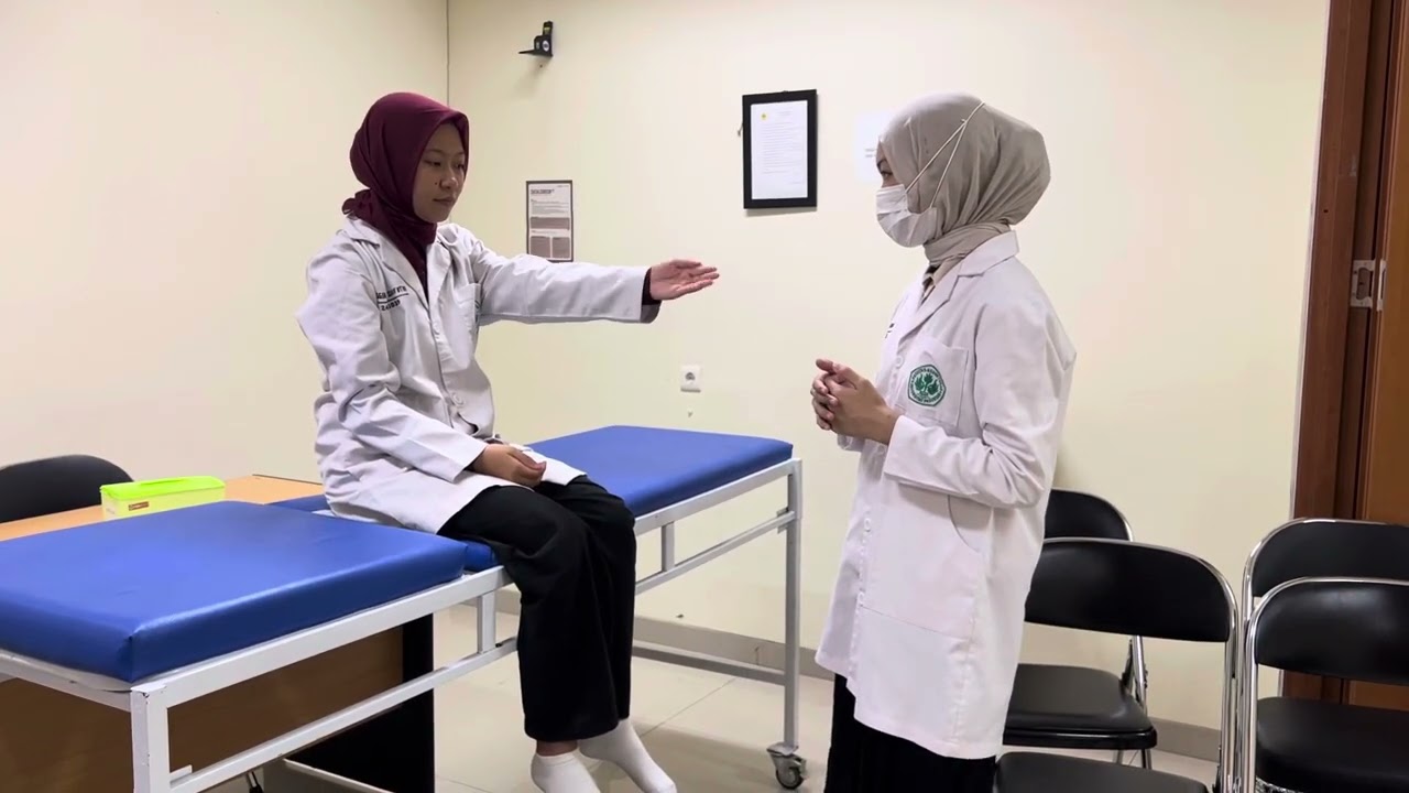
Tes Koordinasi dan Keseimbangan_Nisrina Apriliani Mukhsin_240010018
446
This segment covers lower extremity coordination and balance tests: heel-to-shin test (sliding heel down opposite shin), rebound test (resisting arm movements), Romberg test (standing with feet together, eyes open then closed), Romberg test with feet apart, and tandem gait test (walking with feet one in front of the other). These tests evaluate lower extremity coordination, balance, and proprioception. The examiner stands behind or beside the patient to prevent falls. Results are documented on the medical chart.
Click to watch →
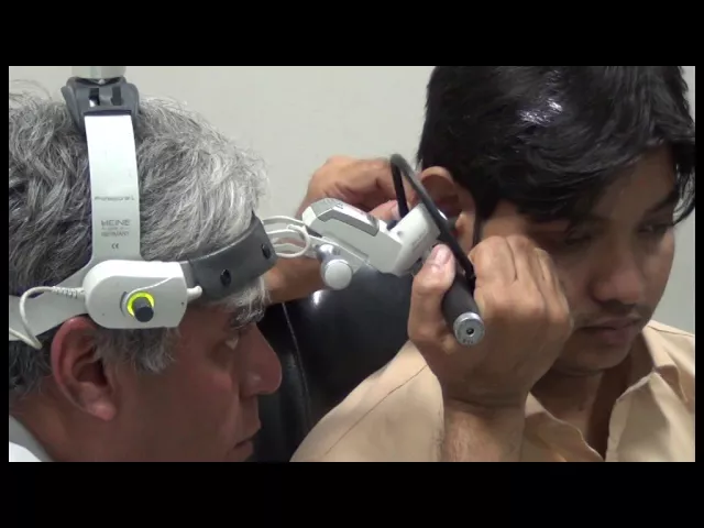
Examination of EAR
39.7K
The Romberg test is controlled by input from the eyes, vestibular system, and proprioception. To perform, the patient stands with feet together and removes their shoes. The physician removes visual input by closing the patient's eyes and observes whether the patient maintains balance or sways. If the patient sways or falls, this indicates dysfunction in the vestibular system, proprioception, or visual system. After the Romberg test, observe the patient's gait with eyes open. Watch whether the gait is straight or if there is deviation to one side. This helps identify vestibular or cerebellar dysfunction.
Click to watch →
Deep Dive
Formulating a clinical differential diagnosis based on abnormal findings such as hyperreflexia, clonus, or focal muscle wasting.

HY USMLE Q #1292 – Neuro
6.6K
Upper motor neuron (UMN) findings include hyperreflexia, hypertonia, Babinski sign, and clonus. Lower motor neuron (LMN) findings include hypotonia, hyporeflexia, fasciculations, and muscle wasting. ALS is characterized by the combination of both UMN and LMN findings, which is a critical diagnostic feature. The presence of both types of findings in the same patient strongly suggests ALS rather than other neurological conditions.
Click to watch →
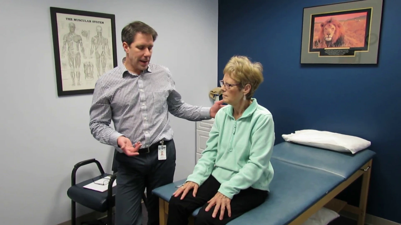
Hyperreflexia, Clonus and a Positive Hoffman's Sign
262K
Upper motor neuron lesions can be identified through specific neurological signs including hyperreflexia (exaggerated reflexes), clonus (sustained rhythmic muscle contractions), and a positive Hoffman's sign (index finger flexion when the middle finger is flicked); these findings collectively indicate damage to the central nervous system and warrant referral to a neurologist or physiatrist for further evaluation and diagnostic testing such as MRI.
Click to watch →
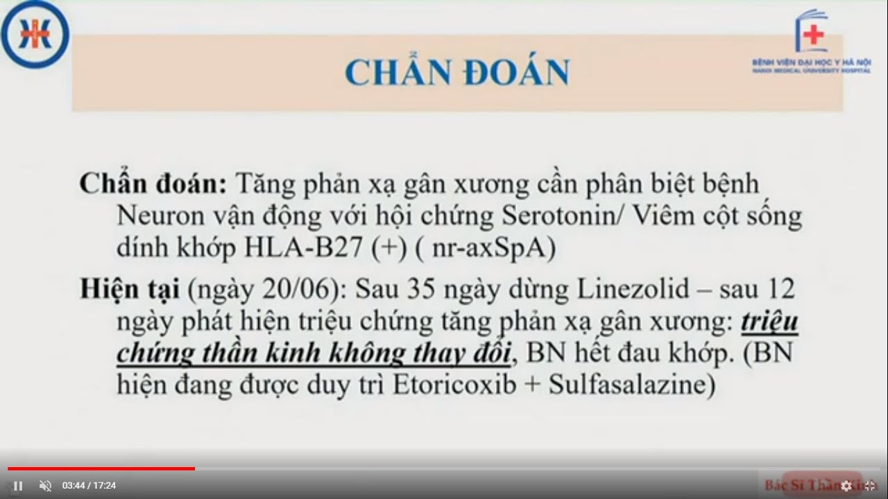
Case theo dõi Hội chứng Serotonin
1.2K
Hyperreflexia in young patients requires differential diagnosis including: (1) physiological hyperreflexia without underlying pathology, (2) drug-induced causes (serotonin syndrome), (3) motor neuron disease, (4) multiple sclerosis, and (5) other neurological conditions. The patient's bilateral symmetric hyperreflexia without other neurological deficits makes motor neuron disease less likely.
Click to watch →

Upper motor neurons | Organ Systems | MCAT | Khan Academy
439.3K
Four key clinical signs indicate upper motor neuron dysfunction: (1) Hyperreflexia - exaggerated muscle stretch reflexes due to supersensitive lower motor neurons; (2) Clonus - rhythmic antagonist muscle contractions triggered by reciprocal reflex activation; (3) Hypertonia - increased muscle resistance during passive movement; (4) Babinski sign - extensor plantar response where toes extend upward when scraping the foot sole. These signs contrast with lower motor neuron signs like hyporeflexia and hypotonia, helping clinicians distinguish between upper and lower motor neuron lesions.
Click to watch →
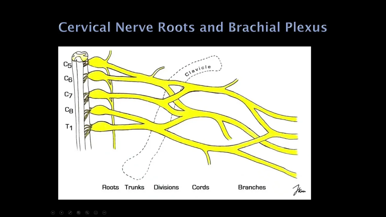
1. Quick Reviews: Neurology Localization
14.9K
Upper motor neuron lesions show hyperreflexia, clonus, Babinski sign, increased tone (spasticity - velocity-dependent), minimal atrophy. Lower motor neuron lesions show areflexia, flaccidity, significant atrophy, and fasciculations. Fasciculations alone are benign but with weakness indicate pathology. Spasticity is velocity-dependent (catch on quick movement); rigidity is non-velocity-dependent (seen in Parkinson's). Paratonia is resistance to passive movement from frontal lobe dysfunction (Alzheimer's).
Click to watch →
Leg Exam
Reflexes
Leg Exam
0:03- 1
Test lower limb strength via lifting and pushing.
- 2
Assess knee flexion, extension, and ankle movement.
- 3
Evaluate toe control and foot inversion/eversion.
Limitations of Standardized OSCE Checklists and the Need for Functional and Quantitative Assessment
While traditional OSCE guides emphasize a rigid, step-by-step approach to lower limb motor exams (tone, power, reflexes, myotomes) for standardized testing, critics argue this method promotes rote memorization over clinical reasoning. In practice, traditional manual muscle testing (using the MRC 0-5 scale) suffers from poor inter-rater reliability and subjective bias, particularly in detecting subtle weakness. Opposing perspectives advocate for incorporating quantitative clinical tools, such as hand-held dynamometry, which provide objective, reproducible data. Furthermore, critics point out that isolated, segmented bedside exams lack ecological validity; they recommend prioritizing functional assessments, such as gait analysis and task-oriented mobility tests (e.g., the Timed Up and Go test), which correlate more accurately with a patient's real-world disability, daily functioning, and rehabilitation outcomes.
Basic neuroanatomy of the lumbosacral plexus and the specific nerve roots (L1-S2) supplying the lower limbs.
The physiological mechanism of the deep tendon reflex arc (myotatic reflex).
Understanding the Medical Research Council (MRC) scale for muscle power grading (0 to 5).
The fundamental differences between Upper Motor Neuron (UMN) and Lower Motor Neuron (LMN) lesions and their clinical signs.
Conducting a comprehensive Lower Limb Sensory Assessment (covering dermatomes, light touch, pinprick, vibration, and proprioception).
Integrating motor and sensory findings to localize neurological lesions (e.g., distinguishing between radiculopathy, peripheral neuropathy, and spinal cord compression).
Performing advanced coordination and gait assessments, including Romberg's test and the heel-to-shin test.
Formulating a clinical differential diagnosis based on abnormal findings such as hyperreflexia, clonus, or focal muscle wasting.
222.6K views2.7Klikes3:34@geekymedicsOriginal Release: 2024-01-04
[Music] so I'm now going to test some of the tone in your legs so if you just let your legs go loose and floppy okay good and just relax your ankle I'm just going to pull back on it okay so I'm going to test the strength in your legs now so if you can lift this leg straight up off the bed and stop me from pushing it down good just relax this and lift this leg up straight off the bed and stop me from pushing it down good and just relax it I'm going to place my hand underneath your leg if you can push it down into to the bed good and stop me from lifting it good great so I'm just going to bend your knee so bring your ankle back towards you good and relax I'm going to bend your knee and bring your ankle back towards you good now if you kick out against me good and kick out against me good and can you bring your toes up towards you and just keep them up and keep them up and can you push down against me good and push down against me good and just bring your big toe up towards you and keep it up good and keep it up good now if you turn your foot in that's great good and turn your foot in good and turn this foot out great and keep it out good and turn this foot out and keep it out okay so I'm just going to tap some of your reflexes just both at your knees and ankles so if you just relax your leg as much as possible so I'm now just going to bend your leg and just let it flop to the side same on the other side so we'll Bend your leg just let it flop to the side I'm now going to test a reflex in your foot I'm just going to use this dull end of this tip it may feel a little bit ticklish okay if you liked this video you'll love our textbook the geeky Medics clinical examination guide summarizes all the key examination skills for your practical exams by now at geeky medics.com [Music]
Up Next

33:31
Sensory System Examination: A Simplified CNS Guide for Graduates
@DradilRanjha
21.3K views•2021-07-15

48:47
Graphic Medicine: Comics for Collaborative Healthcare Communication
@nationalpatientadvocate
189 views•2023-12-04

10:28
Neuroanatomy: Central and Peripheral Nervous System Divisions Explained
@AKLECTURES
136.2K views•2014-09-20

3:05
Stages of Labor and Vaginal Birth | Childbirth Animation
@nucleusmedicalmedia
52.1M views•2017-08-18
Related Study Plans & Knowledge Roadmaps
Structured learning paths in Medicine
medicine
Abnormal LFTs: Diagnostic Workup & Pathology
01Liver Anatomy and Basic Physiology
02Introduction to Liver Function Tests (LFTs)
03Hepatocellular vs. Cholestatic Patterns
+ 3 more learning modules
12h6 modules
Explore Roadmap
medicine
Analyzing Blood Smears: RBC, WBC & Platelets
01Introduction to Blood Components & Smear Preparation
02Normal Morphology of Blood Cells
03Erythrocyte Pathologies & Morphological Variants
+ 2 more learning modules
12h5 modules
Explore Roadmap
medicine
Mechanical Ventilation: Waveforms & Alarms
01Anatomy and Physiology of Breathing
02Introduction to Mechanical Ventilation & Core Modes
03Ventilator Settings and Waveform Interpretation
+ 2 more learning modules
12h5 modules
Explore Roadmap
medicine
Neuro Exam: Localization & Stroke Syndromes
01Foundations of Neuroanatomy and Vascular Supply
02The Neurological Examination: Step-by-Step
03Principles of Neurological Localization
+ 3 more learning modules
18h6 modules
Explore Roadmap