










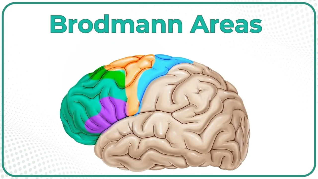
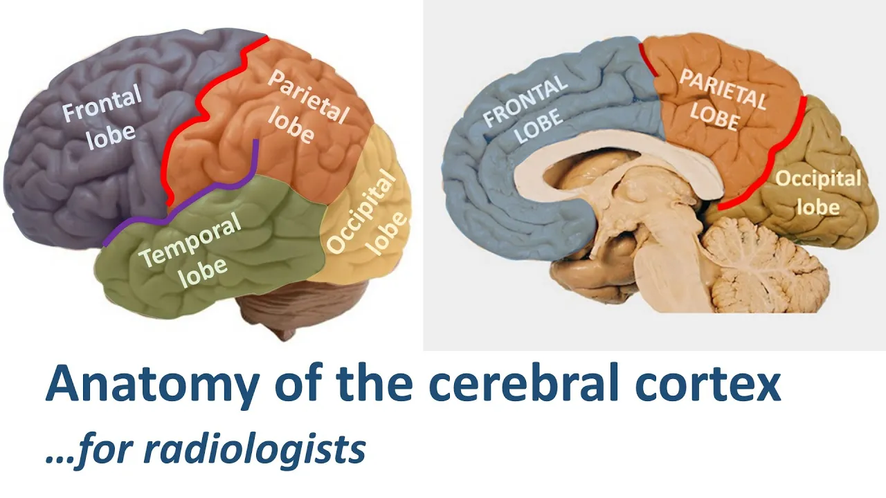



The cerebral cortex is the 4mm thick outer layer of the cerebrum, divided into four visible lobes (frontal, parietal, occipital, temporal) and two hidden parts (cingulate gyrus, insula), with key fissures including the longitudinal fissure (separating hemispheres), central fissure (separating frontal from parietal lobes), and sylvian fissure (separating temporal lobe). The frontal lobe contains the motor cortex (precentral gyrus), premotor cortex, and Broca's area (areas 44-45 in left hemisphere), responsible for motor planning, speech production, and executive functions. The temporal lobe contains the primary auditory cortex and Wernicke's area (area 22 in left hemisphere), critical for language comprehension. The parietal lobe processes somatosensory information through the postcentral gyrus (sensory homunculus) and handles spatial processing. Cranial nerves 1-12 emerge from the brainstem, with nerve 5 (trigeminal) providing facial sensation and motor control, nerve 7 (facial) controlling facial expression and taste, and nerve 8 (vestibulocochlear) responsible for hearing and balance.
Cerebral Cortex & Cranial Nerves: Complete Anatomy Guide
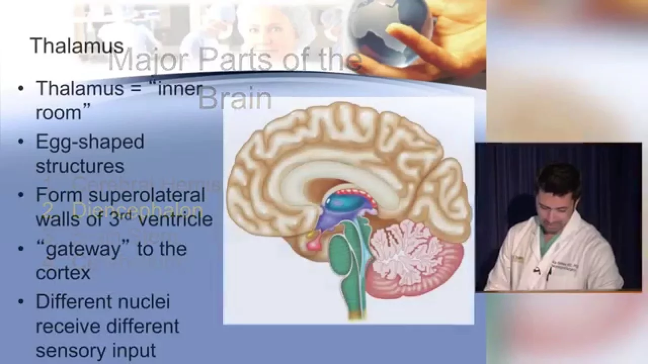
Added:hello in this video we are going to speak about the cerebral cortex and the cranial nerves so this follows the previous one and it focuses exclusively on these two areas the cerebral cortex and the cranial nerves now recall that the cerebrum has several parts so the cerebrum and the first part of that is the top layer that is four millimeters thick and that is called the cortex of the cerebrum to make it simple we say the cerebral cortex please do not confuse the two because there the cerebral cortex is only part of the cerebrum and the cerebrum is the whole entire thing that has right hemisphere left hemisphere and all other parts that we discussed in the previous video so the cerebral cortex is what makes the difference between a human being and and another animal it is the most extensively um the most extensive cortex in in all animals and it is the system where we store information that we that we acquire and that gives us cognition and language and thought and decision making and everything that you can think about that makes us human is mediated by the cortex so you can think about it as a as a thin pancake big and wrapped around the entire brain and it is convoluted into prominent areas that we call gyri one of them is called gyrus and the the valleys in between are called sulci one is sulcus sometimes you also use the terms fissure especially if if it is very very deep a deep groove and the brain is called fissure and uh uh most of them are called sulcus and the plural is suicide in most anatomy books that are not specialized i mean that do not really address the whole thing in detail they tell you oh the brain has four lobes it has more than that okay this is not the brain this is part of the brain this is the cerebrum and it has four visible four visible parts it is divided into four visible parts these divisions are not random they are divided based on the the functions of each one each lobe for example and as you recall each lobe is covered by a part of the bone of the skull it gives the name to each one of these another thing is that instead of the word lobe you could also use the word cortex for example we have the frontal cortex that includes all of this area or you can call it the frontal lobe the parietal cortex or the parietal lobe and the occipital lobe or the occipital um uh the occipital lobe or the occipital cortex and then the temporal lobe or the temporal cortex and these are all named after the bones that cover them parietal bone occipital bone temporal bone and frontal bone so the cortex is divided into six main parts and two of them are visible and two i'm sorry two are invisible and four of them are visible so we're going to discuss these and look at the main landmarks of each one and discuss the functions basic functions of each but to let you know that you always think about the anatomic name and the functional name so for example the occipital lobe is almost exclusively dedicated to visual processing so you are going to hear its functional name as or physiological name as the visual cortex the visual cortex so for example this somatosensory cortex this is a functional name it it is what it does but we call it a different anatomic name we call it the post central gyrus so we'll discuss this in detail and and the comment in this video so these again the six major parts of the cerebral cortex are the frontal lobe parietal lobe occipital lobe and the temporal lobe that you could see on the surface and now there are um there are other parts that are hidden one is called the insula or the insular lobe uh insular cortex and then there's another one that we will we will show you called the singular gyrus singulate gyrus so the main functions of the cortex as a whole is to give you cognitive all kinds of cognitive functions are mediated by or controlled by the cortex logic the language functions emotional processing functions uh sensory functions motor functions visual processing functions so the what again it is what makes us human the before we get into the details about every uh part of the cortex and what it what it has we are going to look at the boundaries that the boundaries that separate one area from another and these uh we call them fissures or fissures are the deep ones and gyra i mean um uh sulci or you know the shallow ones so one of the most important or the most prominent ones is that line that deep valve a groove that separates the right hemisphere from the left hemisphere it is much deeper than this but just because the brain here doesn't have anything to support it it is just flopping down but the the two sit together the right hemisphere the atmosphere sit together just like this and when you open them up you you see that that fissure is very deep so the longitudinal fissure goes from the rostral the far front and rostral end of the cerebrum to the far and at the back the uh the caudal end from the rostral end to the caudal end of the cerebrum oh that is all that you need to say if you are asked what is uh the longitudinal fissure be prepared to identify these fissures on pictures and to also explain what they separate so the longitudinal fissure extends from the rostral end to the caudal end of the the cerebrum and it separates the right hemisphere from the left hemisphere and um so that if you say that this is all you need to to say about a structural i mean a landmark like this then we have um the gyri uh these are prominent areas like little hills and each one has a particular name the central fissure is it is on the center of the head on the center of the brain and it separates the frontal lobe from the parietal lobe that central fissure here is going to have more than one name it will have more than one name it is called the rolandic fissure or rolandi or the fissure of rolandos and again it separates the frontal loop from the parietal lobe and because it is called central the frontal gyrus the gyrus in front of it anterior to it is called the pre central gyrus and the gyrus posterior to it is called the post central gyrus these are anatomic names but the functional names for these are the functional name for the precentral gyrus is the motor cortex and the functional name of the post posterior um uh the post central gyrus that is called the somatosensory cortex or the primary somatosensory cortex so then we have the um so so this center of fissure how do we describe it it is the fissure that extends from the uh longitudinal longitudinal fissure and extends all the way down to the lateral fissure or the sylvian fissure so it and add to this that the central fissure or the rolandic fissure or rolandic sulcus it doesn't matter what you see you call it as long as it's one of these it separates the frontal lobe from the parietal lobe and that's it then the the fissure that separates the temporal lobe from the anterior and the parietal lobes that one when you open it also it just it has a deep groove in in between so that is called the temporal fissure or the sylvian fissure or the fissure of sylvias all of these are names or one more is the lateral fissure all of these are names for the same the same groove that separates the parietal law the parietal and frontal lobes from the temporal lobe then we have the parietal occipital sulcus and that separates the parietal lobe from the occipital lobe you get the clue from the name parietal occipital or i mean parietal occipital it's a this is that boundary between the parietal and and the occipital lobes so now let's look at the major the six major parts of the cerebral cortex number one the cingulate gyrus and then the insula sometimes called the insular cortex insula comes from the word hidden or insulated and then you have the four lobes you have the frontal lobe or the frontal cortex parietal temporal and occipital and remember again instead of the word lobe you can always use the word cortex cingulate cortex insular cortex or insula so the cingulate you are not going to see it from the surface but it is part of the cerebral cortex how do you see it is when you open the longitudinal fissure between the two hemispheres you open that up and then you are going to see that um that um gyrus extending from the the posterior end of the posterior inferior end of the parietal lobe to all the way down to the uh to the prefrontal cortex this is called the prefrontal cortex and it goes right above that white structure that is the corpus callosum that's a different view for the corpus callosum so it extends all on the um that is superior to the corpus callosum and extends again from of the parietal lobe um all the way to the anterior most part of the um of of the prefrontal i'm sorry to the prefrontal cortex but it is all around the corpus callosum it it the functions include uh analysis of thought and also uh it plays a great role in decision making and uh self the regulation of mood and it is also critical for motor planning there are two areas here and the center of it that interact with these two areas for motor motor planning then the insula or insular cortex when you open the the temporal fissure or temporal um or the fissure of the sylvias or lateral fissure you open it up you're gonna find this you know area that is buried uh covered underneath and that is the ancillar cortex now the um this area is specialized in translating your feelings and emotions into things that you physically can feel like when you say someone says something it felt like i was punched in the stomach that physical sensation that you get as a result of your feelings is made possible by the insular cortex it contributes to pleasure the feeling of pleasure it it is also response it's a major part of the system called the brain default mechanism which makes kind of decisions to link memories with your conscious memory it is it contributes to speech it has an extensive connection between it and broca's area brokers areas right above it here and it has connections with it that contribute to verbal uh production to uh production of speech and um in some cases if that link is severed that could lead to mutism uh the um it is also a center for intuition for example when you feel oh something is gonna happen uh if you see someone walking so he's gonna fall he's gonna fall didn't i tell you so i had the intuition so these intuitions are basically calculated by the insular cortex and it uh it communicates with various areas of the brain and it gets like little pieces bits and pieces assembles them together into one complete you know unit of information that will enable you to make judgment even though you do not know why that judgment comes true if your knowledge that you have gathered you know through your life is true then your intuitions will be also accurate so what makes you think that this person is going to fall you are going to judge based on your own experiences that you know he would was walking you know for example with imbalance may be tilted this way and as a result you know that the weight on one foot or one leg is going to exceed the weight and the other significantly and the angle here there you get all these that will give you the logical conclusion that if you put your body on two legs under these circumstances that person cannot sustain the movement they that person will fall so but if the person doesn't have accurate information their intuition will not be always accurate but anyways that area that gives you that intuition is the insular cortex and so again it is involved in motor speech production and it has direct connections with broca's area right above it now the areas that cover the insula all these areas especially the part you know when you put these together back again let's see here the angle the the point where the frontal lobe the parietal lobe and the temporal lobe meet this area these are called the opercula opera means lid means cover so they cover the insula now these are critical to language functions uh language whether um whether verbal production or processing so this is this little area of that cover is called the parietal or perky alum and the area next to it that that's right next to it here from the frontal lobe that angle of the frontal lobe that is called the frontal operculum and the area that is also part of the temporal cortex we call it the temporal operculum and like i said these are critical for language function but before i move on to explain the frontal lobe the lateral fissure especially in the left hemisphere this is the left hemisphere the lateral fissure is the area around which around which our language production and language processing like comprehension and production are all the areas around the temporal fissure are the central hub for the processing of language spoken verbal sign you name it and the other areas around then are cognitive areas and there are areas to connect cognition with language the frontal lobe begins from the central fissure and it is anterior so it is all the part of the brain that is anterior to the central fissure and it is separated from the temporal cortex by the lateral fissure so you need to know describe in words what you know the boundaries of each of these lobes and then you need to focus on certain landmarks certain major areas of every lobe that and you know their function so the major landmarks of the frontal lobe this frontal lobe is your hub for motor activity motor activation motor planning and it's also for um emotional processing for making judgment for decision making uh for work and memory for attention it has numerous numerous functions it is what runs your entire brain your entire body but let's take different parts of it so one the biggest major landmark is the pre pre-central gyrus that is the pre-central gyrus and we call this gyrus the motor cortex or the primary motor cortex it extends from the longitudinal fissure all the way down to the temporal fissure then we have the the pre-motor cortex so pre-motor this is the motor cortex function and the area in front of it this area is sometimes called area number four it is called the pre motor area so there's the motor area in front of it is the pre-motor area right there and in front of the foot of the pre-motor area or the pre-motor cortex lies broca's area the most important area for speech processing for motor motor planning and for motor production and for syntax for grammar this little little gyro small jar right here one area here is called 44 one is 45 together you put them together like this that makes broca's area and broca's area is only found inside of the left hemisphere even though there is a mirror image of it in the other side in the right hemisphere we do not call that area any you know we don't call it bro we only call brokers when the area that is in the left hemisphere and particularly where is it you need to understand first know how to recognize it on a picture second how to describe it in words third to know broad man areas what numbers does it have on the map so the numbers are 44 ba rodman area 44 broadman area 45 and uh because the guy who discovered it and that it it is responsible for syntax uh his name was paul broca we called this broker's area to this day so broken area lies in in the left hemisphere in the frontal lobe of their left hemisphere in the inferior posterior part of the frontal lobe anterior to the foot of the pre-motor cortex yes you need to know all these descriptions in my new detail and thorough detail so exactly just repeat the video if you want repeat that segment and and describe it the way that i described then this segment here of the the pre-motor cortex right here um and the motor cortex and the somatosensory cortex all of them extend continue inside inside of the lat the longitudinal fissure so each area comes down and extends into the into the fissure like i'm going to show you so you could see for example here this is the part of the motor cortex this is part of the somatosensory cortex this is part of the pre-motor area or the the three motor cortex and so it extends all the way to the singular gyrus in both of them so the the you look again is the pre central and the central circus or the romantic fissure or the central fissure whatever you call it and here's that red area here the gyrus is called the motor cortex the function of it and it is also called the pre-central gyrus and the area in front on this area here is called the pre-motor cortex or the pre-motor area the top segment of the pre-motor area the top segment here is called the supplementary motor cortex supplementary motor cortex it is involved in the initiation of speech and in editing speech orders and here is broca's area it's going to be in this and the inferior and the left inferior um posterior part of the frontal lobe in front of the pre-motor the foot of the pre-motor cortex so broke broca's area let's see um a broca's area is air again area 44 and 45 and it is responsible for uh motor planning for speech and it is responsible for the activation uh of the motor motor structures for speech because it it enable it contributes to the script you know you need in order to say one word you need to have the linguistic script you need to to know the sounds of it you need to know that the word have it as one one whole unit and also um know the motor map what's uh what organs of speech are needed to produce that word and each sound in that word so all of this is rehearsed in broca's area before you speak and it's it sends the information directly to the to the um the area the motor cortex to activate your muscles of speech but once it sends the broken area sends the information then it becomes quiet and then the motor cortex activates the muscles you say something and then the broca's area will send additional information so it sends and it pauses sends and it pauses it is not it is not active when you are actually saying the words it is active in between because it is what feeds in so this area broca's era is extensively connected with all kinds of areas here in the prefrontal cortex and in um in in the the motor cortex and so on um then in front of the pre-motor area and in front of broca's area this segment here the anterior most part this area here of the frontal lobe is called the pre frontal cortex the prefrontal cortex so that is the area that is responsible for your decision making for your self-regulation uh for example to determine when should i talk when should i stop what is appropriate what is not uh we know this is against the law this is not so being able to make decisions make judgment um and they have this area also has work and memory skills it has attention capacities and it has self-regulation it has it gives you awareness and it connects to the brain stem and other structures as well it also connects with the hippocampus for memory processing so it basically is the central driver or the central engine of your entire brain and your entire body as far as that it helps you run your life on a conscious level so now here is the longitudinal fissure you could see now that between the two hemispheres now let's take go back here take the motor cortex and the the motor cortex and the uh somatosensory cortex these two and we are gonna kind of separate them and look inside of them so what you have we have here is see for example the um we're looking at this area here from here to here and from here from the uh from the longitudinal fissure all the way down on the other side looking at this sensory cortex that that blue area i mean yeah so this area here is for sensory and this is the area for is for motor functions the you see little a little person like drawn on each one this is called the homunculus homunculus means little man i'll go back help you visualize so in the that picture it is an imaginary situation an imaginary kind of figure it says if it's like having a a man sitting on top of the uh on the superior segment of the motor cortex and the legs are extended between the two hemispheres the legs are extended let me go back here between the two hemispheres like this the legs will be coming down here so the idea of this is that this area this is part of the motor cortex that is between the two between the two hemispheres so this area controls the feet and the ankle and this area controls the knees here and then as you go back here so foot knee torso um waist shoulders and then all the from this point on the the the rest is basically dedicated to the hand and to the face so all of this area here from from about here two-thirds of the whole structure is dedicated to controlling the hands and controlling the face you can think of this whole little area as the map for motor activation you can think about it as the switch board as as every part of your body that you can move it has a button that activates it and it makes makes it move and this way is how it is sequenced how it is organized is if you imagine a little man sitting you know on on the top uh on the top part of the say motor cortex and dangling his feet in between and leaning back and leaning back you know down this way so every part that that's in the in the cortex is activates particular parts of his body so this is a systematic map that goes from the from the toes all the way down to the head the same structure is or a similar structure is found on the other side on the somatosensory cortex but the motor cortex gives you movement movement and movement coordination and so on but the sensory cortex brings you sensation from the areas that you are moving so um let me go back here so this is the sensory homunculus in the same organization and we go from the toes foot ankle and then torso and then chest and then a hand the hand has a has a big big representation because it really does it's very important to our existence and then you have the head and the nose the lips and the mouth and then the tongue and um so all of these structures that have to do with speech now go back again i'm gonna show you where they all lie on this map so here's the tongue and the head and the lips for motor and the same tongue lips and head for sensation so you move your tongue you feel it in the somatosensory cortex and in front of this motor area is it is this is part of the pre-motor area or the planning area motor planning and in front of that the lower area of that motor pre uh motor cortex or motor planning area here you'll find broca's area so broca's area contributes directly to the planning for speech whether motor production or putting stringing words into sentences using grammar and then sending the information to this area where you activate the organs of speech and then from there it goes as i explained so this is called the motor homunculus this is called the sensory homunculus these are just imaginary images that show that are used to represent the map of the motor cortex and how each part of the motor cortex activates a specific part of our body in a systematic way from feet to to the the top of your head and also sensory homunculus is the representation of sensory information as it it comes from different parts of the body from toes all the way down to your head you could see that the the little areas here that dark area on side of the cerebral cortex that is the cerebral cortex that is the the little four millimeter line margin on top uh that is the cerebral cortex and here it is in in dark purple or blue now from each one each all these neurons that live in the cerebral cortex they send down their axons they send axons so all of this white space is white matter made out of axons it's very busy a very busy area these axons make pathways that go down to connect to the lower brain areas including the brain stem and also the spinal cord so all this wiring goes down this system that you see here connecting from the motor cortex and goes down to the brain stem and the spinal cord that is called the pyramidal tract or the pyramidal system all these neurons that when they go from the cerebral cortex to the brain stem and the spinal cord we call them the upper motor neurons upper motor neurons and the ones that go branch out you know from this we call them the lotus lower motor neurons that go down uh from the spinal cord and connect with the legs and and different parts of the body so the um so you see here now here's the medulla oblongata and this major landmark i explained before then the inferior segment of the medullary pyramids um we have the uh pyramidal decussation where the motor fibers from the right hemisphere cross to the contralateral side of the body and the motor fibers of the left hemisphere go down cross over to can to control the contralateral side of the body the pre-motor area lies anterior to the to the motor cortex or to the central gyrus i'm sorry to the pre-central gyrus so some of these of the fibers from the the pre-motor cortex uh it is it lies here some of these fibers here go down as part of the pyramidal tract or the pyramidal pathways that goes down because you need the planning to go together with the actual motor activation and there's also even sensei sensory pathways going with that to monitor the movement that is going to be occurring so the free motor area contributes some fibers to the parameter tract and it is very important for motor planning before you speak you have to to construct your brain will construct a linguistic script of what you're going to say and it will construct a map for your motor activity that you are going to produce and it merges integrates the motor with the linguistic and it reverses it multiple times before you actually activate your muscles of speech so the um the pre-motor area contributes to the motor planning and contributes to the activation of movement and it is also a there is a pathway a major pathway between the pre-motor area the pre-motor area and the parietal cortex the parietal cortex has to do with space and spatial processing and your sensations that you get from all your body so there are pathways here that connect with the pre-motor cortex to to to help in constructing the the spatial motor map so that your muscles will move in a certain space and move in you know in a certain direction and you have to have this this map before you begin activation of the the muscles so the um in addition the pre-motor cortex it serves as a storage site for movements that you do so this way the movements that you learn how to make are stored in that area so that you do not have to relearn them you just only build upon them to improve your performance so these are major functions here of the free motor cortex i needed to know where it is to know exactly what it does and to know that the broca's area lies anterior to the foot of the pre-motor area or the pre-motor cortex so here is the the the the motor cortex here it is not highlighted the area in blue and in purple this is the pre-motor cortex and the area that has all these arrows that is broca's area it just shows you how sends information to the you know planning areas and to the pre-motor area so the supplementary motor cortex you can see it extending between the two hemispheres you can also see here the motor area extending so the um supplementary motor cortex or the supplementary motor area lies it is in some cases it's considered part of the pre-motor cortex and some cases it is considered like as an individual area but it doesn't matter to us no i know it as the supplementary motor area know that it lies on the um on the superior segment of the pre-motor area and it can it is continued between the two hemispheres it contributes to motor planning it contributes to editing of movement um before you you perform it and also uh it contributes to generation generating the script for movement and for linguistic planning you could see in the singular gyrus lies as as we described it before the singular gyrus is between the two hemispheres and the central pieces of the singular gyrus there are two areas here that work with the with the motor planning and the motor areas so the frontal lobe in general just to in terms of the functions has motor functions planning or motor planning functions it has cognitive functions um it is a very busy area the anterior part of the frontal lobe or the frontal cortex up like about here like this area here that is called the prefrontal cortex and it is responsible for a ton of neurocognitive functions neural behavioral functions and regulation of your sensory or regulation of your vision regulation of your emotions and so on so it's responsible for alertness and awareness and in that case it works with the brain stem you know with the reticular formation that i discussed before and together they give you your conscious awareness where you are aware of who you are and as long as you know all your life you feel like your life is like a continuous stream together it it facilitates attention emotional regulation control over emotions for example there's a pathway to pathways one between the prefrontal cortex and the and the emotional system the amygdala and the emotional processing system um in childhood about five or six years of age the pathway from the amygdala extends to the prefrontal cortex why it it gives it it conveys to it the emotions that the person feels and then the prefrontal cortex then is is a decision maker to say what do i do i feel sad what do i do i feel happy what do i do but it if it is not yet you know four or five years i mean five or six years of age that extension from the amygdala goes up i mean extends to go to the prefrontal cortex you know to convey information about emotions but then the person has no control over emotions kids tantrum and they go through a lot of they are not accepting things and they are not using their mind to control their feelings and anger and this and that but then 10 years or 15 years when the person reaches puberty about 14 or 15 then you have a pathway originating in the prefrontal cortex extending down to the amygdala and now it allows control over emotions only this is why only 14 or 15 years of age the person has control over emotions and will be able oh i feel angry no i should be balanced i should control myself this is not the situation to tantrum or the situation to show my myself as this or that so this is just an example of emotional regulation but it works with other areas inhibitory control control it means you you want to do something but you stop yourself you know from doing it uh you want to have a cookie now no it's not good i haven't had my lunch this that so inhibiting verbal behavior inhibiting uh movement and inhibiting um any kind of behavior having control over your behavior but it contributes to your motivation and regulation of motivation uh problem solving judgment and decision-making social communication in terms of what is appropriate to say when is it appropriate to say it is this the context where i should say this joke should i be should i interrupt now because this is an emergency or should i wait because this is not an emergency so it enables you basically to look at the person and build a fear that helps contribute construct a theory of mind about the person but in that case it works with the parietal lobe to to construct that theory of mind social again integration of language and cognitive abilities so you you see something for example you know what it is what it is used for and um so all of these functions and many many more but these are overall the basic functions of the free frontal cortex now let's look more in depth at broca's area broken area again you need to know exactly precisely where it is and what areas and with every little detail that you need to know this critical area so it is found in the left hemisphere and the frontal lobe in the inferior posterior part of the frontal lobe anterior to the foot of the pre-motor cortex you say that you get all the points now the functions it is responsible for motor planning and motor activation it is responsible for syntax say in processing of syntax uh helping you string words into sentences to make sentences uh and it is uh so grammatical processing or syntactic processing motor planning and motor activation it also contributes to the rehearsal of the linguistic script before you say something your frontal part of the frontal lobe working with other areas and working with the temporal cortex establishes makes a script of what you will say just think about yourself going to place a an order at a take out a place um or say at a window what do you go through before you place the order you yourself even think oh i should get this and this and that and do you rehearse and rehearse but your brain does this automatically for you as you speak so that by the microsecond so that as you speak your stream of speech doesn't get interrupted so your ideas are being retrieved and put into linguistic forms and uh in a way that is systematic so that script you have to make a script before you talk and that is uh broca's area is part of that process warning is area and the temporal lobe is part of that process as well so it is contributes to language comprehension as well and it contributes to the rehearsal of speech motor commands it also contributes to phonological memory there are pathways that connected to the temporal cortex particularly to the primary auditory cortex and to wernicke's area and that one pathway goes out of there and can goes to broca's and it makes part of what is known as the arkey with fasciculus the physical the pathway that is arched like an arch and another branch goes to the pre-motor area planning area for speech so this these two pathways are dedicated to motor planning and motor activation for speech and another pathway that goes underneath is called the doors the ventral pathway goes down and goes to the anterior to the frontal lobe it goes to the second area of broca's area and um it is responsible for phonological processing so these are called the dorsal and ventral language strains but i want you to know that uh wernicke's area here i will discuss it in more detail is connected with broca's areas via the arcuate fasciculus that's an important landmark an important pathway so now we've we discussed the main landmarks of the frontal cortex now we speak about the temporal cortex and the temporal cortex consists of three gyri the superior temporal gyrus the medial or the middle temporal gyrus the inferior temporal gyrus that area is particularly critical for language uh comprehension and it is critical for memory processing and auditory processing behind this area where the frog is the this area that you see on the surface is called the auditory association cortex for integration of auditory signals and it helps us connect words with their meaning on the inside behind this wall uh and the middle temporal lobe you are going to find that little structure that we know as the hippocampus that is for memory processing so these two areas uh the auditory association area and the hippocampus work together to help you recognize the meaning of the words that you hear and from there these are sent to wernicke's area and wernicke's area shares these words with broca's area and then they go back and forth exchanging information so that you know what the word is is it a noun is it a verb where should you place it in the sentence and then does this sound right does it not sound and so on so the major landmarks of the temporal lobe or cortex is the primary motor cortex which lies now know exactly these details these are critical communication details the temporal i mean the other primary auditory cortex is the area that is responsible for processing of auditory signals and it lies in the middle superior temporal gyrus that pinkish area here and it it is made up of two areas one area is called broadman area 44 41 and broadman area 42 these two areas and post on the same temporal gyrus the superior temporal gyrus the posterior segment of the superior temporal gyrus behind the primary auditory cortex that is called wernicke's area wernicke's area wernicke's area is broadman area number 22 and it's only found in the left hemisphere what are the functions it it recognizes speech it is assembles [Music] phonemes single sounds and to syllables and words and it works with the primary auditor cortex it works with the association or auditory association cortex this one auditory association cortex that's called area number 21.
they work together in order to match the sounds of a word with the meaning of the word so you have the word frog what does that mean it consists of the sounds and then you assemble the oh frog what is a frog and you send the information to the auditory association cortex in that area and then the auditory association cortex communicates with the hippocampus to get the meaning and it tells you oh frog is an animal that does this and this and this and that you integrate that and then you send the information from broca's from warnick's area out to broca's area and one one copy goes to the pre-motor area so that you plan on how to say the word that you have heard wernicke's area is is critical for for uh associating sounds with their meanings uh with words with their meanings uh for assembling phonemes into individual words for oral expression because before you say something you need to rehearse it and to in order to rehearse it you have to go to wernicke's area for monitoring your own voice and for monitoring your own verbal output am i sequencing the phonemes correctly am i putting them in the right place and then also for comprehending what someone is saying even if you are reading something it is comprehended in this area again keep in mind know the location be able to identify it on on a picture know that the the major pathway that connects wernicke's area with broca's area is called the archeoid fasciculus arc with fasciculus and that fasciculus is a two-way street it broke as i mean wernicke's area sends the formation to broca's broca sends back information to warnikis and they exchange information that they have so that you can receive a message and you can also give a message or reply to it because that requires integration of auditory and verbal functions if someone has damage in this area they have wernicke's aphasia if they have damage in broca's area we call this broca's aphasia the parietal lobe or the parietal cortex that area here is responsible for somatosensory uh like for sensation all kinds of sensation from your body is processed in the parietal lobe and it also processes spatial aspects of the environment say for example my hand you know it's moving where is it moving the space that it is moving in it the angle the the way the direction of the movement and so on so it gives you a information about the structures or the organs and how they are oriented in space the direction of their movement the extent of their movement and so on it is the area that enables you to locate your car in a garage or in a busy area for example you need to to think and follow a map and reverse directions and so on that is the area that allows you to do that i mean the whole cortex now we said that the the line between the pink line here between the motor cortex or the pre moto pre-central gyrus and and the um the post-central gyrus that is the central sulcus or central fissure the that gyrus that lies posterior to posterior to the central fissure that is called the somatosensory cortex but now go back to the central fissure the central fissure divides or separates the anterior the frontal lobe from the parietal lobe so the parietal lobe begins at the central fissure and continues to the line that is the parietal occipital sulcus that is one and it also extends from the longitudinal fissure down to the boundaries of the temporal lobe individual cortex the major landmark in this gyrus and this loop is the post post central gyrus this map that i showed you here that is the sensory homunculus and every segment of the gyrus the the somatosensory gyrus i mean gives you sensation from one particular part of your body so the feet are the parts that are dedicated to the toes and the feet is are in between you know buried within the longitudinal fissure and then as you go back again the person is like like a person lying on his back all the way down and then you can see that a big part is dedicated to the hand and the big part to the face a big part to the tongue and the lips so again here you get the motor activity and here you get the sensation about the movement and any sensation from that organ general functions of the of the parietal lobe it helps you locate objects and also manipulate objects like for example um i do an experiment in the class sometimes where you say you know open your close your eyes open your hand i'm going to put something in your hand if you put something in your hand like this there is no way that you know what it is that you are that you are holding yes you can feel it's a piece of metal because of the the cold but it may be a piece of ice so what helps you i mean if i say to you do not use your finger tips you will not be able to identify this because the areas that have that that have sensory maps or detectors receptors are the fingertips so immediately if you go with your fingertips oh it's a piece of this or that it's a penny it's a quarter your fingertips each one has a sensory center that you can use to read information and collect information about the texture about the size about the thickness whatever it is but these are the areas that can help you read that and you guess the information is sent to your brain using and then finally goes to the parietal cortex and then you'll be able to recognize what you are touching so that is what you know as touch perception goal directed voluntary movements it contributes to the manipulation of objects again to be able to hold something and and and kind of you know shape it in different ways with your hands or or twist it or package it and so on integration of different senses that allows for understanding of a single concept so it enables you to integrate the small pieces of information about one object or one thing and then into one complete whole so ability to attend to more than one object at the same time that is sometimes called dual attention um uh or actually also part of work and memory processing naming objects that is also a function of the parietal lobe and and remember remember that it the biggest land market has is the post central gyrus what does the gyrus do it is it has the map of sensation for all your body that is called the sensory homunculus the parietal lobe enables you to locate words while you are reading and writing for example as you just look and you identify certain words you want to look for a particular thing you can just scan the page and find it ability to draw objects because it has spatial processing abilities and it helps you with a processing directions in space like driving and reversing directions being aware of certain body parts and also surrounding the surrounding space for example a surgeon once said that he was in a hospital and a man said to him you know these people he was complaining about the food that the food was not being uh they didn't give him a snack so and then he was also complaining that uh the guy next to him was always putting his arm on him and bothering him he was totally unaware that this arm that he thought belonged to the neighbor to the other person in the other bed he was totally unaware that this is his own arm that is paralyzed so when the nurses would come in and would find that arm hanging down you know out of the bed they they lift it up and put it on his chest and then he wakes up who's the hell whose arm is this and he takes it and flicks it and he throws it away down and he so that is because of abnormalities in that lobe and he was not able to recognize his that arm as his own arm the ability to focus visual attention and i hand coordination like how do you use your hand to reach for example your mouth or to use your hand to touch some you know particular parts is your hand accurate is your movement accurate touch your nose touch your eye touch this that that is eye hand coordination the primary visual cortex we don't spend too much on it it is the center for processing visual perception and it is located of course in the posterior to the temporal lobe and inferior to the parietal cortex and it is responsible for storage and processing of visual stimuli and it contributes to higher level of visual processing like the depth of something perception of color perception of distance for example you see when you look at something moving away it becomes smaller and smaller and smaller something coming to you becomes bigger and bigger that is what we mean by higher level visual processing and of course reading because you require you need your eyes you need to scan things vertically and you need to scan things horizontally now we look at the cranial nerves and um a lot of them are visual so and and we are going to focus a lot on the ones that are that we use for language and communication so the first one is these are lined up if you split this go down you're going to have a right side and the left side the right side is identical to the left side so here you have cranial nerve one cranial nerve one two two three three but they are um organized only using latin numerals and organized from the very top to the lowest one from one to twelve with one the first one being the old factory which has to do with smell and the twelve it has to do with the tongue that's called the hypoglossal nerve and remember the only reason we call them cranial is that their roots on the spine on the brain stem their roots are inside of the skull that is the only reason and then all each one is going to exit out of the skull to control different structures so the optic nerve optic nerve number two has to do with vision and visual acuity uh the islands basically and the third one is motor it is it controls reaction to light and it moves the eyelids and it moves uh the eyes up and down for vertical scanning visual tracking fixation light reflexes like when see is almost shines light into your eyes the people should constrict and if it doesn't then there's something wrong either the person is drunk or uh has a neural uh like the nerve is damaged or uh maybe the person is sexually aroused that that causes dilation of the pupils um and then the trochlear nerve number four cranial nerve four that is a for downward and lateral movement of the eyes number six is the abducens which is for lateral eye movements so the only just basically uh know this just in the most general terms we are gonna focus a lot on this from this point on you need to know the details and you should go to your textbook to find out more details cranial nerves cranial nerve five and then to the end so five and then seven and and from seven to all the way to twelve so cranial nerve number five is the trigeminal tri means three gemini geminis means twin so you have one side here one side here and now each one has a twin stri means three so the one that has three twins but this each side works independently from the other of course like in other words if one is paralyzed you know the other one it may not be so trigeminal this one has three branches one goes to the the forehead and uh and around the eyes and the nose this is one the other one goes to the maxilla and the other one goes to the jaw so these three see we have ophthalmic that has to do with the eyes and the forehead and nose and then we have the maxillary branch this one and we have the mandibular branch that mandibular branch is the one that is numbed when you go to have your teeth done so when we look at nerves we describe them in terms is this nerve sensory sensory doesn't move things it just gets sensation from the structure and sends it up motor it moves things there are nerves that have both sensory and motor they have branches sensory branch motor branch and there are ones that have mixed it means they have sensory they have motor and they have also um they some have autonomic or autonomous autonomous it means autonomic functions as well so the functional divisions of the trigeminal nerve cranial nerve five number one it is both sensory and motor sensory it carries information from cutaneous means on the skin um from the and proprioception um from the skin and the muscles and the joints of the face and the mouth exclusively face face mouth muscles and the joints like your temporomandibular joint for example uh the joints um you know any joints that you have you know in this um like inside for example so um it it is sensory it provides sensory innervation for the teeth and jaw it feels it sends the information from there so you can feel pain and so on or pressure uh the motor functions of it it activates muscles of mastication muscles of chewing and that rotate and elevate the mandible so that is the motor function so it contributes to swallowing functions and it also has sensory functions that have just explained damage to this nerve is going to cause decreased sensitivity or numbness on the face for example some people have trigeminal neuralgia they they might have severe attacks on the face for example because of this nerve uh they're called trigeminal neuralgia so um also someone might have weakness or wasting of the jaw if they are not receiving innervation they the the jaw will be weak and uh it will start to atrophy the asymmetrical chewing asymmetrical because usually one side becomes impaired and then the the jaw is gonna deviate so when the person tries to chew they are not going to be able to to rotate the jaw you know to crush the food and and prepare it the so again recall a nerve can be sensory or it can be motor or it can be both or it can be sensory motor and it will have an autonomic uh branch as well that controls things that you do not have control over so the cranial nerve number seven that's called the facial nerve facial nerve and that's a mixed nerve it has the sensory branch so you can look at it here for example special sensor is the the green uh general sensory is the blue around your eye can uh ear canal for example uh uh the bronchi bronchial motor is the yellow and the orange here is the visceral motor so it has different branches but the sensory gives you sensation from the skin of the external ear canal here and also taste from the anterior two-thirds of the tongue um so that is the the sensory for motor it activates muscles of the facial expression and for example it activates also the stylohyoid the stapedius muscle that gives you that is the muscle for the acoustic reflex when you hear a very loud sound you it gives you like a little pinch uh beca because of the loudness of the sound uh so uh also the posterior belly of the digastric so you need to know all these muscles that are activated by each nerve autonomic uh branch of it it activates the salivary salivary glands that produce saliva in the mouth lacrimal glands that produce tears in the eyes and blood vessels of the cerebral cortex the blood vessels need to constantly regulate their pressure they are gonna dilate or expand to lower pressure they will constrict to raise pressure so this facial nerve sends branches into these blood vessels that are inside of your cerebral cortex to regulate the level of pressure that you have damage to the cranial or the facial nerve causes what is known as bell's palsy and by the way you see the the uh parroted gland here for saliva uh and you see the submandibular gland on sublingual gland all of these three glands produce saliva while you are chewing so that you you mix your saliva with your food so the facial the facial nerve damage to it causes bill's palsy and that is characterized by uh you know kind of paralysis on one side of the face when you look at this face first now i'll give you one a few seconds to tell me which side is paralyzed is it left side or right side so now i hope you got the answer for yourself so the right side of this man right this man's right side is the weaker side why because it is pulled it is lacks the other side here his his left side is contracted normally you can see the the wrinkles on the forehead that is uh normal functioning you can see his um his uh cheek and his the side if say his nose near it is rounded and and closed um you can see this basically the let his left side is intact and actually it's pulling on the his right side so that lacks flaccid side of the right side you can see the eyebrow how it is drooping down because it cannot contract because all this side of the facial nervous gym paralyzed while the other side is is contracted normally and you can see the difference so the person will have a dry cornea do i because the lacrimal gland cannot be activated it doesn't have nerve input so the person will have a dry eye an ability to close eyes during blinking causes sound to also in addition to that the damn that that these are this is bill's pulse but another problem is uh the the the stapedius muscle will not be will not get innervation and the it will make the the three bones in the middle ear kind of loose so when they vibrate they go slowly and the sound is gonna sound is gonna be booming uh because of uh the the the stape the stapedious muscle that anchors the sleepies and holds it in place and also you know kind of gives some stability it um it is loose it doesn't contract and the person's you hear voice sounds voices as having a booming quality to them the vestibulocochlear nerve that is made out of the cochlear branch and the vestibular branch of the inner ear and together they are combined as they exit and go to the brain stem they combine into the vestibulocochlear nerve and that is you
Up Next

Joseph LeDoux: Our Emotional Brains | Copernicus Lecture 2011
@CopernicusCenter
36.8K views•2011-12-14

Deciphering Neural Circuits Controlling Anorexia in Mice | Richard Palmiter
@AllenInstitute
2.3K views•2011-10-10

Vagus Nerve (CN X): Anatomy, Nuclei & Functions Explained
@Alilamedicalmedia
305.2K views•2022-10-31

How Exercise Benefits Your Brain: Science Explained
@TED
11.4M views•2018-03-21
Related Study Plans & Knowledge Roadmaps
Structured learning paths in Neuroscience