








![[ Nội ] LT Modun Hô hấp ( B1 ) : PP thăm khám CLS](https://i.ytimg.com/vi/zhvCCHtjEUs/sddefault.jpg)



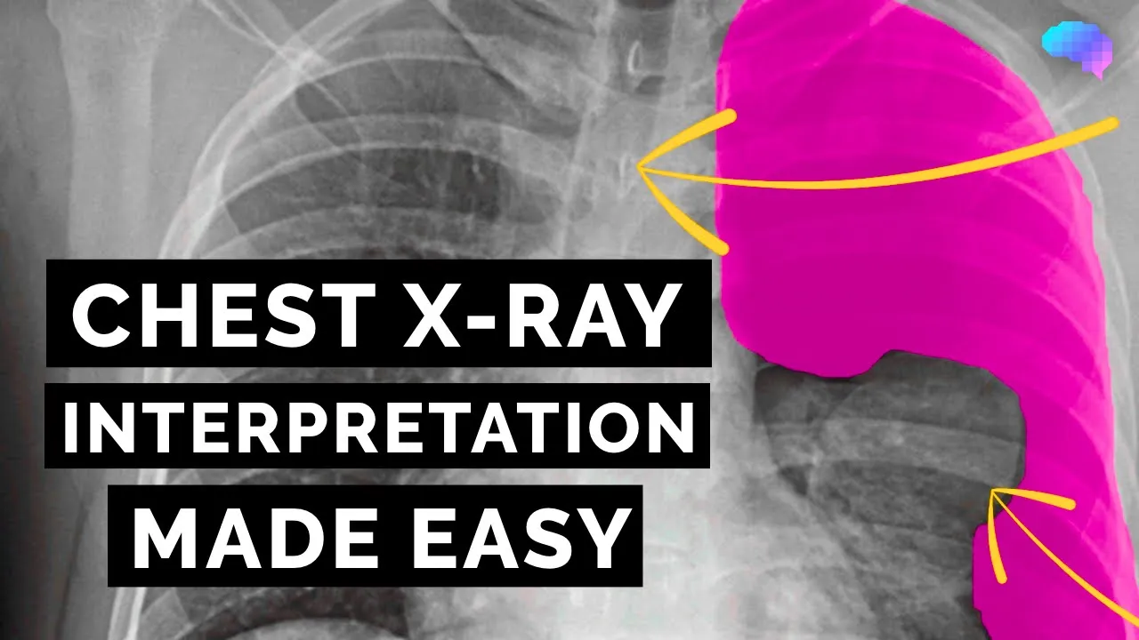







A systematic approach to chest X-ray interpretation involves first assessing technical adequacy (inclusion, penetration, inspiration, and rotation), then systematically evaluating anatomical structures including the trachea, mediastinum, diaphragm, lungs, hilar regions, bones, and soft tissues to identify abnormalities such as pleural effusion, consolidation, fractures, or masses.
Chest X-Ray Interpretation: A Systematic Approach
Added:Normal gross anatomy of the thoracic cavity, including the lungs, heart, mediastinum, pleural spaces, and bony thorax.
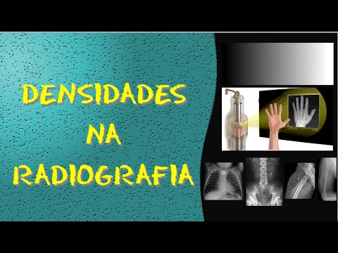
The physical principles of radiography, specifically how different tissue densities (air, fat, soft tissue/fluid, bone) attenuate X-rays and appear on film.
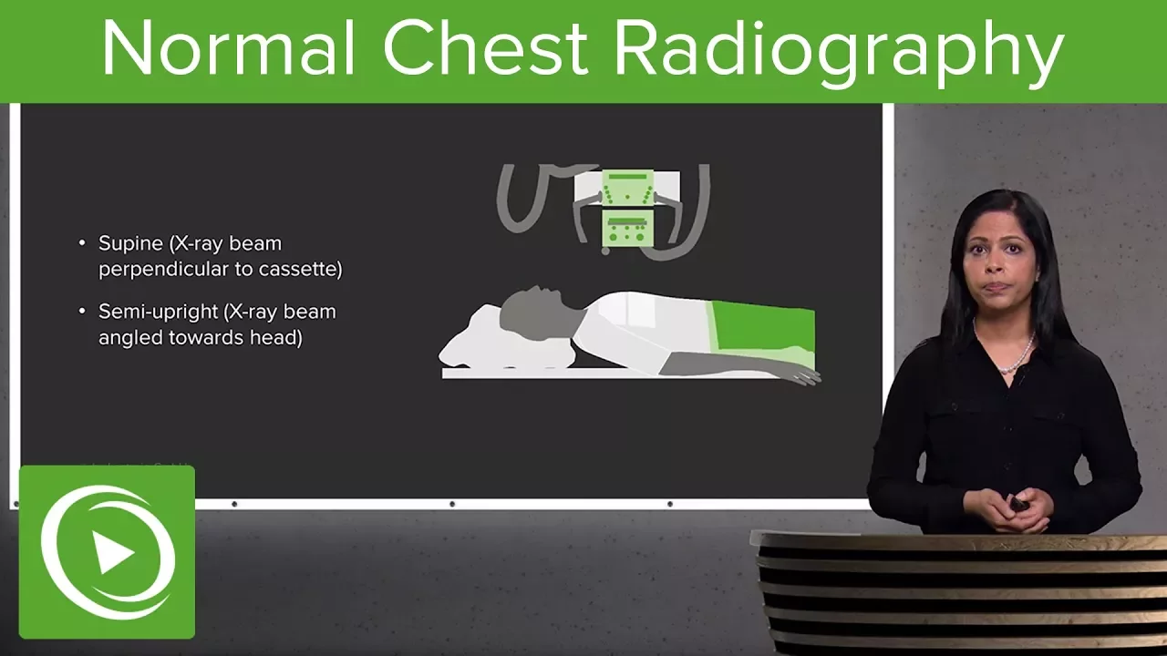
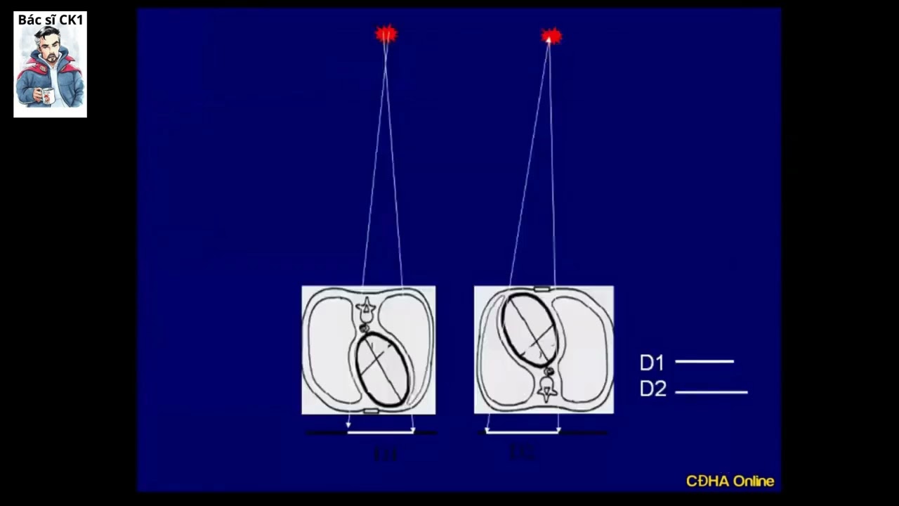
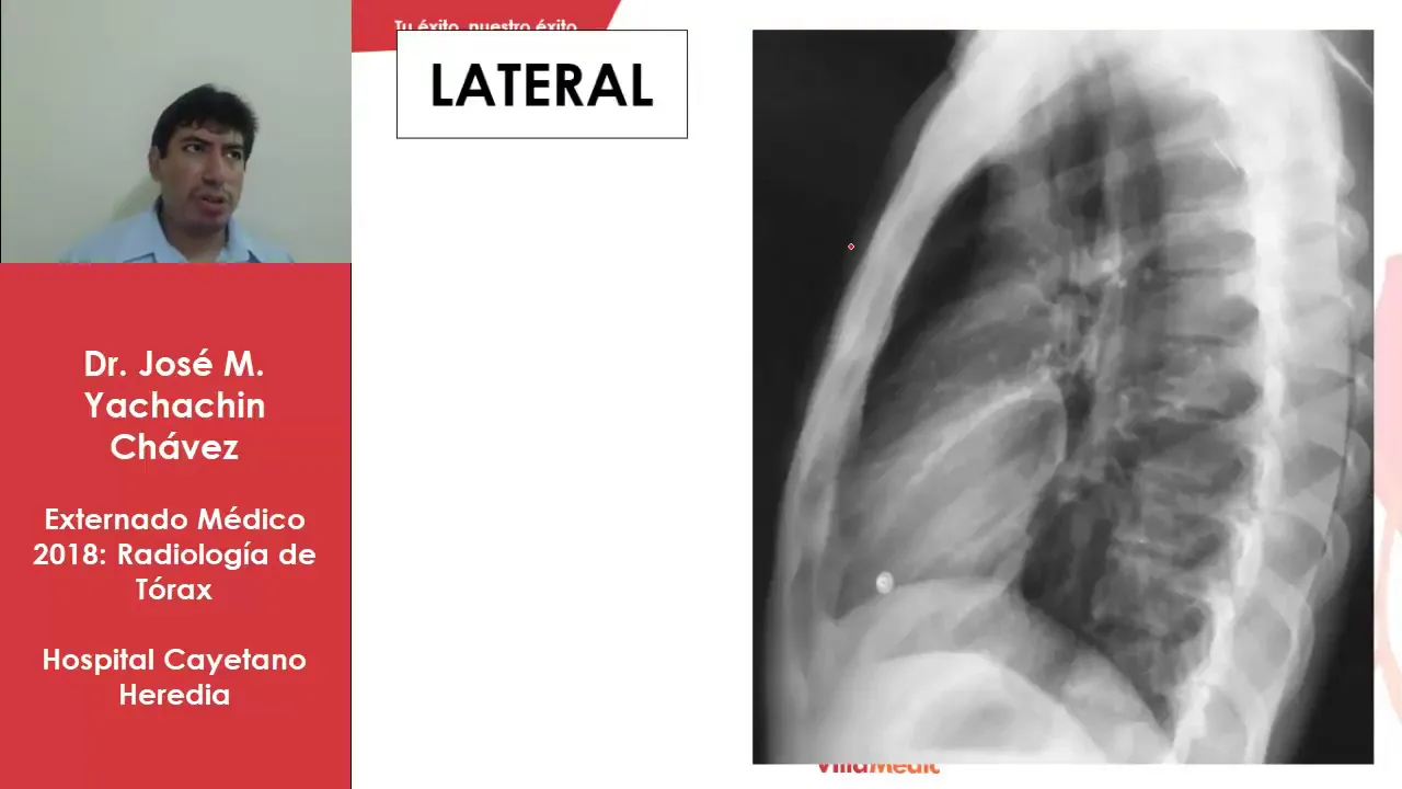
Standard radiographic views of the chest, including the technical and positional differences between Posteroanterior (PA), Anteroposterior (AP), and Lateral projections.
Basic medical terminology related to cardiopulmonary pathology and symptoms (e.g., dyspnea, consolidation, effusion).
Identification and differentiation of complex thoracic pathologies on CXR, such as pneumothorax, pulmonary edema, lobar collapse, and various pneumonia patterns.
The systematic evaluation of medical tubes, lines, and foreign bodies (e.g., endotracheal tubes, central venous catheters, pacemakers) on a chest radiograph.
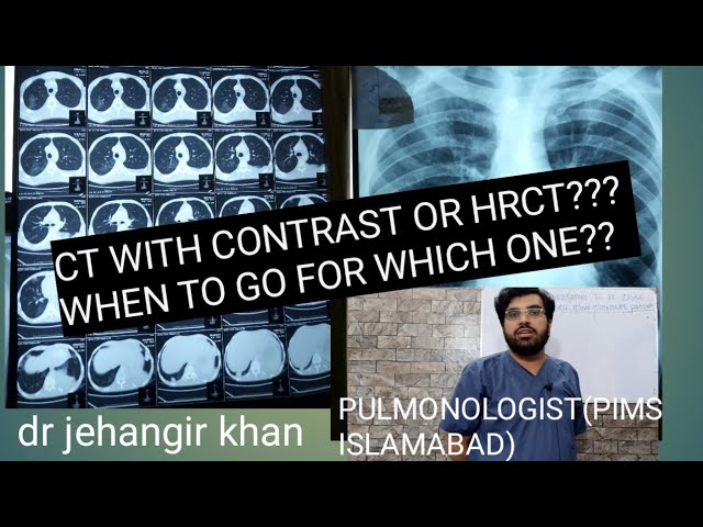
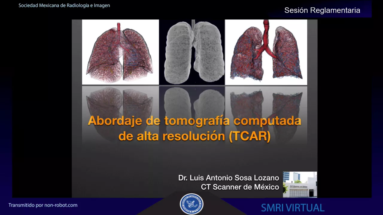
Transitioning to chest Computed Tomography (CT) interpretation and understanding when to order high-resolution CT (HRCT) versus standard chest radiography.
Clinical decision-making algorithms that integrate chest X-ray findings with patient history, physical examination, and laboratory data to guide patient management.
69K views1Klikes6:11Original Release: 2017-07-19
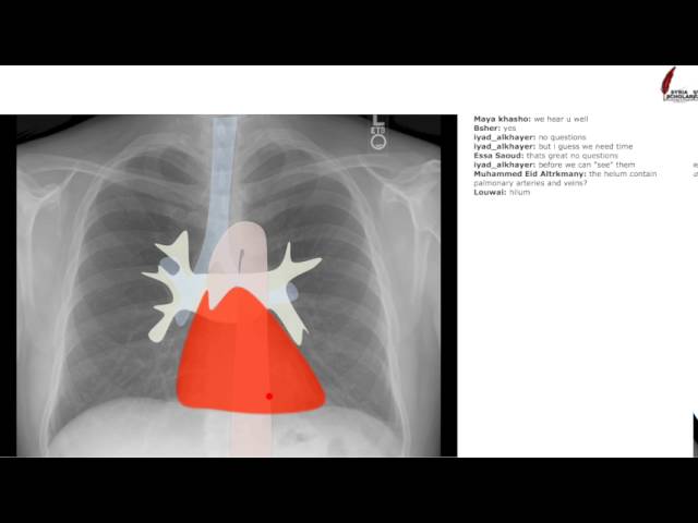
welcome to this tutorial on chess radiograph interpretation we start our assessment of this frontal chest radio graph by determining its projection as is indicated in the top right corner of this image the film was taken in the PA orientation this means that the radaph was taken with the patient standing with their back to the X-ray source and their front against the detector we now assess the film for the four parameters of technical adequacy firstly inclusion for complete assessment of the chest we need to see both apes and both costophrenic angles it takes 2 to 300 mL of plural fluid before blunting of the costophrenic angle can be seen on a PA film even slight obscuration of the costophrenic angle can hide a significant amount of plural fluid now we assess the film for adequate penetration we should be able to see the vertebral bodies through the cardiac silhouette in an adequately penetrated film after this we determine how deeply the patient has breathed in we should expect to see nine or 10 posterior ribs the final parameter we assess is rotation in a non-rotated image the cicular heads should be equidistant from the spinus processes in this example the left clavicular head is slightly closer to the spinus process than the right cicular head this indicates that the patient's body is rotated to their right side rotation to the right means that the right lung will appear less D than the left and therefore blacker in this particular case the degree of rotation is unlikely to make a significant impact having assessed the four parameters of technical adequacy we move on to assessing the film itself we start with the trachea which should be seen in the midline of a non-rotated film we can follow the Contour of the trachea down to the Karina where the trachea bifes into the right and left main bronchi the tra May deviate towards or away from certain pathology a tension pneumothorax or large plural Fusion will push the tra away from the affected side whereas lung collapse will pull the trachea towards it we describe the appearances of lar collapse in a separate video on either side of the trachea we see the right and left parat tral Stripes abnormal thickening in this area can indicate a media styal pathology such as lymph node enlargement in lymphoma the inferior part of the medyum contains the heart on the right the border is formed by the right atrium whilst the left border is formed by the left ventricle the presence of cardiomegaly can be assessed on a PA radaph if the width of the heart is greater than 50% of the width of the lungs and cardiomegaly is present pericard diffusion May mimic the appearances of an enlarged heart with the shape of a water bottle when assessing the media stum we review the parotic line This begins at the atic knuckle and extends infer L the parotic line represents the left hand side of the descending thoracic aota the Contour of the aota may be deviated in the elderly as the aota becomes more ectatic having assessed the media stum we now move on to the diaphragm follow the Contour of the diaphragm from the cardiophrenic to the costophrenic angles as discussed previously blunting of the costophrenic angles can signify a plural affusion we continue along the lateral border of the lungs and make our way into the Apes as we do this we evaluate for plural based pathology and for numores a small apical numor can be hard to spot look for a Pencil Thin Line along the lung edge with an absence of lung markings Beyond this line we now come to the lungs themselves it is crucial to compare the bronchovascular markings on one side to the other when assessing the lungs compare the upper zones then the middle zones and then the lower zones remember that the lungs extend posteriorly below the level of the diaphragm look for abnormal lesions such as metastases or lung nules consolidation or airspace of pacification is another common finding and is confirmed by the presence of air bronchograms the location of consolidation can be determined by assessing which medial or diaphragmatic silhouette has has been obscured for more information please watch our supplementary video on consolidation we now move on to the higher regions the left highum is expected to lie slightly superior to the right particular attention should be paid to the higher regions as malignancy or enlarged nyph nodes can be overlooked in this area for all the visualized ribs follow the Contour of their upper and lower borders discontinuities in a ribs Contour can indicate a fracture assess the height and alignment of the vertebrae loss of height can indicate trauma or bony metastases malalignment of the spine can signify scoliosis the bones of the shoulder joint are often seen on chest films and the presence of fractures should be noted below the diaphragm we have the liver on the right and the stomach on the left air contained within the stomach is a normal finding however finding free air under the diaphragm is suggestive of a perforated abdominal viscus or recent abdominal surgery the chest wall breast tissue shoulders and lower neck can often be seen on a chest film subcutaneous empyema may be visible as gas locules in these areas this concludes your assessment of the normal chest Raph during this video we have demonstrated a standard approach to chest radiograph interpretation if you would like more detail on chest Anatomy then please visit visit our website
Up Next

31:28
PhD Research: 'Free Economy' Metaphors in HK Political Discourse
@englpolyu
281 views•2022-09-13

12:54
Triumph of Orthodoxy Icon: Byzantine Art & History Explained
@BenCallan
2.1K views•2024-08-06

9:49
FastAPI vs Flask vs Django: Choosing the Right Python Web Framework
@TechWithTim
302.5K views•2024-05-26

1:42
Game of Thrones Opening Credits: A Cinematic Analysis
@gameofthrones
46.3M views•2011-04-18
Related Study Plans & Knowledge Roadmaps
Structured learning paths in General & Interdisciplinary Studies
general-studies
Elevator Engineering: History, Physics & Cities
01From Hoists to Safety Brakes: The Dawn of Vertical Lift
02Hydraulic vs. Traction: The Engineering Physics of Elevators
03Modern Speed, Safety, and Algorithms
+ 2 more learning modules
12h5 modules
Explore Roadmap
general-studies
Rubber: Material Science, History & Geopolitics
01Origins of Natural Rubber & Mesoamerican History
02The Chemistry of Natural Rubber & Vulcanization
03The Geopolitics of the Rubber Boom & Colonization
+ 2 more learning modules
12h5 modules
Explore Roadmap
general-studies
Containerization: Evolution, Logistics & Trade
01Ancient Trade & Bulk Cargo: Amphorae to Barrels
02The Industrial Revolution & Cardboard Packaging
03The Containerization Revolution: Malcom McLean
+ 2 more learning modules
12h5 modules
Explore Roadmap
general-studies
Textile Tech: Evolution, Labor & Global Impact
01Pre-Industrial Textiles: Handloom & Cottage Industry
02Mechanization: The Spinning Jenny to the Power Loom
03The Factory System & Global Labor Transformation
+ 2 more learning modules
10h5 modules
Explore Roadmap