Oligodendrocytes are glial cells in the central nervous system derived from neural stem cells, named from Greek words meaning 'cells with few branches'; they produce myelin sheaths by wrapping their plasma membrane around axons multiple times, creating a fatty lipid coating that insulates axons and enables faster, more efficient nerve impulse transmission, with each oligodendrocyte potentially myelinating multiple axons and also influencing neurons and other glia through substance exchange.
Oligodendrocytes: Structure and Function | Neurobiology
Added:Deep Dive
Basic structure of a neuron, specifically the anatomy and function of the axon.

Anatomy of the Neuron
362
The axon transmits nerve impulses away from the cell body. Key structures include: (1) Axon hillock—the cone-shaped junction between axon and cell body, (2) Initial segment—where the first action potential is generated, (3) Axolemma—the axon's cell membrane, (4) Axoplasm—the cytoplasm inside the axon. The axon ends as branching telodendria, allowing innervation of multiple structures. At telodendria ends are axon terminals (synaptic bulbs) containing neurotransmitters like acetylcholine, dopamine, and serotonin for signal transmission to other neurons.
Click to watch →

BASIC STRUCTURE OF NEURONS | Coordination and Control
212
The axon is a long, thin, tail-like extension that attaches to the cell body. It carries electrical impulses (information) away from the cell body to other neurons, muscles, or glands. Many axons are insulated with a fatty substance called myelin, which causes impulses to jump from one node to another (saltatory conduction) rather than traveling in a straight line. Myelin also provides protection to the axon.
Click to watch →

Neuron or Nerve Cell
245K
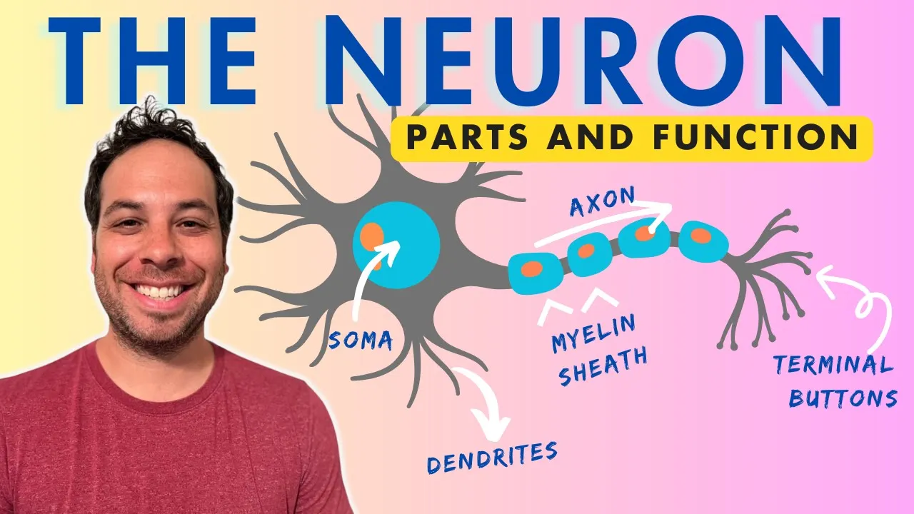
The axon is a single long and cylindrical projection whose primary function is to conduct nerve impulses away from the cell body. The plasma membrane of a neuron is called neurolemma. The axon is covered by myelin sheath and possesses nodes of Ranvier, which may form terminal arborizations at their ends.
Click to watch →

Structure of Neuron
171
The axon is the third component of a neuron. It is a long, single process originating from the cell body or soma. The axon carries signals beyond the neuron to the next neuron. The entire axon gives the shape of a nerve fiber and is known as the axis cylinder of the nerve.
Click to watch →
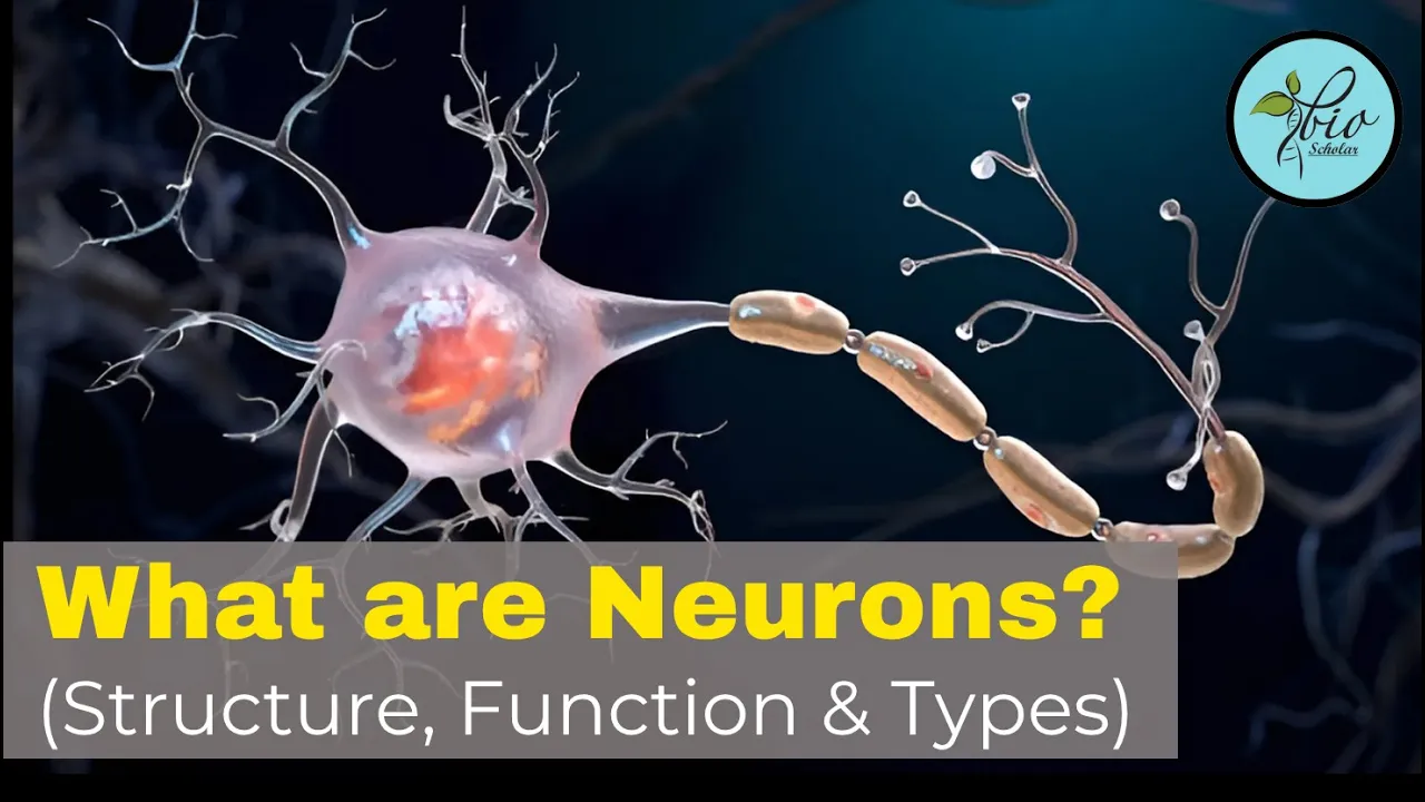
Structure and function of neuron| Types of neurons | Neuron Class 10 | Bio Scholar
501
The axon is a long slender projection extending from the cell body, surrounded by the axolemma (membrane separating axoplasm from external environment). The axoplasm contains microtubules, neurofibrils, rough endoplasmic reticulum, and mitochondria. The axon's main function is to transmit action potentials away from the cell body to other neurons, muscles, or glands.
Click to watch →
Deep Dive
The fundamental mechanism of action potential generation and propagation.

Action Potential | Part 1/3
652.8K
An action potential is a rapid, propagating electrochemical change in the membrane of excitable cells (neurons and muscle cells) that serves as a signaling mechanism; it begins when a stimulus causes sufficient sodium influx to reach the threshold potential (-60 mV), triggering voltage-gated sodium channels to open and depolarize the membrane toward sodium's equilibrium potential (+65 mV), followed by the closure of inactivation gates and opening of voltage-gated potassium channels that repolarize the membrane back toward potassium's equilibrium potential (-85 mV), with the depolarizing current spreading locally to trigger adjacent membrane segments, creating a wave of depolarization followed by repolarization that travels along the neuron toward the central nervous system.
Click to watch →

ADMITERE LA MEDICINĂ: Potențialul de acţiune
19.2K
Action potentials follow the all-or-nothing principle: when membrane potential reaches threshold, voltage-gated sodium channels open, allowing sodium influx due to electrical and concentration gradients. This depolarizes the membrane. Potassium then exits, repolarizing the cell. During the absolute refractory period, sodium channels become inactivated and cannot respond. The sodium-potassium pump restores original ion distribution. Sodium and potassium channels have passive action (voltage-gated), while the sodium-potassium pump has active action (using ATP to move ions against gradients).
Click to watch →

أحياء 2008 | الفصل الثاني | الحصة 5 | انتقال السيال العصبي | د. محمد القاضي
1.7K
Action potential is the electrical signal generated in neurons when stimulated. It consists of four phases: (1) Depolarization - sodium ions enter through leak channels, raising membrane potential from -70mV to -55mV threshold; (2) Rising phase - voltage-gated sodium channels open rapidly, causing potential to rise to +30mV; (3) Repolarization - potassium ions exit, returning potential toward -70mV; (4) Hyperpolarization - potassium channels remain open, dropping potential to -90mV. The sodium-potassium pump then restores resting potential. Action potentials propagate sequentially along the neuron: when an action potential occurs at one point, it depolarizes the adjacent membrane region, bringing it to threshold and triggering a new action potential. This process repeats along the axon from cell body to axon terminals. The direction is unidirectional because each region enters a refractory period after firing, preventing backward propagation.
Click to watch →

Проведення нервового імпульсу | Фізіологія №4
256
Action potentials are generated when threshold stimuli cause membrane potential to reach critical levels, opening voltage-gated sodium channels. Sodium influx depolarizes the membrane, creating local circuit currents that spread to adjacent regions. These currents depolarize neighboring areas to threshold, triggering sequential sodium channel opening. Refractoriness prevents backward propagation by rendering recently depolarized regions unresponsive. The refractory period limits maximum firing rates: neurons can fire ~500 times/second, while cardiomyocytes can contract ~200 times/minute.
Click to watch →
Nervous Control & Coordination 01 | Cells of Nervous System & NerveI Impulse Conduction | 11 | NEET
1.3M
Action potentials follow the all-or-none principle, requiring threshold stimulation (~-55 mV). Depolarization occurs when voltage-gated Na+ channels open, allowing rapid Na+ influx that reverses membrane polarity to ~+30 mV. Repolarization follows as voltage-gated K+ channels open, allowing K+ efflux that restores negativity. Hyperpolarization briefly overshoots resting potential before returning. Propagation occurs through current flow in extracellular fluid, with saltatory conduction in myelinated axons dramatically increasing speed. Refractory periods (absolute and relative) prevent immediate re-firing.
Click to watch →
Deep Dive
The anatomical distinction between the Central Nervous System (CNS) and the Peripheral Nervous System (PNS).
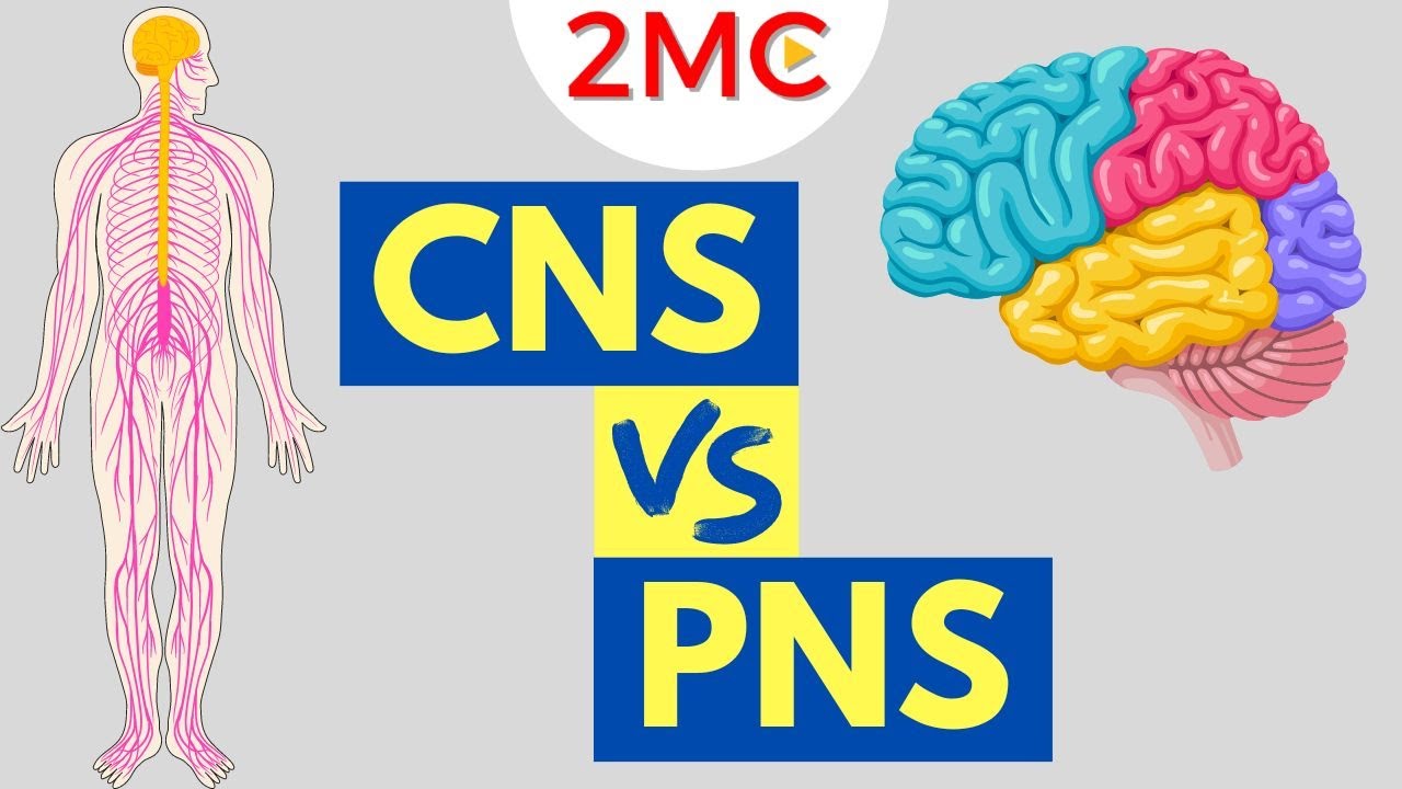
Central Nervous System vs Peripheral Nervous System | CNS & PNS Structure and Function
299.7K
The central nervous system (CNS) consists of the brain and spinal cord, with the brain containing the cerebrum (responsible for higher functions like memory, emotion, and reasoning), cerebellum (movement and balance), and brainstem (regulatory processes like breathing and blood pressure), while the peripheral nervous system (PNS) includes all nerves outside the CNS and is divided into the somatic nervous system (controlling voluntary movement and sensation) and the autonomic nervous system (controlling automatic functions like heart rate and digestion), which itself has two branches—the sympathetic branch that increases organ activity and the parasympathetic branch that decreases organ activity.
Click to watch →

Foundational Neuroscience - Neuroanatomy & Neurophysiology
392
The nervous system is divided into two main parts: the Peripheral Nervous System (PNS), which includes everything outside the bone (muscles, sensory organs, internal organs), and the Central Nervous System (CNS), which includes the spinal cord and brain. The CNS is encased in bone for protection, representing a fundamental anatomical distinction in brain organization.
Click to watch →

Nervous System Introduction for Anatomy and Physiology (CNS and PNS)
820
The nervous system can be divided anatomically into the Central Nervous System (CNS) and Peripheral Nervous System (PNS). The CNS is contained within a body cavity and shares cerebrospinal fluid, consisting of the brain and spinal cord. The PNS carries information to and from the CNS. The CNS acts as an integration center, performing processing or thinking, much of which is unconscious (like monitoring blood pressure, oxygen levels, and balance).
Click to watch →
Introduction to Neuroanatomy - Learn the Basics - Neuroanatomy Playlist
377.9K
The central nervous system (CNS) consists only of the brain and spinal cord. The peripheral nervous system (PNS) includes all other nerves, including cranial nerves and spinal nerves. This fundamental distinction is essential for understanding neuroanatomy.
Click to watch →
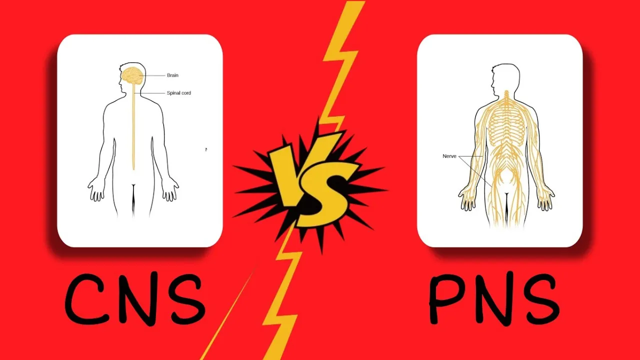
DIFFERENCE BETWEEN CENTRAL AND PERIPHERAL NERVOUS SYSTEM (CNS & PNS)
469
The Central Nervous System (CNS) consists of the brain and spinal cord. The Peripheral Nervous System (PNS) consists of all nerves that extend from the brain and spinal cord, including all neurons located outside the brain and spinal cord.
Click to watch →
Deep Dive
An introduction to glial cells (neuroglia) as the supporting cells of the nervous system.

Nervous & Muskuloskeletal System | NORCET 8 | RRB Nursing Superintendent | Aarogyam Marathon
69.8K
Neuroglia (glial cells) are supporting cells of the nervous system. Unlike neurons, neuroglia can be divided, which is why tumors can arise from neuroglia. In adults, the most common neuroglial tumor is glioblastoma, while in children, medulloblastoma is most common. Neuroglia provide structural and functional support to neurons throughout the nervous system.
Click to watch →

O que as pessoas precisam saber sobre - #anatomiafacil a polilaminina
8.2K
Neuroglia (glial cells) are the support cells of the nervous system, derived from the Latin word 'glia' meaning 'glue' or 'colla' (cola). While the human brain contains approximately 100 billion neurons, it contains even more glial cells—about 1 trillion. For every neuron, there are approximately 10 glial cells. Neuroglia constitute about 1% of the total cellular component of the body. These cells provide structural and functional support to neurons throughout the nervous system.
Click to watch →
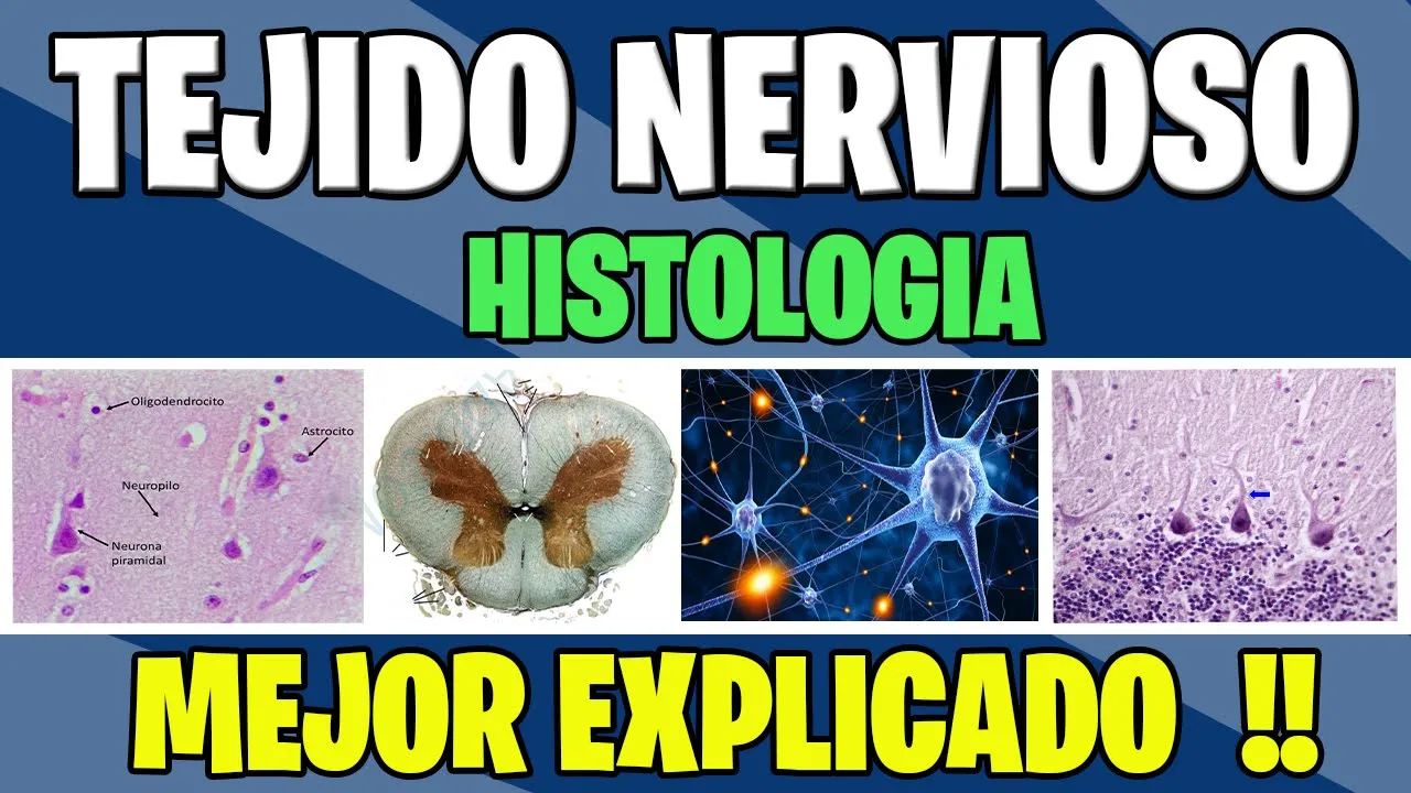
TEJIDO NERVIOSO COMPLETO | Histologia
195.3K
Neuroglia (glial cells) are support cells 5-10 times more abundant than neurons, providing essential structural and functional support. Most derive from ectoderm except microglia from mesoderm. Functions include structural support, neuronal isolation, potassium concentration maintenance, neurotransmitter removal, developmental guidance, blood-brain barrier formation, nutrition, and repair. Types include microglia (phagocytic, clear debris in lesions), astrocytes (largest, star-shaped, with intermediate filaments), and ependymal cells (lining ventricles, producing cerebrospinal fluid). Astrocytes have two types: protoplasmic (gray matter, abundant cytoplasm, blood-brain barrier regulation) and fibrous (white matter, finer processes, neurotransmitter confinement).
Click to watch →
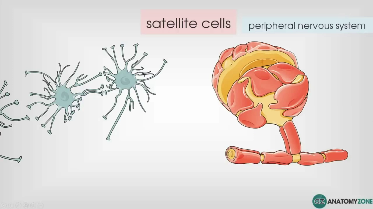
Глиальные клетки.
18.4K
Glial cells (neuroglia) are supporting cells of the nervous system that do not directly process information but provide essential support functions. There are six main types: astrocytes (star-shaped cells in the CNS that form the blood-brain barrier and support neurons), satellite cells (similar to astrocytes in the PNS, surrounding neuron cell bodies), Schwann cells (PNS cells that form myelin sheaths around axons), oligodendrocytes (CNS cells that form myelin sheaths), microglia (immune cells that perform phagocytosis), and ependymal cells (produce and circulate cerebrospinal fluid).
Click to watch →

Anatomy & Physiology Chapter 11 Part A: Nervous System & Nervous Tissue Lecture
187.1K
Nervous tissue comprises neurons and neuroglia (glial cells). Glial cells are smaller, more numerous (about ten times neurons), and make up half the brain mass. Astrocytes provide star-shaped support, anchoring neurons to capillaries and controlling chemical environments. Microglial cells monitor neurons and transform into macrophages to clear debris. Ependymal cells line CNS cavities with cilia that circulate cerebrospinal fluid. Oligodendrocytes form myelin sheaths in the CNS.
Click to watch →
Prerequisite Knowledge
- Concept 01Basic structure of a neuron, specifically the anatomy and function of the axon.
- Concept 02The fundamental mechanism of action potential generation and propagation.
- Concept 03The anatomical distinction between the Central Nervous System (CNS) and the Peripheral Nervous System (PNS).
- Concept 04An introduction to glial cells (neuroglia) as the supporting cells of the nervous system.
Subsequent Learning
- Step 01The biophysics of saltatory conduction and the role of the Nodes of Ranvier in signal acceleration.
- Step 02Pathology of demyelinating diseases, specifically the etiology and symptoms of Multiple Sclerosis (MS).
- Step 03Comparative neurobiology of CNS myelination (oligodendrocytes) versus PNS myelination (Schwann cells), including axonal regeneration capacity.
- Step 04The role of activity-dependent myelination in neuroplasticity, learning, and cognitive development.
Structure
Function
Structure
0:01Playing Section
- 1
Oligodendrocytes are CNS glia extending processes to wrap myelin.
- 2
Each cell myelinates multiple axons, with segments from different cells.
Beyond Passive Insulation: Oligodendrocytes as Active Metabolic Partners and Plasticity Regulators
While traditional neurobiology frames oligodendrocytes primarily as passive providers of myelin insulation to accelerate signal propagation, an emerging paradigm shift challenges this view. This alternative perspective argues that oligodendrocytes are active, dynamic partners in neuronal survival and cognitive function. Beyond structural insulation, they provide crucial metabolic support to axons by directly transferring energy substrates, such as lactate, which are essential for sustaining axonal mitochondria during high-frequency firing. Furthermore, myelination is now understood to be highly dynamic rather than static. Oligodendrocytes exhibit activity-dependent plasticity, remodeling myelin thickness and node spacing in response to learning and environmental stimuli to fine-tune neural circuit timing. This positions oligodendrocytes not merely as cables of the brain, but as active modulators of synaptic plasticity and information processing.
Deep Dive
The biophysics of saltatory conduction and the role of the Nodes of Ranvier in signal acceleration.

Motor neurons | Muscular-skeletal system physiology | NCLEX-RN | Khan Academy
352K
Myelinated axons use saltatory conduction, where electrical signals jump from node to node rather than traveling continuously along the entire axon. The spaces between myelin segments are called nodes of Ranvier. This jumping mechanism dramatically increases signal transmission speed while preventing signal degradation over long axon lengths.
Click to watch →

Action Potential in Neurons Explained: Neurology Lecture
605.7K
Myelinated axons have nodes of Ranvier—gaps between Schwann cell myelin sheaths rich in voltage-gated sodium channels. Action potentials appear to jump from node to node (saltatory conduction), dramatically increasing speed while reducing energy consumption. Only these nodes undergo significant ion flux and action potential generation.
Click to watch →
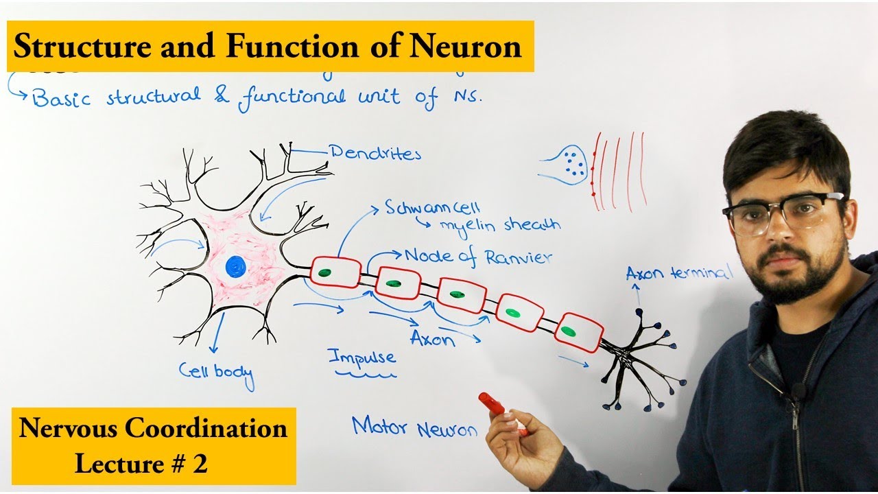
Neuron |How nervous system works|
327.9K
The myelin sheath is interrupted at regular intervals by gaps called nodes of Ranvier. During saltatory conduction, action potentials jump from one node to the next rather than traveling continuously along the entire axon. This jumping mechanism dramatically increases the speed of signal transmission compared to unmyelinated axons.
Click to watch →
Neuron Structure and Function: Parts of a Nerve Cell
521.9K
The nodes of Ranvier are tiny gaps between segments of myelin along the axon. Instead of traveling continuously through the myelinated axon, the action potential 'bounces' or jumps from node to node in a process called saltatory conduction. This mechanism dramatically increases the speed of signal transmission.
Click to watch →

สรุปชีวะ ระบบประสาท EP.1 (เซลล์ประสาท)
509K
Nodes of Ranvier are the gaps between myelin segments along a myelinated axon. At these nodes, the action potential is regenerated, allowing the signal to 'jump' from node to node in a process called saltatory conduction. This jumping mechanism is much faster than continuous conduction along unmyelinated axons, with myelinated axons transmitting impulses at speeds up to 120 meters per second compared to 2 meters per second in unmyelinated axons.
Click to watch →
Deep Dive
Pathology of demyelinating diseases, specifically the etiology and symptoms of Multiple Sclerosis (MS).
Multiple Sclerosis Made Easy- Pathophysiology,symptoms,Treatment.
117.1K
Multiple sclerosis (MS) is a demyelinating disease characterized by damage to the insulating myelin sheaths surrounding nerve cells in the brain and spinal cord. This damage impairs communication within parts of the nervous system, leading to a variety of physical, mental, and sometimes psychiatric symptoms. Common manifestations include double vision, blindness in one eye, muscle weakness, impaired sensation, and coordination difficulties. MS presents in multiple forms: relapsing forms involve new symptoms occurring in isolated attacks, while progressive forms involve gradual symptom accumulation. Between attacks, symptoms may fully resolve, but permanent neurological deficits often persist, particularly as the disease advances. The exact cause remains unclear, but the underlying mechanism is believed to involve immune-mediated destruction of myelin or failure of myelin-producing cells. Proposed contributing factors include genetic predisposition and environmental triggers such as viral infections. Diagnosis is based on clinical signs, symptoms, and supporting medical tests. There is no known cure for MS. Treatments aim to restore function after an attack and prevent new episodes. Medications used are only modestly effective and may cause side effects or poor tolerance. Physical therapy is recommended to support functional ability. Many patients seek alternative treatments despite insufficient evidence. Prognosis varies; better outcomes are typically associated with female sex, early disease onset, a relapsing course, and low initial attack frequency. Life expectancy is, on average, 5 to 10 years lower than in the general population.
Click to watch →

Multiple Sclerosis | Etiology, Pathophysiology, Types of MS, Clinical Features, Diagnosis, Treatment
463.9K
Multiple Sclerosis (MS) is a chronic, immune-mediated demyelinating disease of the central nervous system characterized by an autoimmune response where autoreactive T-cells and inflammatory cytokines attack myelin sheaths surrounding axons, leading to disrupted neural signal transmission; the disease primarily affects young adults (ages 20-40), with environmental factors like low vitamin D levels and viral infections (Epstein-Barr virus, HHV-6) combined with genetic susceptibility genes such as HLA-DR2 contributing to its development; clinically, MS presents with diverse neurological deficits including optic neuritis, bilateral internuclear ophthalmoplegia, pseudobulbar palsy, and spinal cord involvement, manifesting through four main subtypes: relapsing-remitting (90% of cases with flare-ups and partial recovery), secondary progressive (progressive deterioration after initial relapsing phase), primary progressive (continuous decline without relapses), and progressive-relapsing (continuous deterioration with non-recovering flare-ups); diagnosis relies on MRI showing characteristic white matter lesions in peri-ventricular regions, brainstem, and spinal cord, supported by visual evoked potentials and lumbar puncture findings; treatment includes acute management with high-dose corticosteroids and plasmapheresis, plus disease-modifying therapies such as interferon beta, glatiramer acetate, monoclonal antibodies, and natalizumab to suppress immune activity and prevent future relapses.
Click to watch →

Central Nervous System Physiology Rapid Revision 🧠🧠🧠
137.6K
Multiple sclerosis is an autoimmune disorder where oligodendrocytes (myelinating cells of the CNS) are destroyed, leading to demyelination of CNS neurons. This disrupts nerve conduction and causes the various neurological symptoms of MS. The destruction of oligodendrocytes is the primary pathological feature.
Click to watch →

Рассеянный склероз - причины, симптомы, диагностика, лечение
136.5K
Multiple sclerosis is a leading central nervous system disease affecting brain and spinal cord. Myelin, the protective sheath around axons, enables faster nerve impulse transmission. MS involves demyelination where immune cells attack and destroy myelin, disrupting neural communication. The blood-brain barrier normally protects neurons, but activated B-lymphocytes target barrier cells, allowing T-cells to breach it. This triggers Type IV hypersensitivity where T-cells produce cytokines (IL-1, IL-6, TNF, IFN-gamma) that expand vascular walls, recruit B-lymphocytes and macrophages, and destroy myelin. Attacks occur in waves with initial remyelination, but over time damage becomes irreversible. Risk factors include female gender, HLA-DR2 gene, infections, and vitamin D deficiency, explaining higher prevalence in polar regions.
Click to watch →

Esclerosis múltiple 1 : Aspectos generales y enfermedades desmielinizantes
28K
MS pathophysiology involves a complex cascade: foreign antigens (possibly viral) sensitize T lymphocytes, which breach the blood-brain barrier by disrupting endothelial membranes. Activated lymphocytes enter the CNS and bind to CD4 cells, triggering B lymphocytes to produce antibodies. The disease has both inflammatory and degenerative components, with the inflammatory phase (Big Bang) followed by neurodegeneration. Etiological factors include Epstein-Barr virus infection (10x more frequent in MS patients), vitamin D deficiency, and smoking (causing nitric oxide toxicity, altered immunity, and increased blood-brain barrier permeability). Genetic factors include major histocompatibility complex genes. The interplay between genetic predisposition and environmental triggers remains incompletely understood.
Click to watch →
Deep Dive
Comparative neurobiology of CNS myelination (oligodendrocytes) versus PNS myelination (Schwann cells), including axonal regeneration capacity.

Chapter 12 part 1
140
Myelin sheaths are multi-layered lipid-protein coverings that insulate axons and increase nerve impulse conduction speed. In the CNS, oligodendrocytes produce myelin around multiple axons; in the PNS, Schwann cells produce myelin around single axons. The neurolemma (Schwann cell outer layer) enables PNS regeneration but is absent in CNS myelination. Nodes of Ranvier are gaps between myelinated segments. Key CNS vs PNS differences include: terminology (ganglion vs nucleus, nerve vs tract); white matter (myelinated axons) vs gray matter (cell bodies); spinal cord (gray matter core) vs brain (gray matter outer layer); and regeneration capacity (PNS regenerates, CNS has limited regeneration).
Click to watch →
Lecture 4: Christine Bandtlow - Myelination in the CNS and PNS
3.5K
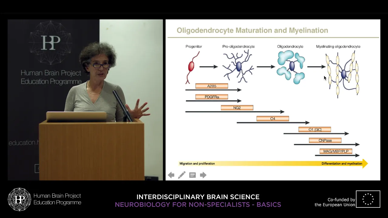
Oligodendrocytes and Schwann cells differ fundamentally in their myelination capabilities. Oligodendrocytes can myelinate multiple axons simultaneously using multiple processes, while Schwann cells myelinate only one axon each, requiring multiple cells per axon. Axon diameter determines myelination status, with only axons above approximately 1 micrometer diameter receiving myelin sheaths. Dendrites and small-diameter axons like mossy fibers remain unmyelinated. In the PNS, some axons form Remak bundles wrapped by single non-myelinating Schwann cells, a phenomenon absent in the CNS.
Click to watch →

Anatomy & Physiology HELP! -- Study Session #120 -- Neuroglial Cells
139
The PNS contains Schwann cells that form myelin sheaths and enable nerve regeneration after injury, unlike CNS oligodendrocytes. Satellite cells protect neuronal cell bodies by enveloping them. The fundamental difference between CNS and PNS repair capacity lies in whether myelination uses oligodendrocytes (non-regenerative) or Schwann cells (regenerative), explaining why peripheral nerve injuries typically heal better than central nervous system injuries.
Click to watch →

TGT/PGT Biology Classes #4 | Biology For DSSSB/BPSC TRE 4/EMRS/KVS/NVS/UP LT Grade | Deepank Sir
3.1K
The nervous system consists of the central nervous system (CNS) and peripheral nervous system (PNS). In the PNS, Schwann cells produce myelin sheaths, with each Schwann cell covering only one segment of an axon. In the CNS, oligodendrocytes produce myelin sheaths, with each oligodendrocyte capable of myelinating multiple axons. Nerve regeneration occurs primarily in the PNS because Schwann cells contain neurolemma, the site of regeneration. Each Schwann cell covers only one axon segment, allowing for regeneration. In contrast, oligodendrocytes in the CNS can myelinate multiple axons simultaneously, which limits regeneration capability.
Click to watch →

MED LEVEL1 NSI NAS PRACTICAL Ganglia, Neuroglia,degeneration
165
Oligodendrocytes are the most numerous glial cells in the CNS, originating from neural stem cells. They exist in both gray and white matter and support myelination of axons. Each oligodendrocyte myelinates multiple segments of axons within a single ganglion. Schwann cells (myelinating cells in the PNS) are flat cells that myelinate single segments of nerve fibers. Both are myelinating cells but differ in structure and location. Schwann cells are important for nerve regeneration, while oligodendrocytes do not participate in regeneration.
Click to watch →
Deep Dive
The role of activity-dependent myelination in neuroplasticity, learning, and cognitive development.
2022 MCHRI Symposium: Neurodevelopment
4.7K
Evidence supports that myelination is activity-dependent rather than hardwired. Transcriptomic comparisons between four-month-olds with visual experience and prenatal samples show increased myelination gene expression. Animal studies demonstrate that depriving monkeys of visual input prevents face-selective response emergence. This suggests visual experience accelerates myelination in higher-order visual areas, creating an interplay between structural changes and functional developments in infant brain maturation.
Click to watch →

Cam Neuro - Thóra Káradóttir -Activity dependent myelination: mechanism for learning & regeneration?
1.5K
The brain's white matter, comprising approximately half the brain volume, consists of oligodendrocytes wrapping myelin sheaths around axons. This architecture maximizes neural connectivity with minimal conduction delays, providing maximum computational power. Myelin sheaths lower axonal capacitance and increase resistance, enabling faster action potential propagation. Recent evidence reveals myelination is not uniform along axons—alternating unmyelinated and myelinated segments allow differential speed adjustments, enabling synchronized firing across neurons of different lengths. Myelination begins late in human development, starting in anterior brain regions shortly after birth and continuing through childhood and adolescence. Oligodendrocyte precursor cells (OPCs) are born during early development when neurons have already projected to their targets and become active. OPCs migrate throughout the brain, proliferate, and differentiate into myelinating oligodendrocytes. These OPCs possess voltage-gated sodium channels and receive synaptic inputs from glutamatergic neurons, expressing NMDA, AMPA, and kainate receptors, establishing direct neural-oligodendrocyte communication pathways. Myelination is regulated by two distinct mechanisms: activity-independent myelination occurring autonomously, and activity-dependent myelination requiring neural activity and glutamate receptor signaling. OPCs exhibit significant heterogeneity in their properties across different brain regions and developmental stages, transitioning through naive, primed, and quiescent states. Myelin plasticity supports learning and skill acquisition—professional pianists show corpus callosum thickness correlating linearly with practice hours, and mouse studies demonstrate that motor skill learning requires new myelinating oligodendrocytes.
Click to watch →

Researcher Dave Siever, sheds light on new breakthrough in healing the brain!?
1.1K
This section covers oligodendrocyte precursor cells and myelination processes. OPCs establish axon-glial synapses and differentiate into myelinating oligodendrocytes in response to neuronal activity. Enriched environments automatically increase OPC proliferation and myelination throughout the hippocampus, cortex, and white matter. Activity-dependent myelination explains why learning thickens myelin while inactivity thins it. Stimulation at 25 Hz and 300 Hz promotes greater myelination than 5 Hz stimulation. The corpus callosum transmits signals in 30 ms (myelinated) versus 333 ms (unmyelinated), demonstrating how myelination speed affects neural communication. Myelinated nerves support faster conduction velocities and better phase coherence between brain regions.
Click to watch →

Michelle Monje - Brain-Body Interactions
754
Human infants undergo extraordinary neurodevelopment in their first year, producing approximately 500 billion synapses daily, 14,000 new hippocampal neurons per day, and 127 million cerebellar granule neurons per day. Myelin development is primarily postnatal, beginning just before birth with minimal myelin in central brain regions, progressing from the brain's center toward the poles over the first year and continuing for approximately three decades. Myelination follows predictable topographical and chronological patterns, with basic neural circuits underlying sensation and movement myelinating first, followed by higher-order circuits supporting cognition during adolescence and young adulthood. The spatial and temporal pattern of glioma genesis maps onto the pattern of developmental myelination, suggesting that the cell of origin for many gliomas lies in the oligodendroglial lineage. Research demonstrates that neuronal activity in cortical projection neurons elicits rapid and robust proliferation of oligodendrocyte precursors, generating mature oligodendrocytes that alter circuit dynamics and improve behavioral outcomes. Molecular mechanisms involve brain-derived neurotrophic factor (BDNF) signaling through TrkB receptor, which is required for activity-dependent OPC proliferation. Activity-dependent myelin plasticity manifests in two forms: generation of new oligodendrocytes forming new myelin internodes on previously unmyelinated axons, and remodeling of existing myelin by mature oligodendrocytes changing internode geometry. Healthy activity-dependent myelination promotes coordinated circuit functions by synchronizing inputs between neural network nodes, enhancing attention, memory, and learning. However, in cognitive impairment syndromes such as cancer therapy-related brain fog and post-COVID cognitive dysfunction, myelin homeostasis and plasticity become impaired due to neural-immune interactions. In generalized epilepsy syndromes, aberrantly increased neuronal activity leads to aberrantly increased myelination within the seizure network, contributing to epilepsy progression by promoting hypersynchrony between nodes.
Click to watch →

MCHRI Seminar Series (3/1/21): Maladaptive Myelination in Pediatric Epilepsy
159
Myelination spans over 30 years in humans, beginning postnatally and continuing throughout life. The concept that neuronal activity regulates axon myelination emerged in the 1990s, proposing neurons control myelination extent through experience-dependent mechanisms. Using in vivo optogenetics, researchers demonstrated that stimulating cortical projection neurons rapidly increases oligodendrocyte precursor cell proliferation, generating new oligodendrocytes that alter myelin ultrastructure and improve motor function. A key molecular mechanism involves BDNF-TrkB signaling: genetic models with impaired BDNF secretion or TrkB receptor deletion from oligodendrocyte precursor cells completely lose activity-regulated myelination responses. Loss of this myelination impairs attention and short-term memory function, demonstrating myelin plasticity supports cognition by promoting network synchrony through coordinated neural circuit function.
Click to watch →
Structure
Function
Structure
0:01- 1
Oligodendrocytes are CNS glia extending processes to wrap myelin.
- 2
Each cell myelinates multiple axons, with segments from different cells.
Beyond Passive Insulation: Oligodendrocytes as Active Metabolic Partners and Plasticity Regulators
While traditional neurobiology frames oligodendrocytes primarily as passive providers of myelin insulation to accelerate signal propagation, an emerging paradigm shift challenges this view. This alternative perspective argues that oligodendrocytes are active, dynamic partners in neuronal survival and cognitive function. Beyond structural insulation, they provide crucial metabolic support to axons by directly transferring energy substrates, such as lactate, which are essential for sustaining axonal mitochondria during high-frequency firing. Furthermore, myelination is now understood to be highly dynamic rather than static. Oligodendrocytes exhibit activity-dependent plasticity, remodeling myelin thickness and node spacing in response to learning and environmental stimuli to fine-tune neural circuit timing. This positions oligodendrocytes not merely as cables of the brain, but as active modulators of synaptic plasticity and information processing.
Basic structure of a neuron, specifically the anatomy and function of the axon.
The fundamental mechanism of action potential generation and propagation.
The anatomical distinction between the Central Nervous System (CNS) and the Peripheral Nervous System (PNS).
An introduction to glial cells (neuroglia) as the supporting cells of the nervous system.
The biophysics of saltatory conduction and the role of the Nodes of Ranvier in signal acceleration.
Pathology of demyelinating diseases, specifically the etiology and symptoms of Multiple Sclerosis (MS).
Comparative neurobiology of CNS myelination (oligodendrocytes) versus PNS myelination (Schwann cells), including axonal regeneration capacity.
The role of activity-dependent myelination in neuroplasticity, learning, and cognitive development.
111.1K views527likes2:56@khanacademymedicineOriginal Release: 2013-09-24
In this video, I want to talk about oligodendrocytes.
Oligodendrocytes are glia of the central nervous system, derived from neural stem cells and named from Greek words for cells with a few branches.
To show the structure of oligodendrocytes, let me first draw a few neurons.
I'll just draw their somas and axons and I'll leave off their dendrites.
I'll draw the somas for a couple of oligodendrocytes, one here and one here.
And each oligodendrocyte will extend a few processes, maybe up to a few dozen processes each, towards the axons of neurons.
And the structures at the end of these oligodendrocyte processes will be the myelin sheath for neurons that have a myelin sheath on their axons.
And you can see that each oligodendrocyte can be creating segments of myelin sheath for the axons of multiple neurons.
And the different segments of myelin sheath can be from different oligodendrocytes on any particular neuron's axon.
This material of myelin is composed mostly of lipid, which is the same kind of substance that makes up fat.
So this is kind of a fatty sheath around some of the axons of certain neurons.
So let's take a little closer look at this over here.
And what we'll do is we'll kind of cut through the soma and the process in one of these segments of myelin sheath, kind of like this.
And then we're going to look at it end-on, like we're looking down from the end of the axon.
So here, I'll draw the axon.
And we've cut it.
So we're looking at it end-on.
So we're looking down the tube of the axon.
And the myelin sheath is just the membrane at the end of the process of the oligodendrocyte, that's wrapped very thinly around the axon many, many, many times, like a roll of tape.
And I like to think of these like the rubber coating on a wire, kind of insulating the axon.
And we'll get into how information is transmitted along axons in other videos.
But basically, this makes the transmission of information faster and more efficient.
And the myelin sheath is still connected by the process to the soma of the oligodendrocyte to maintain it.
So each oligodendrocyte process forms one segment of myelin on an axon.
And each oligodendrocyte may myelinate multiple axons.
In addition to this function of creating the myelin sheath, oligodendrocytes also appear to influence neurons and other glia, and vice versa, through exchange of a variety of substances.
There are also some variably-shaped nonmyelinating oligodendrocytes in parts of the central nervous system.
But their function is not yet entirely clear.
Up Next

49:18
Forensic Autopsy Techniques in Homicide & Drowning Cases
@FilmRiseTrueCrime
336.3K views•2021-06-17

48:47
Graphic Medicine: Comics for Collaborative Healthcare Communication
@nationalpatientadvocate
189 views•2023-12-04

13:10
PV Loops Explained: Contractility, Ea, and Preload Effects
@khanacademymedicine
40.5K views•2012-11-28

3:05
Stages of Labor and Vaginal Birth | Childbirth Animation
@nucleusmedicalmedia
52.1M views•2017-08-18
Related Study Plans & Knowledge Roadmaps
Structured learning paths in Medicine
medicine
Abnormal LFTs: Diagnostic Workup & Pathology
01Liver Anatomy and Basic Physiology
02Introduction to Liver Function Tests (LFTs)
03Hepatocellular vs. Cholestatic Patterns
+ 3 more learning modules
12h6 modules
Explore Roadmap
medicine
Analyzing Blood Smears: RBC, WBC & Platelets
01Introduction to Blood Components & Smear Preparation
02Normal Morphology of Blood Cells
03Erythrocyte Pathologies & Morphological Variants
+ 2 more learning modules
12h5 modules
Explore Roadmap
medicine
Mechanical Ventilation: Waveforms & Alarms
01Anatomy and Physiology of Breathing
02Introduction to Mechanical Ventilation & Core Modes
03Ventilator Settings and Waveform Interpretation
+ 2 more learning modules
12h5 modules
Explore Roadmap
medicine
Neuro Exam: Localization & Stroke Syndromes
01Foundations of Neuroanatomy and Vascular Supply
02The Neurological Examination: Step-by-Step
03Principles of Neurological Localization
+ 3 more learning modules
18h6 modules
Explore Roadmap