Microfluidic bioprinting enables the creation of vascularized tissue constructs by using a dual-layer concentric printhead where a smaller core needle is inserted into a larger sheath needle, allowing simultaneous deposition of bioink and cross-linking solution through separate channels connected to a dual-channel syringe pump, which facilitates the engineering of complex vascular structures for cardiac, liver, skin, and cancer tissue applications.
Microfluidic Bioprinting for Vascularized Tissue Engineering
Added:Deep Dive
Fundamentals of Tissue Engineering: Understanding the triad of cells, scaffolds, and signaling molecules used to reconstruct functional biological tissues.

Tissue engineering | Technique | Procedure | Bio science
63.9K
Tissue engineering is a medical technique that repairs or replaces damaged tissues such as bones, cartilage, blood vessels, skin, and bladder by developing transplantable tissues and organs. The three essential components are: (1) Cells - primarily adult stem cells which are preferred over embryonic stem cells due to their tissue-forming capacity despite slow division rates; (2) Scaffold - synthetic materials like metals, ceramics, and polymers that provide mechanical strength and structural support for cell growth; (3) Signaling molecules - bioactive molecules including growth factors, hormones, and genetic proteins that act as nutrient media for cells. Understanding the hierarchical organization of the human body (organelles → cells → tissues → organs → organ systems) is fundamental to grasping how tissue engineering manipulates cellular processes to regenerate damaged body parts.
Click to watch →
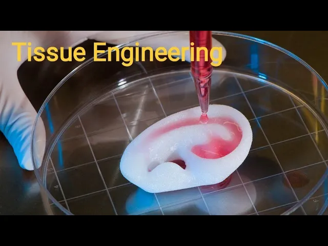
Tissue Engineering / components of tissue engineering.
2.1K
Tissue engineering is a scientific field developing biological substitutes to replace diseased or damaged human tissue. It integrates three core components: stem cells (which can divide repeatedly and differentiate into multiple cell types), scaffolds (support structures made from polymers, ceramics, metals, or composites), and environmental factors (oxygen, pH, temperature, nutrients, growth factors). The process involves cell culture, scaffold incubation, tissue formation, and implantation. Scaffolds are classified as fibrous, porous, hydrogel, microspheres, bioceramic, or composites. Applications include skin, cartilage, heart, and bone tissue engineering. The field combines cell biology, material science, and engineering to restore organ function through native tissue rehabilitation or functional tissue substitutes.
Click to watch →

M. Pikuła - Advanced technologies in tissue regeneration
122
Tissue engineering is a new scientific field based on biotechnology and molecular biology, integrating chemistry, molecular biology, and clinical medicine. It comprises three essential elements: cells, scaffold (biomaterials), and growth factors. Cells require both mechanical support and signaling molecules to survive and function; without growth factors, transplanted cells undergo apoptosis. Different cell sources include autologous cells (from the same patient), allogeneic cells (from donors), and cell lines. Key growth factors include BMP, PDGF, and EGF, which can be bound to scaffolds or synthesized as peptides for controlled cell stimulation. Vascularization is critical for transplantation success, as tissues require adequate blood supply to receive oxygen and nutrients.
Click to watch →

Tissue Engineering Video
946
Tissue engineering is an interdisciplinary field that creates functional tissues to regenerate, repair, or replace damaged tissues and organs through three essential components: cells (which form the functional matrix), scaffolds (which provide structural support for tissue growth), and signals (which direct cell growth and differentiation). The process involves extracting donor tissue, isolating cells, culturing them under controlled conditions, seeding cells into scaffolds, adding growth factors and stimuli, allowing tissue growth, and finally implanting the new tissue into the human body.
Click to watch →

Matériaux et santé : ingénierie tissulaire, toxicité
937
Tissue engineering is a pluridisciplinary field combining life sciences, materials science, and engineering sciences, coined by Professor Langer who received the 2011 Innovation Award from The Economist. It serves as an alternative to tissue and organ transplantation for large defects and as a model for in vivo experimentation. The field consists of three essential components: cells, biomaterial scaffolds (organic/inorganic, degradable/non-degradable), and active substances like growth factors. Applications span skin, corneal, muscle, organ, blood vessel, and vocal cord tissue engineering. The process involves cell harvesting, culture, seeding onto scaffolds, bioreactor culture, and differentiation into functional tissue.
Click to watch →
Deep Dive
Basic Principles of Microfluidics: Familiarity with fluid dynamics at the micro-scale, specifically laminar flow, shear stress, and diffusion behavior.

Introduction of Microfluidics - Creative Biolabs
5.5K
Microfluidics is a technology for precise control and manipulation of micro-scale fluids, also known as lab-on-a-chip or microfluidic chip technology. It integrates sample preparation, reaction, separation, and detection processes into micron-scale chips for automatic analysis. The historical development traces back to 1959 when Richard Feynman proposed MEMS using semiconductor technology for miniaturization. In 1990, Manz introduced the miniaturized total analysis system (μTAS). Key technical principles involve constructing fluid channels and reaction chambers on micrometer scales using MEMS processing. Flow is driven through micro mechanical pumps, electro hydraulic pumps, and electro osmotic flow, while detection employs methods like laser-induced fluorescence, electrochemistry, and mass spectrometry.
Click to watch →
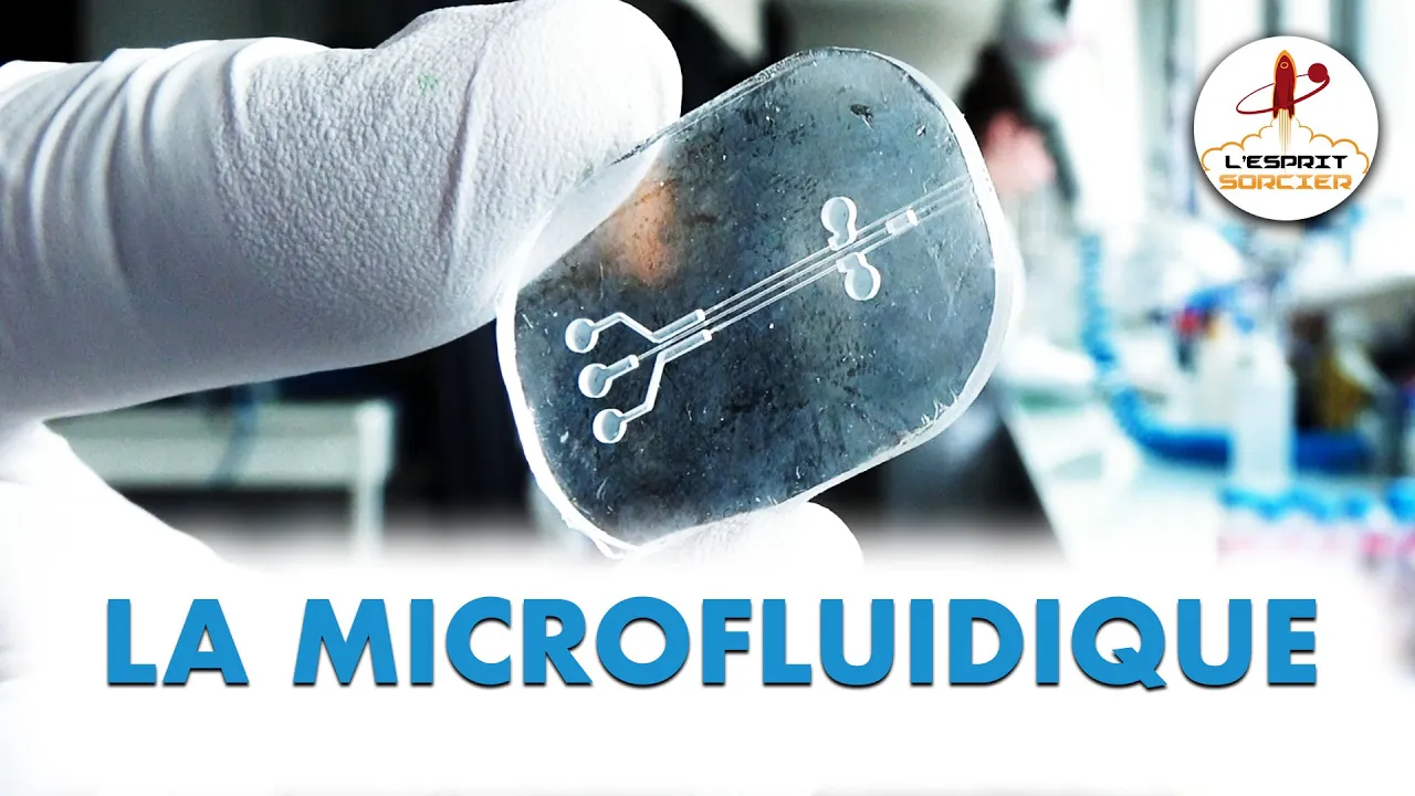
La microfluidique - L'Esprit Sorcier
131.2K
Microfluidics is an emerging interdisciplinary field at the intersection of physics, biology, and chemistry that studies fluid flow at the microscopic scale. Originating from microelectronics technology in the 1990s, it has grown into a major research area with tens of thousands of researchers worldwide. At the microscale, fluids flow without turbulence, heat transfer becomes more efficient, and gravity becomes negligible. Microfluidic chips function as miniature laboratories containing microchannels 10-100 times smaller than a human hair, fabricated from transparent plastic. These devices enable precise fluid sorting, separation, mixing, and droplet generation at thousands per second. Applications include pregnancy tests, inkjet printers, and emerging uses in cosmetics, food industry, and renewable energy. In biology, microfluidics enables organ-on-a-chip technology for studying human organs like kidneys and intestines, reducing animal testing while allowing researchers to manipulate specific cell types to understand disease mechanisms.
Click to watch →

WEBINAR REPLAY - Concepts of Microfluidics - Fluigent
8.3K
Microfluidics is the science of manipulating fluids in microliter to picoliter volumes using micrometer-sized channels, where surface forces dominate over gravitational forces due to the scaling effect (capillary forces scale with length^1 while gravitational forces scale with length^3), enabling unique phenomena such as laminar flow (where fluids move in parallel layers without disruption, allowing precise control of mixing and diffusion) and droplet generation (using designs like flow focusing, T-junctions, or coflow systems with surfactants to stabilize droplets), which has revolutionized applications in single-cell analysis, organ-on-a-chip devices, and point-of-care diagnostics.
Click to watch →
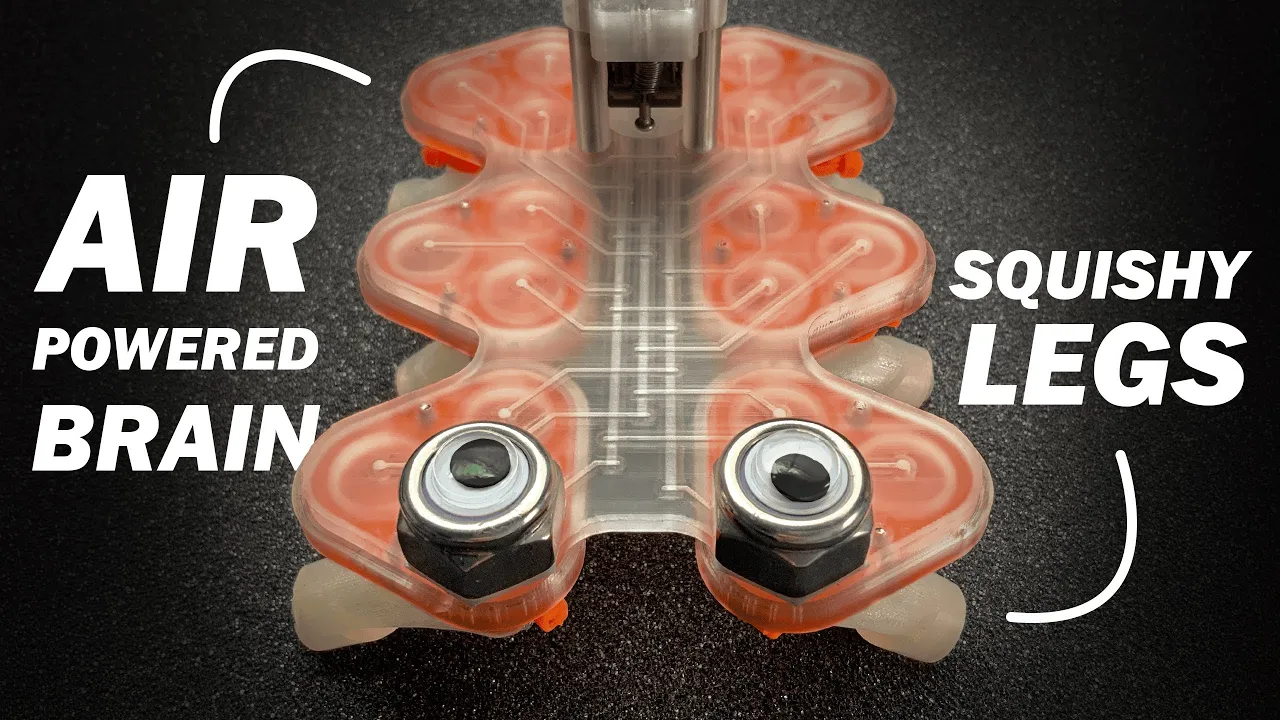
Can You 3D Print a Robot’s Brain Out of Air?
43.9K
Microfluidics combines micrometer-scale channels with fluid flow control, serving as a fluidic equivalent to electronic circuits. The field emerged from observing rolling diaphragm membranes in air pumps—pulling vacuum contracts them while pushing air stiffens them. Key applications include inkjet printers firing precise ink droplets and pregnancy tests using paper capillary action. The core motivation is replacing slow manual pipetting with automation, potentially shrinking entire labs onto single chips. For soft robotics, this means replacing external bulky setups with integrated fluidic control systems inside the robot's body. The transition from simple static channels to dynamic fluidic logic components represents a fundamental shift in how we think about autonomous machine control.
Click to watch →
Game-Changing Microfluidics | Podcast Episode 1
2.7K
Microfluidics manipulates fluids within channels of tens to hundreds of micrometers, where viscous forces dominate over inertial forces, creating predictable laminar flow. Unlike turbulent flow in everyday systems (like garden hoses), microfluidic channels lack turbulence, making diffusion the primary mixing mechanism. This predictable behavior enables precise control of fluid flow and mixing. Applications include point-of-care diagnostics using lab-on-a-chip devices for rapid biomarker detection from tiny blood samples, and chemical synthesis with exceptional control over reaction conditions. The advantages include faster results, lower reagent consumption, and reduced costs compared to conventional methods.
Click to watch →
Deep Dive
Introduction to Bioprinting and Bioinks: Knowledge of different printing modalities (extrusion, droplet, light-based) and the rheological requirements of hydrogel-based bioinks.

5ta Edición del Simposio de Biomateriales Poliméricos
221
3D bioprinting represents one of the most disruptive technological advances of recent decades, part of additive manufacturing that constructs three-dimensional objects from digital models by layer-by-layer deposition. While the concept originated in the 1980s, its biomedical application is recent, driven by convergence of engineering, biology, materials science, and biomedicine. The number of publications has multiplied since 2010, with global market expansion from prototyping to customized medical devices and functional biological structures. Bioink, the working material, is a mixture of cells, biomaterials, and biochemical factors providing structural support and biological environment. Bioinks must meet biological criteria (cell compatibility, no immunological reactions, cell viability during printing), physical criteria (optimal viscosity, thixotropic behavior, shape retention), chemical criteria (non-toxic components, controlled solidification, biodegradability), functional criteria (accurate deposition, microvasculature reproduction), and practical criteria (reproducibility, shelf life, economic viability).
Click to watch →

Introduction to Bioinks
2.2K
Bioink is the most vital ingredient in bioprinting, serving as a bioprintable formulation that provides a suitable microenvironment for encapsulated cells to survive, grow, differentiate, and migrate. For effective bioprinting, bioinks must possess several key properties: ideal printability (with appropriate viscosity and shear-thinning behavior for extrusion-based printing or rapid gelation for inkjet printing), biocompatibility to support cell viability and function, controlled biodegradation kinetics matching ECM production rates, viscoelasticity to mimic natural tissue behavior, oxygen and nutrient permeability for cell survival, and the ability to support tissue regeneration and remodeling. Hydrogels are commonly used as bioink materials because they provide a water-rich environment conducive to cell function.
Click to watch →
Introduction to Bioprinting
510
Bioprinting is defined as 3D printing with cells and/or active components, distinguishing it from traditional 3D printing of inert scaffolds. Bioinks must contain cells or bioactive components serving biological purposes like promoting growth and differentiation. Three main techniques exist: inkjet printing (thermal/piezoelectric droplet ejection), extrusion-based printing (force-driven deposition), and laser-based methods. Inkjet bioprinting uses thermal (bubble formation via heating) or piezoelectric (ceramic actuator pressure) mechanisms to eject droplets. Key advantages include low cost, accessibility, support for cell viability, and straightforward multi-material fabrication. However, significant challenges exist: pressure bubble creation affects cell viability, shear stress causes poor cell development, and low-viscosity requirements limit material choices. Research leverages these disadvantages through genetic engineering and drug delivery during printing. Applications include cell patterning, organoid production, blood vessel creation, and in situ printing. Materials range from hydrogels to cell suspensions.
Click to watch →
ليه العلماء بينتجوا الأعضاء البشرية في الفضاء في محطة الفضاء الدولية
35K
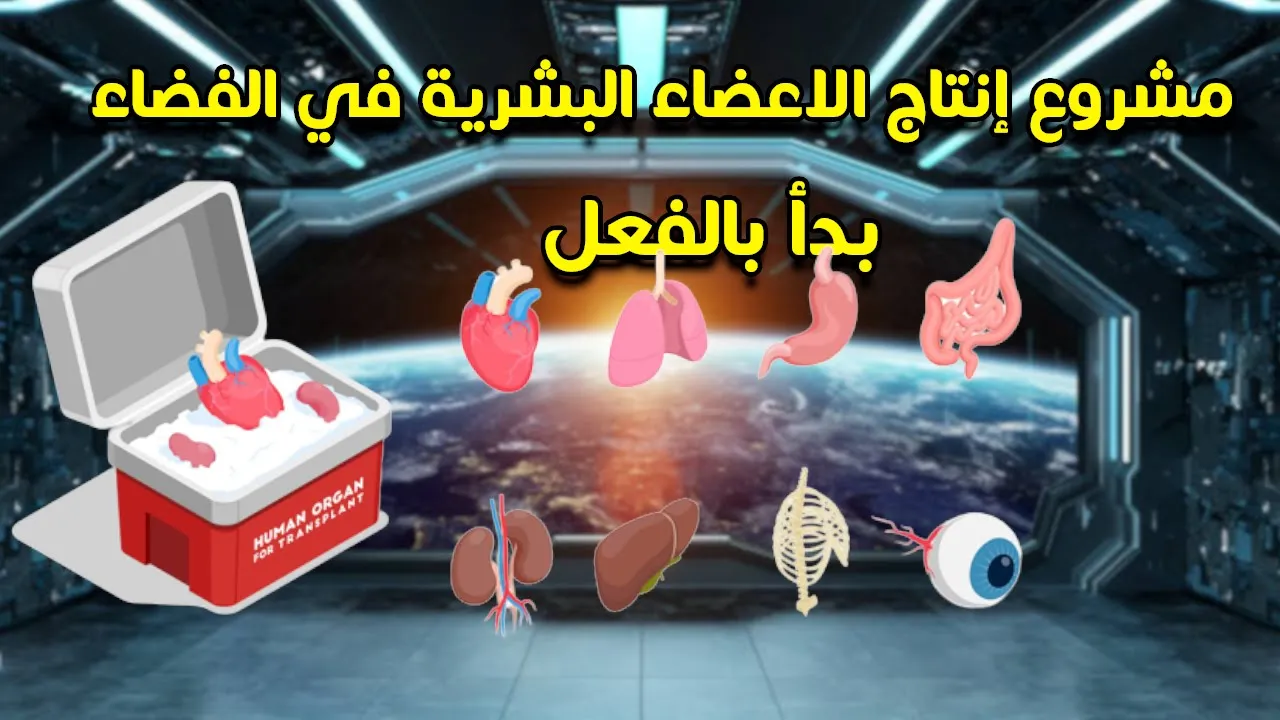
3D bioprinting is a revolutionary technology that uses 3D printers with bio-ink containing living cells to create tissues and organs. Bio-ink is a printable gel with hydrogen peroxide that protects cells during printing. The technology addresses the critical organ shortage problem where available organs far fewer than needed. Dr. Andrew Morgan, a former military physician who joined NASA in 2013, pioneered this approach after witnessing slow transplant recovery rates. The microgravity environment of space is essential because tissues printed on Earth break apart due to gravity, requiring temporary scaffolding that space eliminates.
Click to watch →
Lifeink Collagen Bioinks for 3D Bioprinting
6.2K
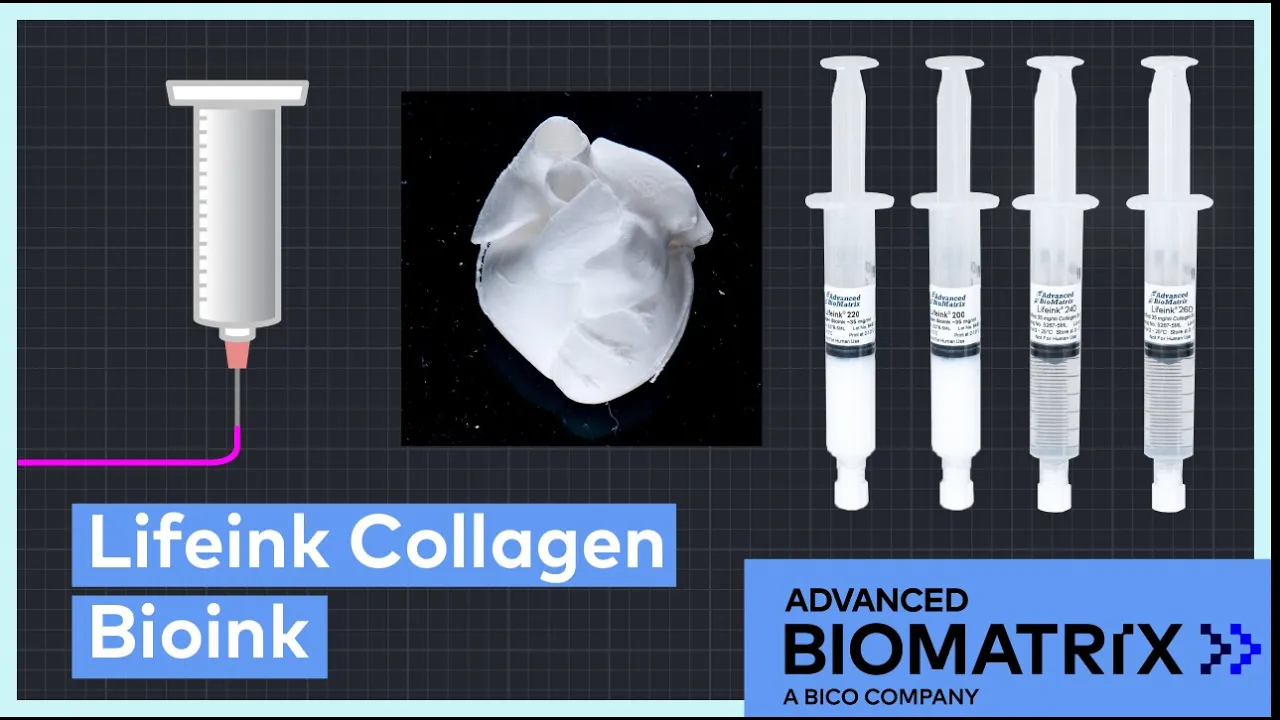
Lifeink bioinks are pure collagen solutions (35 and 70 mg/ml) designed for extrusion-based 3D bioprinting, overcoming traditional challenges of collagen's high viscosity and temperature sensitivity; they come in pH-neutral and pH-acidic variants, with the acidic formulation requiring FRESH printing (Freeform Reversible Embedding of Suspended Hydrogels) using gelatin microparticle slurry to achieve higher resolution, followed by incubation at 37°C where cells remodel the collagen into living tissue.
Click to watch →
Deep Dive
Vascular Biology and Anatomy: Understanding the structure of blood vessels, the role of endothelial cells, and the diffusion limit (approx. 200 micrometers) that necessitates vascularization in thick tissues.

10th Biology Chapter 1 Objective 2026 | जैव प्रक्रम (परिवहन) महत्वपूर्ण Objective | मैट्रिक परीक्षा
179
Plants transport water and minerals through xylem (unidirectional upward flow) and organic nutrients through phloem (bidirectional flow). Gas exchange occurs through stomata controlled by guard cells. The human heart has four chambers (two atria and two ventricles). The right side handles deoxygenated blood, while the left side handles oxygenated blood. The pulmonary artery carries deoxygenated blood to lungs, while pulmonary veins return oxygenated blood to the heart. The pacemaker is the SA node.
Click to watch →
Class-11,NEET Biology,CH-6 Anatomy of Flowering Plants,Dicot and Monocot Root
101K
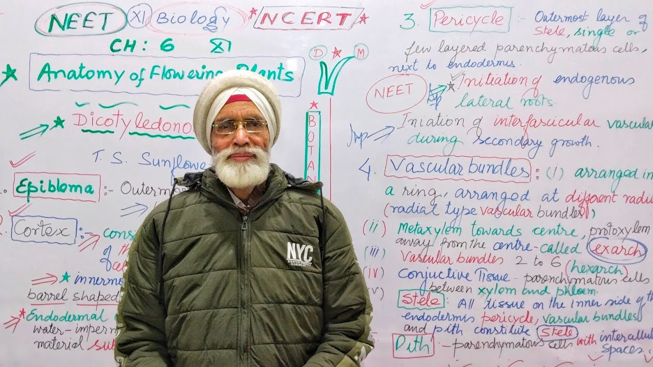
Vascular bundles in dicot roots are arranged radially (at different radii) with xylem and phloem alternating. Protostele (first-formed xylem) is away from center, while metastele (later-formed xylem) is toward center. Conjunctive tissue fills gaps between xylem and phloem. Pericycle gives rise to interfascicular cambium during secondary growth, enabling radial expansion. This arrangement allows efficient water and nutrient transport while supporting the plant's ability to increase in girth over time.
Click to watch →
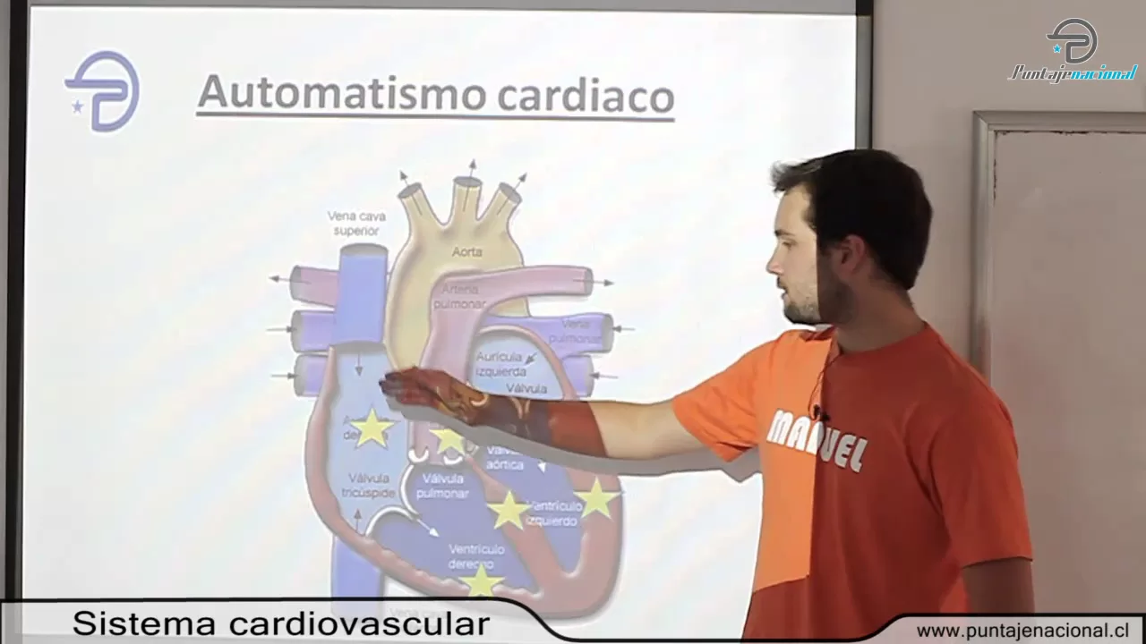
BIOLOGIA MC -- Procesos y funciones vitales -- Sistema cardiovascular
184.1K
The vascular system consists of tubes containing blood, organized hierarchically: arteries (largest, elastic walls, carry blood away from heart), arterioles (regulate blood flow via constriction/dilation), capillaries (microscopic, enable tissue exchange), and veins (return blood to heart with one-way valves). All vessels share three layers: tunica externa (connective tissue), tunica media (elastic/muscular), and tunica intima (endothelium). Arteries withstand high pressure from cardiac pumping, while veins accommodate larger blood volumes with thinner walls.
Click to watch →

Cerebral Vascular Anatomy And Imaging
268.8K
This educational video by Dr. Igor Rybinnik MD from Rutgers Robert Wood Johnson Medical School teaches medical students about cerebral vasculature, including the anatomy of the circle of Willis, vascular imaging modalities (MRA, CTA, conventional angiogram), and the clinical syndromes associated with each major cerebral artery territory (ACA, MCA, PCA, SCA, AICA, basilar artery, PICA), illustrated through a case study of an 83-year-old patient with acute left PCA stroke causing right homonymous hemianopia and cognitive deficits.
Click to watch →

Vascular Anatomy (3 of 4): Venous Drainage -- Head and Neck Anatomy 101
66.8K
The venous drainage of the head and neck involves three main systems: the dural venous sinuses (superior sagittal, inferior sagittal, straight, transverse, and sigmoid sinuses), the pterygoid plexus (collecting from the infratemporal fossa), and the external jugular vein (formed by the retromandibular and facial veins); importantly, all these venous systems drain into the internal jugular vein, which carries blood directly to the heart without valves, making infections in the face potentially catastrophic as they can spread retrograde through the venous system to the cranial sinuses and systemic circulation.
Click to watch →
Prerequisite Knowledge
- Concept 01Fundamentals of Tissue Engineering: Understanding the triad of cells, scaffolds, and signaling molecules used to reconstruct functional biological tissues.
- Concept 02Basic Principles of Microfluidics: Familiarity with fluid dynamics at the micro-scale, specifically laminar flow, shear stress, and diffusion behavior.
- Concept 03Introduction to Bioprinting and Bioinks: Knowledge of different printing modalities (extrusion, droplet, light-based) and the rheological requirements of hydrogel-based bioinks.
- Concept 04Vascular Biology and Anatomy: Understanding the structure of blood vessels, the role of endothelial cells, and the diffusion limit (approx. 200 micrometers) that necessitates vascularization in thick tissues.
Subsequent Learning
- Step 01Perfusable Bioreactors: Studying how to dynamically culture vascularized constructs using fluid flow to promote endothelial cell alignment and vessel maturation.
- Step 02Organ-on-a-Chip and Physiological Modeling: Applying vascularized microfluidic constructs to simulate human organ systems for drug screening and disease modeling.
- Step 03In Vivo Integration and Surgical Anastomosis: Investigating how pre-vascularized engineered tissues connect to a host animal's circulatory system upon transplantation.
- Step 04Scaling Up to Whole-Organ Engineering: Exploring the computational, biological, and manufacturing challenges of bioprinting complex, multi-lineage organs like the liver, kidney, or heart.
Vascular Bioprinting
Vascular Bioprinting
0:00Playing Section
- 1
Method generates vascularized tissue constructs.
- 2
Uses a dual-layer concentric microfluidic printhead.
- 3
Technique applies to cardiac, liver, and skin tissues.
Developmental Self-Assembly and Angiogenesis-Driven Vascularization
While microfluidic bioprinting offers precise spatial control over larger vessel geometries, critics argue it faces severe resolution limits, struggles to replicate capillary-level networks (under 10 micrometers), and subjects cells to damaging shear stresses. An opposing paradigm prioritizes biological self-assembly and developmental biology-driven vascularization. Instead of printing artificial channels, this 'bottom-up' approach relies on the intrinsic signaling of cells (such as endothelial cells and pericytes) to spontaneously self-organize into functional microvascular capillaries (vasculogenesis) and sprout into surrounding tissues (angiogenesis). Proponents of self-assembly argue that biologically-driven networks are more physiologically accurate, dynamic, and capable of natural remodeling over time compared to rigid, mechanically printed synthetic structures.
Deep Dive
Perfusable Bioreactors: Studying how to dynamically culture vascularized constructs using fluid flow to promote endothelial cell alignment and vessel maturation.

Thermo Scientific DynaDrive SUB: Perfusion Application and Customer Experience Highlights
529
This comprehensive presentation covers the development and evaluation of next-generation single-use bioreactors. The industry has shifted from first-generation systems designed for less intense fed-batch production toward high-density perfusion applications, which face oxygen transfer limitations. Thermo Fisher developed the Dino Drive to address needs for intensified cell culture support, flexible multi-product facilities, and larger single-use bioreactors (larger than 2,000L). Key innovations include: cuboid shape using corners as baffles to prevent vortexing; multi-impeller drivetrain improving mixing by 62 times; drill hole sparger providing linear performance across all scales; 10:1 to 20:1 turn-down ratio enabling streamlined cell trains; and lift mechanisms for ground-level loading. In-house evaluation demonstrated 200 million cells/mL at 4 vbd perfusion rate with 80%+ viability, superior CO2 stripping compared to alternatives, and KLa values exceeding 25 inverse hours. Scale-up validation showed consistent performance from 50L to 5,000L. Financial analysis revealed up to 25% cost reduction in cost per gram compared to 2,000L reactors, driven by reduced batch numbers and cycle times.
Click to watch →
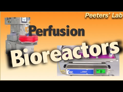
Perfusion bioreactors
12K
Perfusion bioreactors are continuous culturing systems that retain cells within the reactor while continuously harvesting the desired product, enabling higher cell concentrations and product yields through prolonged operation periods (weeks to months), reduced exposure to toxic byproducts, and milder operating conditions with less shear stress; however, these systems face challenges including high medium consumption, complex control requirements, limited commercial availability, and the need for well-characterized processes, making them particularly suitable for vaccine production and precision medicine applications despite requiring significant capital investment and operational expertise.
Click to watch →

3D Perfusion Bioreactor
22K
A 3D perfusion bioreactor creates in vitro cell culture conditions that closely mimic in vivo environments by using three-dimensional polymer scaffolds combined with continuous nutrient perfusion; the assembly process involves connecting four major components—a peristaltic pump, four media reservoirs, four bioreactor chambers, and four CO2 humidifiers—using color-coded tubing matched to pressure-stamped letters on bottle caps, followed by a two-step cell seating protocol including 3-hour static incubation to allow cell adhesion before initiating low-flow perfusion at 0.2 cm/min mean velocity for 24 hours to optimize initial cell seating efficiency.
Click to watch →

NEET 2025 Biology: Biotechnology Principles & Processes | One Shot | New NCERT 360° | Seep Pahuja
268.3K
Two main types of bioreactors are used: (1) Simple stirred tank bioreactor - has a curved base, agitator with flat blade impeller, pH control system, and steam sterilization, (2) Sparged stirred tank bioreactor - similar to simple type but with sparging system that creates bubbles to increase oxygen transfer efficiency. The sparged type is better for aerobic cultures requiring high oxygen levels.
Click to watch →
Биореактор | Как это сделано
36.8K
Bioreactors are high-tech vessels used in food, chemical, pharmaceutical, and cosmetic industries for cultivating microorganisms and cell cultures. They differ from fermenters in terminology (fermenter for bacterial cultivation, bioreactor for cell culture), physical dimensions (fermenters have 3:1 height-to-diameter ratio, bioreactors 1.5:1-2:1), impeller design (turbine for high-speed mixing in fermenters, marine screw with inclined blades for gentle mixing in bioreactors), and aeration systems (ring diffusers with larger openings in fermenters, porous sintered metal diffusers producing small bubbles for better gas solubility in bioreactors).
Click to watch →
Deep Dive
Organ-on-a-Chip and Physiological Modeling: Applying vascularized microfluidic constructs to simulate human organ systems for drug screening and disease modeling.

Microfluidics _Huh, 2020
670
Organ-on-a-chip technology uses microfluidic engineering to create microengineered models of human tissues and organs by mimicking their structural features (such as the three-layer barrier between lung tissue and capillaries) and environmental conditions (like mechanical deformation during breathing), enabling researchers to study complex physiological processes, model diseases, and test drug responses in ways that traditional petri dish cultures cannot achieve.
Click to watch →
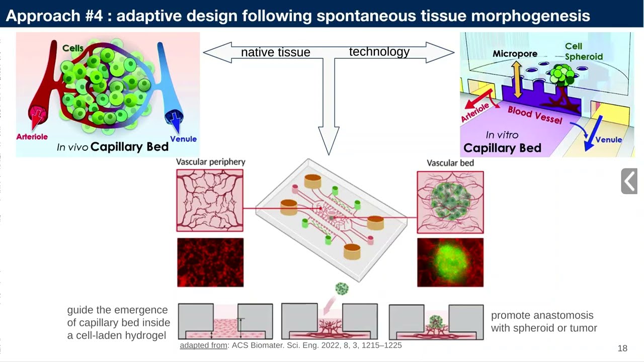
Mifobio 2023 - Mathieu HAUTEFEUILLE - A056-Beginner’s guide to the fabrication and useof..
166
Organ-on-chip systems, also called microphysiological systems, aim to mimic physiological and pathological conditions in vitro for better disease study and drug testing. The FDA Modernization Act 2.0 now legally recognizes these models for drug evaluation if proven physiologically relevant. Cells sense both biochemical and mechanical signals from their environment, with mechanosensing and mechano-transduction pathways affecting gene expression within hours or days. Traditional static culture on stiff substrates causes cells to lose native markers, making organ-on-chip approaches essential for preserving cell identity and function.
Click to watch →

Jos Joore - Physiologically relevant tissue models using a high throughput Organ on a Chip platform
1.1K
The OrganoPlate® is a microfluidic culture platform that enables long-term, membrane-free 3D co-culture models in a standard microtiter plate format, supporting high-throughput screening of physiologically relevant tissue models including neuronal networks, perfused tubular structures, and co-cultures of different cell types for drug testing and disease modeling applications.
Click to watch →

Translational Sciences with Dr Joni Rutter
117
Organ-on-a-chip technology creates miniature models of human organs using flexible materials that mimic natural biological functions. Lung-on-a-chip devices use flexible membranes with vacuum systems to simulate breathing motions, enabling drug interaction testing with living lung cells. Advanced systems integrate multiple cell types (heart, neurons, muscles) on single chips to study complex interactions. These platforms provide more accurate models of human physiology compared to traditional animal models, accelerating drug development and disease research while reducing reliance on animal testing. The technology enables researchers to observe how cells respond to various compounds and conditions in a controlled, human-relevant environment.
Click to watch →
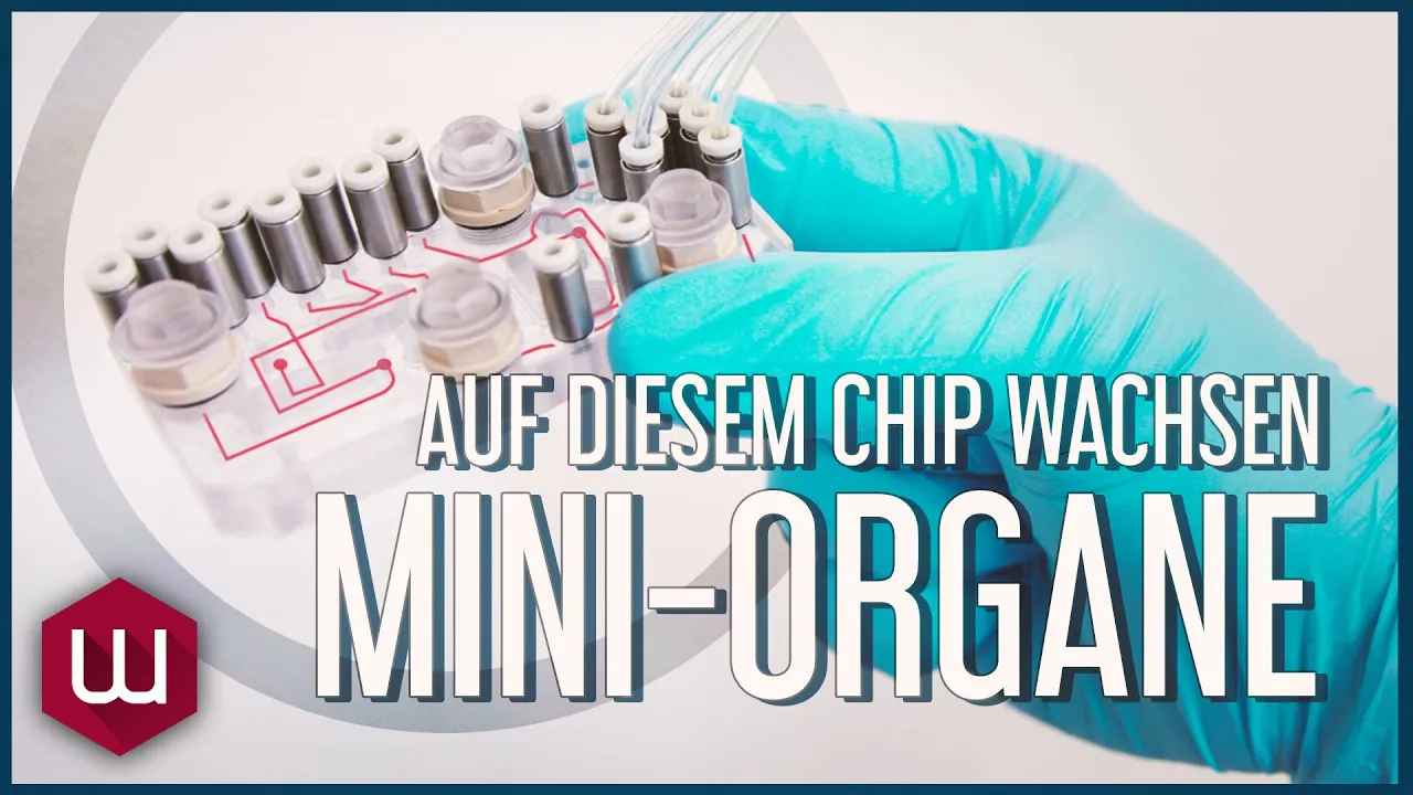
Auf diesem Chip wachsen Mini-Organe
44.4K
Organ-on-a-chip technology creates miniaturized, functional organ models on microfluidic chips that simulate human physiology, enabling personalized drug testing without animal experiments; these chips can simulate multiple organs (currently 16 of 34 human organs, including liver, kidney, and pancreas) connected by nutrient solutions pumped by heart-simulating devices, with the ultimate goal of creating individual biochips from each patient's blood to test medications before clinical trials.
Click to watch →
Deep Dive
In Vivo Integration and Surgical Anastomosis: Investigating how pre-vascularized engineered tissues connect to a host animal's circulatory system upon transplantation.

'Get Out Of My Way,' the Doctor Snapped — Until the Bleeding Army Ranger Begged for the Combat Nurse
144
Vascular anastomosis is the surgical connection between a vein graft and the damaged artery, requiring microscopic precision. The surgeon passes a needle through the vein graft and then through the artery wall, looping the thread and pulling it tight against the bleeding tissue. Multiple knots are thrown to secure the connection. The artery surges with pressure as the graft expands, filling with rhythmic pulse. The suture line is checked for leaks, and if none are found, the anastomosis is successful. This procedure represents the culmination of the surgical team's efforts to save the patient's limb.
Click to watch →
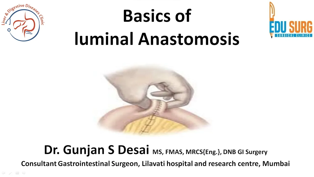
Intestinal anastomosis - layers, nomenclature, phasis of healing, Lembert stitch, and Connell stitch
46.9K
Anastomotic success depends on multiple factors: disease factors (steroids, chemotherapy, immunosuppression, anemia, low albumin, sepsis, contamination, Crohn's, smoking, alcohol, radiation, malnutrition) and patient factors (emergency cases, elderly, diabetics). Surgeon factors include gentle tissue handling, adequate exposure, and standardized technique. Anastomosis configurations include end-to-end, end-to-side, and side-to-side. Functional anastomosis is antiperistaltic (opposite peristaltic direction) versus isoperistaltic. Mode of creation includes hand-sewn (interrupted/continuous) or stapled (linear/circular). Mesenteric orientation must match to prevent ischemia. In high-risk patients, covering stoma protection may be indicated instead of primary anastomosis.
Click to watch →
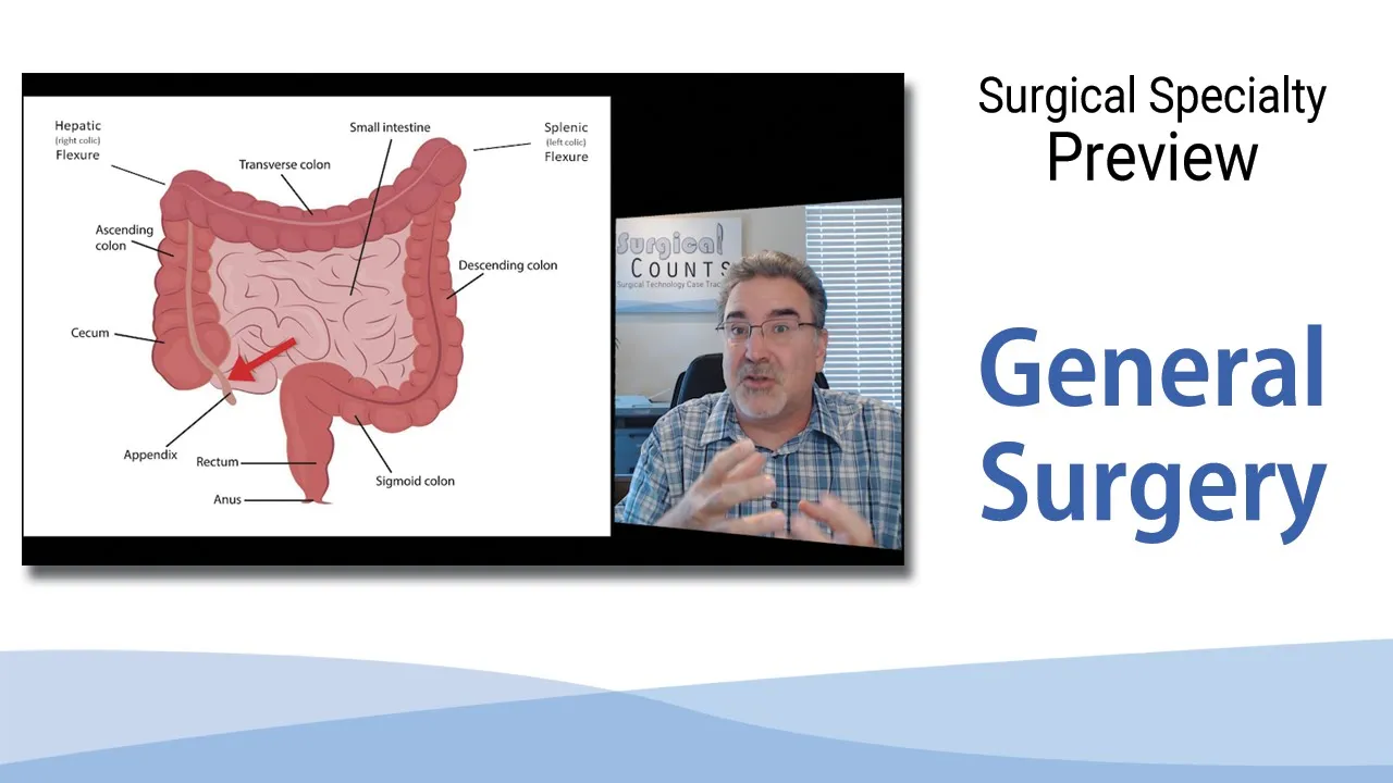
Surgical Technology Preview - General Surgery
91.1K
Anastomosis connects two tubes so their lumens communicate, enabling fluid flow. End-to-end anastomosis joins cut ends (common in colon resections), while side-to-end anastomosis attaches one tube to another (used in vascular surgery). Ectomy denotes surgical removal of part or all of an organ, such as gastrectomy (stomach removal). Ostomy creates an opening—medical terminology follows a pattern: one term indicates opening to outside (colostomy), two terms indicate connection between structures (gastrojejunostomy connects stomach to small intestine).
Click to watch →

Security Stops Black Passenger at the Gate — Then the Plane Won’t Take Off Without Him
4.7K
Anastomosis is the surgical technique of connecting blood vessels between the donor and recipient hearts. The surgeon sutures the left atrium, right atrium, pulmonary artery, and aorta to create a complete circulatory connection. After implantation, the transplanted heart must be monitored closely. The cardiac monitor tracks heart rhythm and pressure. If the heart does not start beating properly, internal defibrillation may be required to shock it into rhythm. The moment of truth is when the heart begins beating on its own, indicating successful transplantation.
Click to watch →
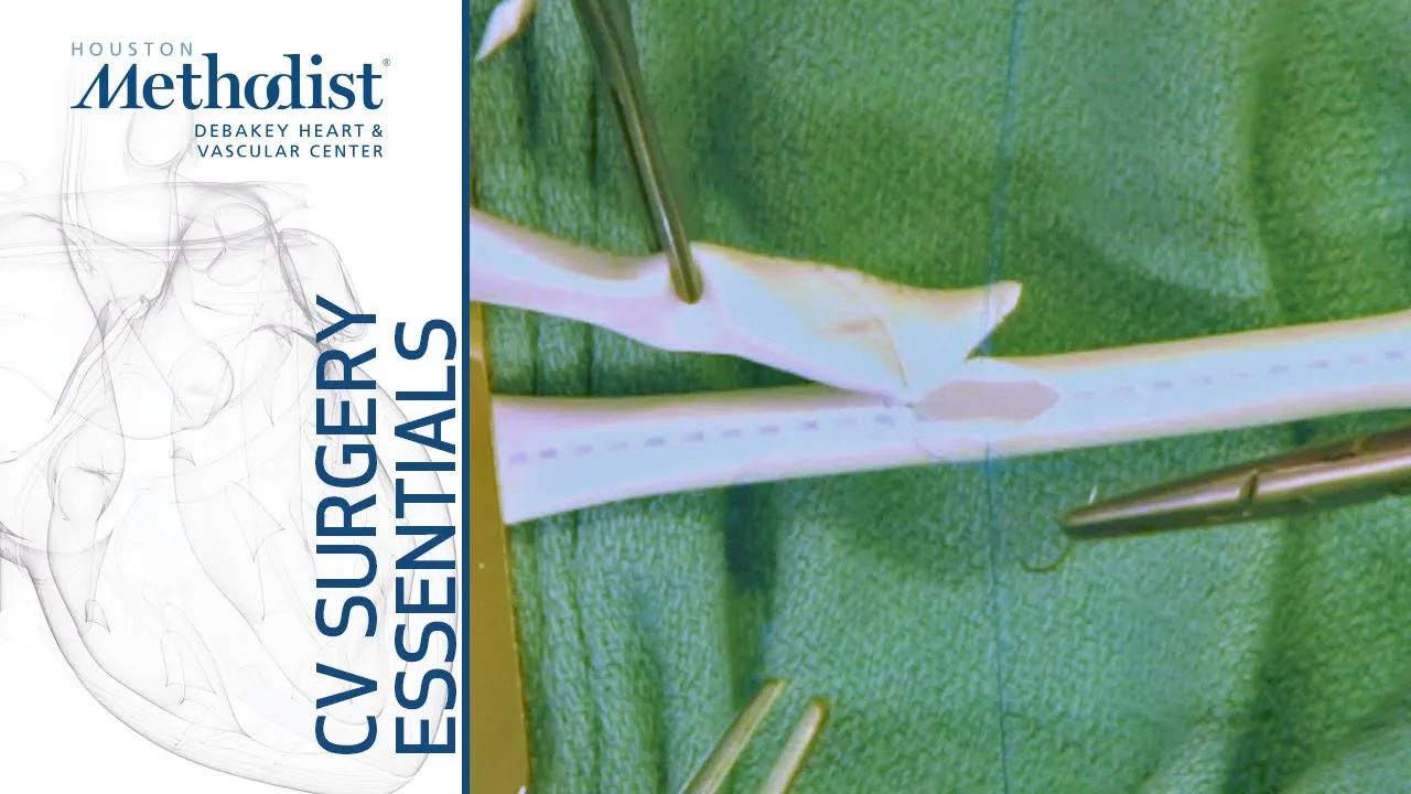
End to Side Anastomosis (Eric K. Peden, MD, M. Mujeeb Zubair, MD)
68.1K
End-to-side anastomosis is a surgical technique where a graft is attached to a recipient vessel by suturing one end of the graft to the side wall of the vessel; the procedure involves preparing grafts at similar angles, placing stay sutures to hold the vessel open, suturing at right angles with even spacing while keeping all action within the anastomotic field, and securing the suture line with a slip knot and multiple throws.
Click to watch →
Deep Dive
Scaling Up to Whole-Organ Engineering: Exploring the computational, biological, and manufacturing challenges of bioprinting complex, multi-lineage organs like the liver, kidney, or heart.

Lecture Session om Stamceller
845
Scaling up organ engineering requires industrialization to reduce costs and increase throughput. Current processes are highly manual and require skilled personnel. The goal is to build 'factories' that can produce thousands of blood vessels, requiring automation, robustness, simplicity, low cost, and full clinical compliance. This transition from laboratory to industrial production is essential for clinical translation.
Click to watch →
Frontier Science #16 - Whole Organ Engineering w/ Gordana Vunjak-Novaković - Prof. @ Columbia | BIOS
1.2K
Whole organ engineering combines biomaterial scaffolds, bioreactors, and stem cells to create functional human tissues and organs for regenerative medicine and disease modeling. The fundamental principle is providing cells with an instructed environment that mimics native tissue conditions, including appropriate mechanical, electrical, and biochemical signals. This approach enables applications ranging from recovering rejected donor lungs to creating patient-specific bone and cartilage grafts, as well as developing multi-organ chip platforms for drug testing and disease research. The field bridges engineering and biology, requiring interdisciplinary collaboration between engineers, clinicians, and scientists to translate discoveries into clinical benefits.
Click to watch →
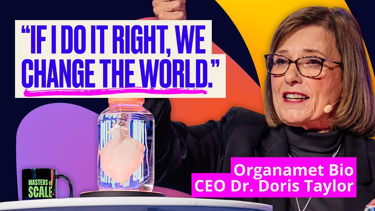
The journey to build a human heart (with Dr. Doris Taylor) | Masters of Scale Summit 2025
9.6K
Scaling up bioengineered organs from small animal models to human-sized versions presents enormous challenges. Heart cells do not divide, making it difficult to obtain sufficient quantities. Progress was slow, moving from grape-sized to kiwi-sized hearts over 14 years. This technology has since been adopted by hundreds of labs worldwide to create various organs including liver, kidney, lung, and pancreas, transforming what was considered impossible into common practice.
Click to watch →

Tissue Engineering - Dr. Alan Russell
6.3K
Whole organ engineering represents the frontier of regenerative medicine, involving decellularization of entire organs while preserving the extracellular matrix scaffold. This approach preserves the pre-existing vascular and structural networks, then repopulates them with new cells. The city analogy applies: buildings are demolished while infrastructure remains, then new structures are built on the existing foundation. This technique has shown promise for generating functional organs, though challenges remain in creating thick tissues with adequate vascularization and integrating cultured organs into recipient bodies.
Click to watch →

Boyang Zhang Speech at the Fall 2017 Networking Event
144
Current tissue engineering produces small-scale tissues that are too small for clinical applications. Two main directions exist for advancement: scaling up to produce larger, more complex tissues with extensive vascular networks, or scaling down to reduce costs and enable high-throughput applications.
Click to watch →
Vascular Bioprinting
Vascular Bioprinting
0:00- 1
Method generates vascularized tissue constructs.
- 2
Uses a dual-layer concentric microfluidic printhead.
- 3
Technique applies to cardiac, liver, and skin tissues.
Developmental Self-Assembly and Angiogenesis-Driven Vascularization
While microfluidic bioprinting offers precise spatial control over larger vessel geometries, critics argue it faces severe resolution limits, struggles to replicate capillary-level networks (under 10 micrometers), and subjects cells to damaging shear stresses. An opposing paradigm prioritizes biological self-assembly and developmental biology-driven vascularization. Instead of printing artificial channels, this 'bottom-up' approach relies on the intrinsic signaling of cells (such as endothelial cells and pericytes) to spontaneously self-organize into functional microvascular capillaries (vasculogenesis) and sprout into surrounding tissues (angiogenesis). Proponents of self-assembly argue that biologically-driven networks are more physiologically accurate, dynamic, and capable of natural remodeling over time compared to rigid, mechanically printed synthetic structures.
Fundamentals of Tissue Engineering: Understanding the triad of cells, scaffolds, and signaling molecules used to reconstruct functional biological tissues.
Basic Principles of Microfluidics: Familiarity with fluid dynamics at the micro-scale, specifically laminar flow, shear stress, and diffusion behavior.
Introduction to Bioprinting and Bioinks: Knowledge of different printing modalities (extrusion, droplet, light-based) and the rheological requirements of hydrogel-based bioinks.
Vascular Biology and Anatomy: Understanding the structure of blood vessels, the role of endothelial cells, and the diffusion limit (approx. 200 micrometers) that necessitates vascularization in thick tissues.
Perfusable Bioreactors: Studying how to dynamically culture vascularized constructs using fluid flow to promote endothelial cell alignment and vessel maturation.
Organ-on-a-Chip and Physiological Modeling: Applying vascularized microfluidic constructs to simulate human organ systems for drug screening and disease modeling.
In Vivo Integration and Surgical Anastomosis: Investigating how pre-vascularized engineered tissues connect to a host animal's circulatory system upon transplantation.
Scaling Up to Whole-Organ Engineering: Exploring the computational, biological, and manufacturing challenges of bioprinting complex, multi-lineage organs like the liver, kidney, or heart.
623 views8likes2:00@JoVEJournalOriginal Release: 2022-08-27
[Music] thank you [Music] the overall goal of this microfluidic bioprinting methodology is to generate a vascularized tissue construct this method can solve key questions in biofabrication of fast grass tissues the main advantage of this technique is that it is versatile in generating a three-dimensional shape controlled vascular bat for engineering vascular tissues through a secondary cell sitting process though this protocol provides insight into engineering vascularized cardiac tissues it can also be applied to many other tissue types such as liver skin and even cancers General individuals new to this method May struggle because setting up the buy printer may not be simple to start this procedure construct a dual layer concentric microfluidic printhead by inserting a smaller blunt needle serving as the core into the center of a larger blunt needle serving as the sheath make sure that the core needle is protruding from the outer shell approximately one millimeter after that insert a 23 gauge needle in the barrel of the central needle in the reverse Direction make a hole on the side of the barrel of the outer needle and insert a matching size metal connector seal with epoxy glue Mount the extruder onto the head of a bioprinter using a polymethyl methacrylate or pmma holder next for injection of the bio Inc and the cross-linking solution through two PVC tubes individually connect the inlets of the print head to a dual Channel syringe pump
Up Next
2:45
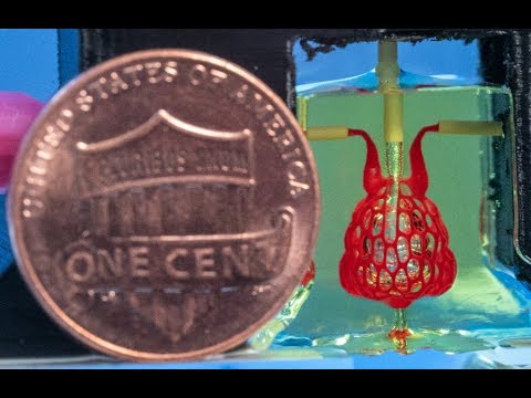
3D Printing Replacement Organs: Bioprinting Breakthrough
@riceuniversity
73.4K views•2019-05-02

12:54
Triumph of Orthodoxy Icon: Byzantine Art & History Explained
@BenCallan
2.1K views•2024-08-06

2:00
Organ-on-Chip Fabrication with Integrated Electrodes for TEER | Protocol Preview
@JoVEJournal
377 views•2022-05-27

1:42
Game of Thrones Opening Credits: A Cinematic Analysis
@gameofthrones
46.3M views•2011-04-18
Related Study Plans & Knowledge Roadmaps
Structured learning paths in General & Interdisciplinary Studies
general-studies
Elevator Engineering: History, Physics & Cities
01From Hoists to Safety Brakes: The Dawn of Vertical Lift
02Hydraulic vs. Traction: The Engineering Physics of Elevators
03Modern Speed, Safety, and Algorithms
+ 2 more learning modules
12h5 modules
Explore Roadmap
general-studies
Rubber: Material Science, History & Geopolitics
01Origins of Natural Rubber & Mesoamerican History
02The Chemistry of Natural Rubber & Vulcanization
03The Geopolitics of the Rubber Boom & Colonization
+ 2 more learning modules
12h5 modules
Explore Roadmap
general-studies
Containerization: Evolution, Logistics & Trade
01Ancient Trade & Bulk Cargo: Amphorae to Barrels
02The Industrial Revolution & Cardboard Packaging
03The Containerization Revolution: Malcom McLean
+ 2 more learning modules
12h5 modules
Explore Roadmap
general-studies
Textile Tech: Evolution, Labor & Global Impact
01Pre-Industrial Textiles: Handloom & Cottage Industry
02Mechanization: The Spinning Jenny to the Power Loom
03The Factory System & Global Labor Transformation
+ 2 more learning modules
10h5 modules
Explore Roadmap