Bioengineers have developed an open-source 3D bioprinting technique that creates living tissues with entangled vascular networks, enabling nutrient and oxygen delivery to cells within soft gel matrices; this breakthrough addresses the critical challenge of maintaining cell viability in large tissue blocks by mimicking the body's natural vascular architecture found in organs like the lung, where airway distension helps boost blood mixing in surrounding vascular structures.
3D Printing Replacement Organs: Bioprinting Breakthrough
Added:Deep Dive
Basic principles of additive manufacturing (3D printing), particularly layer-by-layer material deposition.

Первые шаги в 3D печати: от модели до детали
394.3K
All 3D printers operate on two fundamental principles: layer-by-layer construction and additivity. In layer-by-layer construction, volumetric geometry is replaced by a sequence of flat layers, with material gradually added to the growing object until reaching the desired size and shape. Additivity means material is progressively added rather than removed or reshaped. These principles apply not only to FFF printers but to all 3D printing technologies including Direct Metal Deposition.
Click to watch →
noc19-me24 Lec 1-Introduction to Rapid Manufacturing (Part 1 of 3)
25.2K
Layer-based technology is the fundamental principle underlying additive manufacturing, involving the composition of three-dimensional physical objects from many layers of mostly equal thickness. Each layer is contoured according to corresponding three-dimensional data and placed on top of the preceding layer. This layer-by-layer construction method distinguishes additive manufacturing from other manufacturing approaches and enables complex geometries impossible with conventional methods. The workflow involves automated mesh fixing, slicing, and hatching of 3D CAD data. Photo polymerization processes use UV lasers to cure liquid photo polymer in dot, line, and layer patterns. Key parameters include slice distance (vertical spacing between layers) and hatch distance (horizontal spacing between adjacent hatches), which can be optimized for time efficiency or structural strength.
Click to watch →
Prof. Murugaiyan Amirthalingam | Additive Manufacturing - 3D printing, principles and practices
703
Additive manufacturing operates on fundamental principles of material deposition and layer-by-layer construction. The process begins with a digital 3D model that is mathematically sliced into cross-sectional layers. Each layer is then fabricated by selectively depositing or fusing material according to the design specifications. Common techniques include fused deposition modeling (extrusion), selective laser sintering/melting, and binder jetting. A critical principle is that each subsequent layer bonds to the previous one, creating a unified structure. The technology enables features like internal channels, lattice structures, and topology-optimized geometries that cannot be manufactured through traditional methods. Applications span rapid prototyping, customized medical implants, tooling production, and complex aerospace components. The technology's ability to produce functional parts directly from digital files without tooling significantly reduces time-to-market and enables mass customization at competitive costs.
Click to watch →

8 Additive Manufacturing (3D Printing) Technologies - MJF, FDM, SLS, SLA, DMLS, DLS, SLM, Polyjet.
1.4K
All additive manufacturing processes build 3D objects by depositing or fusing material layer by layer. The fundamental principle involves slicing a 3D model into 2D cross-sections and constructing each horizontal layer sequentially. The build platform moves incrementally after each layer is completed, allowing subsequent layers to be added on top. This systematic approach enables the creation of complex geometries that would be difficult or impossible to manufacture using traditional subtractive methods. The layer-by-layer methodology allows for precise control over the final object's geometry, internal structures, and material distribution throughout the build.
Click to watch →

Materials and Additive Manufacturing with Dr. Jennifer Wolk
2.5K
Additive manufacturing, also known as 3D printing, is a revolutionary manufacturing process that builds objects by depositing material layer by layer in precise locations, unlike conventional manufacturing which removes material through milling or drilling; this technology enables the creation of complex geometries and internal structures that would be extremely difficult or impossible to produce using traditional methods, opening up new possibilities in design and engineering.
Click to watch →
Deep Dive
Fundamentals of tissue engineering and biomaterials, including the role of hydrogels and bioinks as cellular scaffolds.
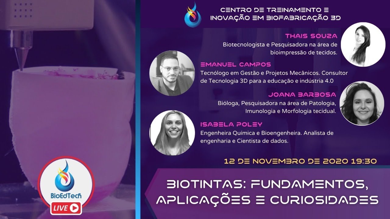
Biotintas: fundamentos, aplicações e curiosidades.
564
This comprehensive section covers the foundational materials and principles of 3D bioprinting. Gelatin serves as both an adhesion material (1:10 ratio with water and propolis for bacterial prevention) and a hydrogel. Hydrogels are defined as three-dimensional polymeric structures that hold large quantities of water, including edible gelatin and contact lenses. Bioinks differ from hydrogels as they are organic polymeric materials (like PLA and PCL) that don't carry cells. Cell selection is critical—researchers must determine what tissue structure they want to create, which determines the appropriate cell type (cell lines from banks or primary cells). Stem cells, particularly induced pluripotent stem cells (iPSCs), offer remarkable potential because they can theoretically generate any tissue type through differentiation, though working with them is challenging due to their sensitivity and high resource requirements.
Click to watch →
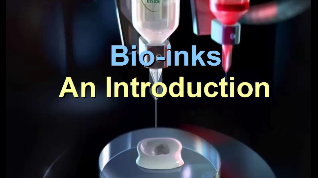
Hydrogel Bioinks in 3D Bioprinting: Key Properties and Types
7K
Hydrogel bioinks are biopolymer-based materials (such as collagen, gelatin, hyaluronic acid, alginate, and nanocellulose) that provide temporary support to cells during 3D bioprinting while they produce their own extracellular matrix. These bioinks must balance printability with biocompatibility, exhibiting key properties including shear-thinning viscosity for smooth printing, specific gelation processes (ionic, thermal, photocrosslinking, or enzymatic), and biological interactions that support cell survival and proliferation. Bioinks are categorized into four types: matrix bioinks for cell encapsulation, curing bioinks for photopolymerization, sacrificial bioinks for temporary support and complex geometry creation, and support bioinks for permanent structural reinforcement. The choice between natural and synthetic bioinks involves trade-offs between biological fidelity and process control.
Click to watch →

Hydrogels in Tissue Engineering
6.2K
Hydrogels are three-dimensional hydrophilic networks formed through polymerization of monomers, either via chain-growth or step-growth mechanisms, with crosslinks (chemical or physical) enabling them to swell in water while maintaining structural integrity; these materials are particularly valuable in tissue engineering because their high water content, flexible three-dimensional structure, and ability to mimic the extracellular matrix make them ideal scaffolds for supporting cell growth and regeneration, with biodegradable versions created by incorporating degradable linkages like ester bonds or enzyme-sensitive components, and injectable forms developed through physical crosslinking triggered by temperature changes or chemical crosslinking via free radical polymerization.
Click to watch →

13. Tissue Engineering Scaffolds: Processing and Properties
62.2K
Tissue engineering scaffolds are porous biomaterials designed to temporarily substitute for the extracellular matrix, enabling cells to attach, migrate, proliferate, and differentiate while the scaffold degrades at a controlled rate matched to new tissue formation; successful scaffold design requires balancing biocompatibility, appropriate pore structure (typically 20-500 microns depending on tissue type), mechanical integrity, and degradation kinetics, with common materials including natural polymers like collagen and glycosaminoglycans, synthetic polymers such as PGA, PLA, and PLGA, and hydrogels, processed through techniques like freeze-drying, salt leaching, electrospinning, or 3D printing.
Click to watch →

Публичная лекция «Гидрогели — из чего делают органы в пробирках»
332
This comprehensive section covers the foundational knowledge of hydrogels and their applications in tissue engineering. Hydrogels are materials consisting of over 95% water that maintain their shape due to complex polymer structures with large pores. The human body's extracellular matrix functions as a natural hydrogel, providing structural support and water balance. Natural hydrogels like collagen and hyaluronic acid have limitations including lack of cell-specificity and potential immune reactions. Synthetic hydrogels offer precise control over signaling molecules and mechanical properties but require complex synthesis. Four critical criteria for synthetic hydrogels include biodegradability, mechanical support, signaling molecules, and soluble signaling molecules like growth factors. The section explains how multi-component hydrogels using polymers like star PEG and sugars like heparin form polymer networks with controlled mechanical properties through component concentration variations.
Click to watch →
Deep Dive
The anatomy and physiology of vascular systems, specifically how blood vessels deliver nutrients and oxygen to sustain living tissue.
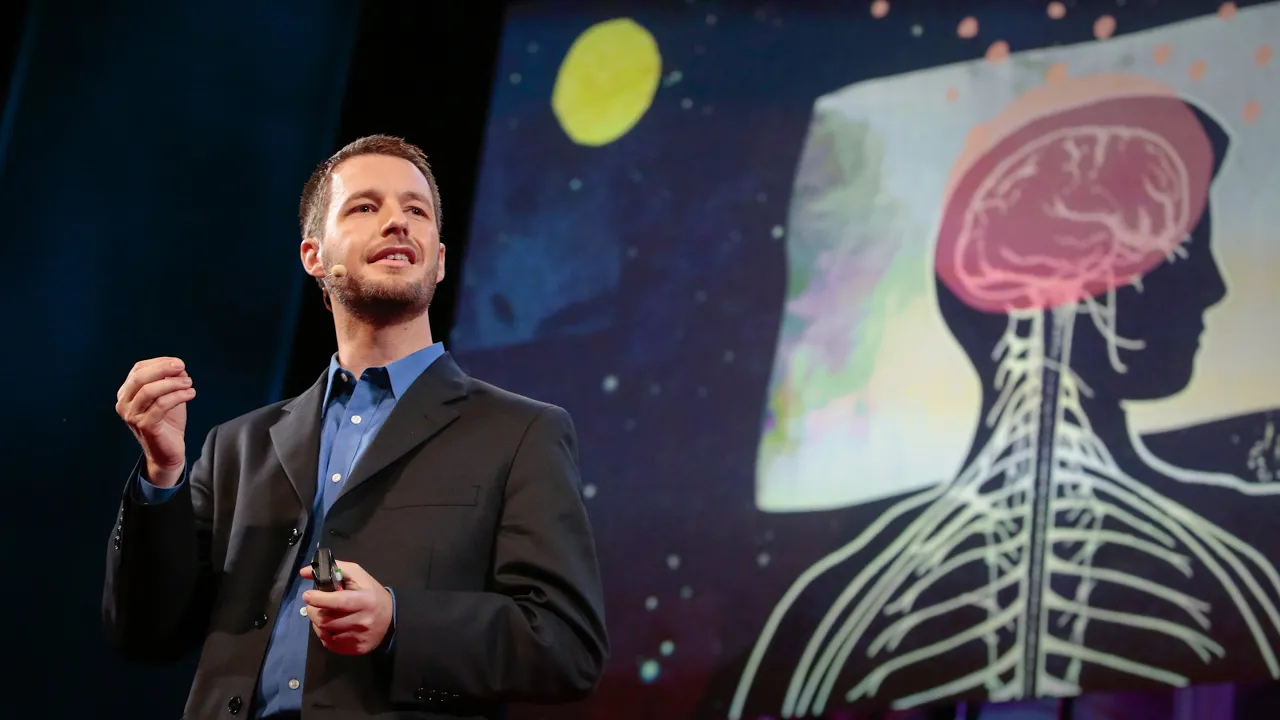
One more reason to get a good night’s sleep | Jeff Iliff
1.7M
The circulatory system solves the problem of delivering nutrients and oxygen to all cells in the body by sending blood vessels throughout every organ. In the brain, blood vessels form a complex network that extends from the surface down into the tissue itself, ensuring that each and every cell receives the necessary nutrients and oxygen to maintain its function.
Click to watch →

LIVE특강_면역과글리칸_김준영원장260223
174
Blood vessels deliver oxygen and nutrients throughout the body. When vessels are damaged, oxygen and nutrient supply is impaired. More seriously, waste products accumulate: after delivering nutrients, waste is generated; after delivering oxygen, carbon dioxide is produced. This waste must be properly processed and eliminated.
Click to watch →

Human Anatomy & Physiology Part-1 by Avadutika Ananda Bratisha Acharya #anandamarga #prout
2.9K
Blood vessels deliver oxygen and nutrients to body tissues. The cardiovascular system ensures that all cells receive the necessary substances for survival and function. Without adequate blood supply, tissues cannot function properly and may become damaged. This delivery system is essential for maintaining tissue health.
Click to watch →
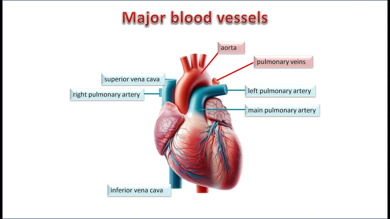
The Vascular System. Blood vessels: structure and function of arteries, veins and capillaries.
1.2K
Blood vessels form the circulatory system that transports blood from the heart to all body parts. They supply oxygen and nutrients to organs and tissues while removing carbon dioxide and metabolic waste. The heart acts as a pump, pushing blood through vessels comprising the mammalian circulation. Oxygenated blood travels from the left ventricle to most organs (systemic circulation) and to the brain (cerebral circulation). Blood vessels branch into narrower vessels until becoming capillaries where gas and nutrient exchange occurs. Deoxygenated blood enters the venous circulation, returning to the right atrium.
Click to watch →

Basic Anatomy & Physiology 13 | BLOOD VESSELS Reference Seeley's
9.4K
Blood vessels form the conduit system of the cardiovascular system, divided into pulmonary and systemic circuits. Pulmonary vessels transport blood from the right ventricle through the lungs for oxygenation and return to the left atrium. Systemic vessels distribute oxygenated blood from the left ventricle to all body tissues and return deoxygenated blood to the right atrium. Blood vessels perform five essential functions: carrying blood, facilitating nutrient/gas/waste exchange at tissue level, distributing and collecting substances, regulating blood pressure, and directing blood flow to appropriate tissues. The three main types—arteries (carry blood away from heart with thick elastic walls), veins (carry blood toward heart with thinner walls and valves), and capillaries (smallest vessels where exchange occurs)—work together in a hierarchical network from arteries to arterioles to capillaries to venules to veins.
Click to watch →
Deep Dive
Basic cell biology concepts, such as cell viability, cell-extracellular matrix interactions, and stem cell differentiation.

Praktikum On Air - Struktur Sel
6.2K
Cells are the smallest structural and functional units of living organisms, serving as units of heredity (containing DNA) and growth (based on Virchow's principle that all cells arise from pre-existing cells). Cells are classified into prokaryotic (lacking membrane-bound nucleus, e.g., bacteria) and eukaryotic (possessing membrane-bound nucleus, e.g., plants, animals, fungi). Plant cells differ from animal cells by having rigid cell walls, chloroplasts for photosynthesis, and large vacuoles for storage. Cell viability is determined by nuclear activity and metabolic processes.
Click to watch →
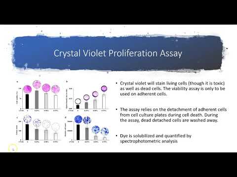
Cell Viability
3.1K
Cell viability refers to the measurement of living, healthy cells within a population that are undergoing appropriate replication and division. It is quantified by tracking the increase in cell number over a specific time period. Healthy cells actively proliferate and grow until they reach confluence (no more space), while damaged cells (due to drugs, viral infections, bacterial infections, or toxins) enter growth arrest or die.
Click to watch →
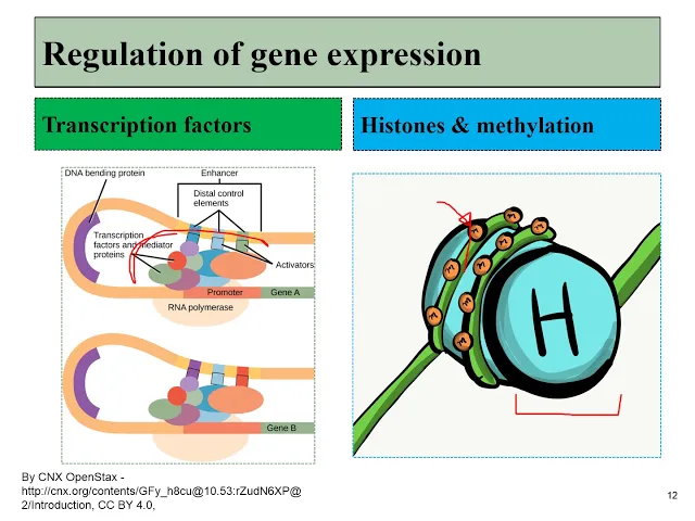
01 cell biology review
667
Cellular differentiation is the process where cells become specialized despite having identical DNA, regulated by signals that alter gene expression patterns. As cells differentiate, their cell fate becomes more limited. Stem cells have broad potential, but their descendants become restricted. The extracellular matrix includes ground substance (a gel of polysaccharides and proteins that attracts water) and fibers like collagen and fibronectin. Hyaluronic acid is a major polysaccharide in ground substance with dental applications. Cellular junctions include cell adhesion molecules, integrins, desmosomes (anchoring junctions between cells), and hemidesmosomes (anchoring cells to extracellular matrix).
Click to watch →

GENERAL PATHOLOGY II CHAPTER 1 II CELL AS A UNIT OF HEALTH & DISEASE II PART 4 II ROBBINS PATHOLOGY
12K
Stem cells are undifferentiated cells capable of self-renewal (producing two identical stem cells) and asymmetric division (producing one stem cell and one differentiated cell), serving as the foundation for tissue regeneration and maintenance throughout life; embryonic stem cells from the blastocyst's inner cell mass are totipotent with unlimited self-renewal capacity, while tissue stem cells in adult organs like the liver, brain, and skin produce only same-tissue cell types; the extracellular matrix provides mechanical support, controls cell proliferation, and establishes tissue microenvironment through fibrillar proteins (collagen and elastin), water-hydrated gels (proteoglycans), and adhesive glycoproteins (fibronectin, laminin, integrins), with collagen synthesis involving hydroxylation, glycosylation, triple helix formation, and cross-linking by lysyl oxidase and vitamin C.
Click to watch →
What is cell viability?
10.1K
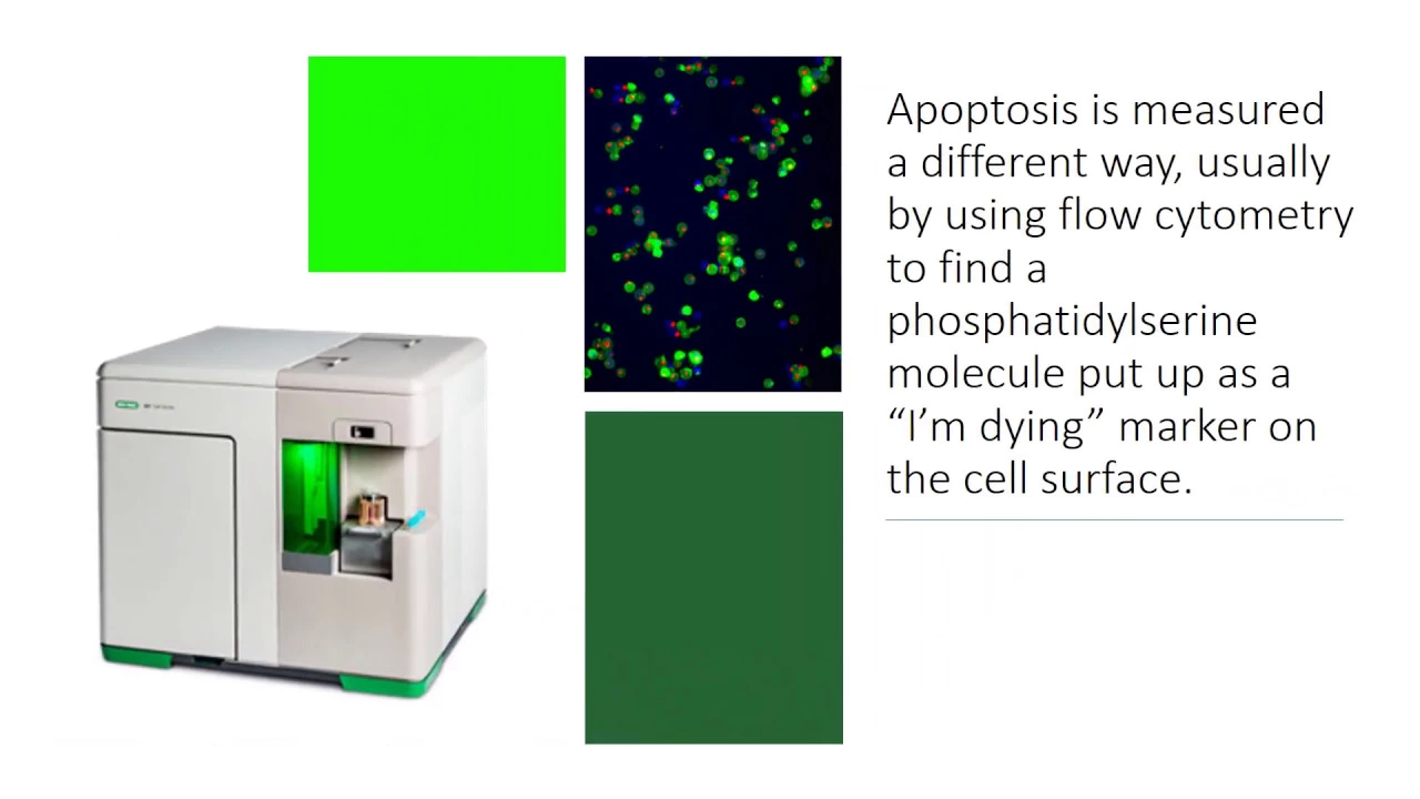
Cell viability, commonly defined as the percentage of living cells, is fundamentally incomplete for assessing cell health; comprehensive evaluation requires four essential tests: (1) dye exclusion assays (like trypan blue) measuring membrane integrity, (2) apoptosis detection via flow cytometry identifying cells undergoing programmed death, (3) proliferation assays demonstrating colony formation capability, and (4) functional assays such as tri-lineage differentiation proving stem cells can develop into osteoblasts, adipocytes, and chondrocytes. Relying solely on simple viability counts fails to reveal whether cells are truly healthy, functional, and capable of therapeutic repair.
Click to watch →
Prerequisite Knowledge
- Concept 01Basic principles of additive manufacturing (3D printing), particularly layer-by-layer material deposition.
- Concept 02Fundamentals of tissue engineering and biomaterials, including the role of hydrogels and bioinks as cellular scaffolds.
- Concept 03The anatomy and physiology of vascular systems, specifically how blood vessels deliver nutrients and oxygen to sustain living tissue.
- Concept 04Basic cell biology concepts, such as cell viability, cell-extracellular matrix interactions, and stem cell differentiation.
Subsequent Learning
- Step 01The challenges of biocompatibility and host integration when transplanting bioprinted organs into living organisms (in vivo testing).
- Step 02Advanced perfusion bioreactor technologies used to mature and sustain 3D-printed organs in laboratory settings.
- Step 03Ethical, legal, and regulatory pathways (such as FDA approval processes) for translating bioprinted tissues to clinical human trials.
- Step 04The collaborative impact of open-source science in accelerating biomedical engineering breakthroughs and hardware development.
Tissue Structure
Scalable Tissues
Tissue Structure
0:16Playing Section
- 1
Investigates how human tissue structure dictates function.
- 2
Demonstrates airway distension boosts blood mixing in lungs.
The Biological Complexity and Hype-Reality Gap in Bioprinting
While bioprinting vascular networks is a major engineering milestone, critics argue that the enthusiasm surrounding 3D-printed organs overlooks immense biological hurdles. Replicating the physical structure of blood vessels is vastly different from replicating the dynamic, metabolic, and endocrine functions of complex organs like the liver or kidney. Printed tissues often lack the cellular density, heterogeneous cell types, and mature extracellular matrix necessary for long-term viability. Furthermore, severe challenges remain regarding long-term immune integration and the potential for printed materials to trigger inflammatory responses or tumorigenesis. Consequently, many researchers argue that alternative approaches—such as xenotransplantation (using genetically modified animal organs) or decellularization (re-populating donor organ scaffolds with stem cells)—are far more realistic and closer to clinical viability than building highly complex organs from scratch using 3D printers.
Deep Dive
The challenges of biocompatibility and host integration when transplanting bioprinted organs into living organisms (in vivo testing).

Simulation #112 Dr. Melanie Matheu - 3D Printing Organs and Tissues
987
Successful biofabricated organs require four key factors: resolution for capillaries, architectural complexity, printing speed to preserve cell viability, and biocompatibility. Materials combine FDA-approved polymers with natural extracellular matrix components (collagen, hyaluronic acid, fibronectin) that cells recognize and integrate with. Once implanted, cells produce their own extracellular matrix and remodel the tissue over time. The goal is to create structures that the body recognizes as self, similar to skin grafts where cells from one body part are transplanted to another. This approach minimizes immune rejection and promotes long-term integration.
Click to watch →

Mod-01 Lec-14 Lecture-14-Introduction to Biomaterials
1.6K
Biocompatibility is defined as the ability of a material to perform with an appropriate host response in a specific application, requiring no inflammation, toxicity, or adverse effects. Materials must be biochemically compatible (non-toxic), non-irritating, non-allergenic, non-carcinogenic, and biomechanically compatible (matching elastic modulus to prevent stress shielding). Stress shielding causes surrounding bone atrophy when implants carry excessive load. In vitro tests cannot replace in vivo testing because they cannot detect inflammation, immune responses, multiple cell type interactions, or dynamic tissue remodeling. In vivo testing protocols include subcutaneous implantation (fibrous capsule assessment), extract testing, and direct bone implantation. Animal model selection depends on application: rabbits for bone replacements, guinea pigs for sensitivity testing.
Click to watch →

Estudos em Dispositivos Médicos, Importação de Produtos, Regulatório 4.0 | Passarini SUMMIT 2022
170
In vivo biocompatibility testing includes: (1) Irritation tests (primary and repeated) for local effects; (2) Sensitization tests (LLNA for mice, maximized for guinea pigs) for allergic reactions; (3) Pyrogen testing for fever-inducing contaminants; (4) Systemic toxicity tests (acute, subacute 28 days, subchronic 90 days, chronic 6-12 months). The choice of animal (rats vs. rabbits) depends on implant type and test duration requirements. Testing must be planned before animal selection.
Click to watch →

Биопринтинг: как учёные печатают органы из стволовых клеток. Вероника Усатова
850
Bioprinting can be performed in vitro (laboratory setting) or in vivo (within living organisms). In vivo bioprinting requires real-time tracking to compensate for tissue movement from breathing and heartbeat. While simple applications like skin grafts have been used clinically, complex organs like the brain present greater challenges. The technology faces barriers including regulatory approval, manufacturing scalability, cost, and the need for extensive safety testing before routine clinical use.
Click to watch →

Zukunft der Ersatzteilmedizin | Projekt Zukunft - Interview
575
While 3D bioprinting of organs like skin has been demonstrated, creating complex internal organs such as livers or kidneys remains challenging due to two main obstacles: finding biocompatible materials that allow cells to settle without triggering immune rejection or inflammation, and enabling cells to communicate and form functional networks within the printed structure; experts estimate this technology may become viable in 15-30 years, though significant scientific questions remain unresolved.
Click to watch →
Deep Dive
Advanced perfusion bioreactor technologies used to mature and sustain 3D-printed organs in laboratory settings.

3D Perfusion Bioreactor
22K
A 3D perfusion bioreactor creates in vitro cell culture conditions that closely mimic in vivo environments by using three-dimensional polymer scaffolds combined with continuous nutrient perfusion; the assembly process involves connecting four major components—a peristaltic pump, four media reservoirs, four bioreactor chambers, and four CO2 humidifiers—using color-coded tubing matched to pressure-stamped letters on bottle caps, followed by a two-step cell seating protocol including 3-hour static incubation to allow cell adhesion before initiating low-flow perfusion at 0.2 cm/min mean velocity for 24 hours to optimize initial cell seating efficiency.
Click to watch →
XVIVO is Revolutionizing Organ Transplantation with Advanced Perfusion Technologies
51.1K
XVIVO Perfusion has developed perfusion technology that circulates oxygenated solutions through donor organs outside the body, extending heart preservation time from the traditional 4-hour ice storage limit to approximately 12 hours in clinical trials, thereby reducing the carbon footprint of organ transportation by enabling commercial airline travel instead of private jets and potentially increasing transplant success rates.
Click to watch →

Perfusion bioreactors
12K
The bioprocessing industry is transitioning from batch to continuous production driven by precision medicine demands and advances in continuous culturing technology. Perfusion bioreactors, first developed in the 1980s for unstable protein production, enable continuous harvesting of pharmaceuticals while retaining cells within the reactor. This approach addresses batch limitations including product instability and toxic byproduct accumulation. Cells are retained using membranes, filters, or settlers, enabling prolonged operation lasting weeks to months. The technology provides milder operating conditions with reduced shear stress, benefiting sensitive mammalian and plant cell lines. Despite early promise, adoption was delayed due to complex retention system requirements until recent technological improvements made perfusion systems more commercially viable.
Click to watch →

Владимир Миронов: Вся правда о 3D-биопринтинге
843
Creating vascularized tissues is essential for larger organ survival, involving hollow cell spheroids that fuse to form continuous vascular channels. Bioreactors provide mechanical stimulation and controlled environmental conditions for tissue maturation, enabling cells to develop mechanical properties necessary for function. Computer-aided design creates digital models converted into printer instructions, enabling complex patient-specific tissue structures. Multi-dispenser bio-printers use multiple nozzles to deposit different materials simultaneously. Different organs present varying complexity levels - skin and cartilage are relatively simple and can be created without vascularization, while the heart, lungs, liver, and kidney require extensive vascular networks. The heart requires a complex conduction system for electrical signaling, while the kidney contains approximately 24 different cell types organized into functional units called nephrons.
Click to watch →

8:00 ИСТОЧНИКИ СТВОЛОВЫХ КЛЕТОК ПЕЧЕНИ И ПОДЖЕЛУДОЧНОЙ ЖЕЛЕЗЫ
178
3D bioprinting involves computer modeling, printing with bio-inks (hydrogels with proteins and polysaccharides), and bioreactor maturation. Scaffold technology uses biodegradable porous supports. Fibrin-based printing uses two-nozzle systems where thrombin cleaves fibrinogen to form fibrin bonds. Spheroid-based printing uses pre-formed cell aggregates (70%+ cell volume) for efficiency. Cell adhesion occurs through receptor interactions, electrostatic forces, and collagen matrix binding.
Click to watch →
Deep Dive
Ethical, legal, and regulatory pathways (such as FDA approval processes) for translating bioprinted tissues to clinical human trials.
3D Bioprinting Human Tissues and the Path Towards Translation
178
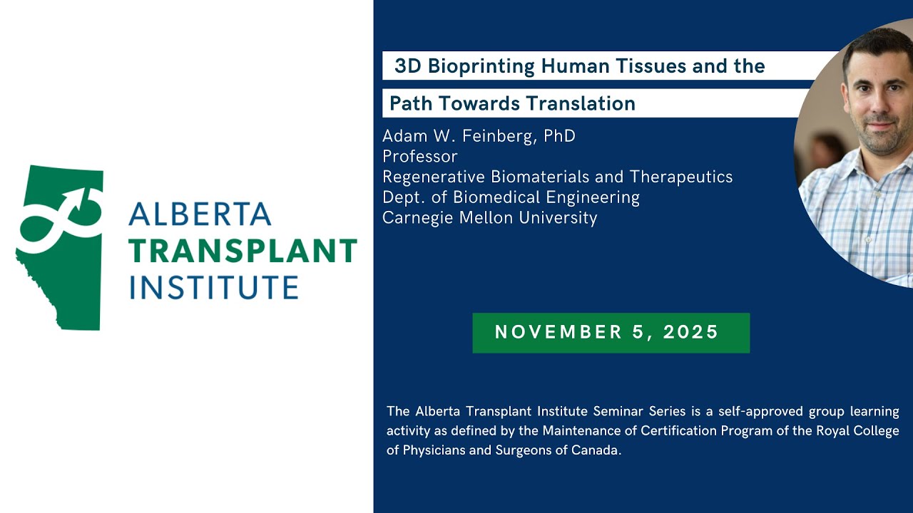
This section addresses the translation of bioprinted technologies from laboratory to clinical application. Bioprinted cardiac conduits have been implanted in mice by suturing directly into the inferior vena cava, achieving 100% patency and progressive vascularization over 6 months. Ultrasound tracking shows intrinsic beating and neo-tissue remodeling with proper sarcomere structure. The section discusses clinical pathways, noting the first FDA-approved trial for 3D bioprinted ear implantation occurred in 2022, with expectations for acellular scaffolds entering clinical trials soon. For solid organs, phase I/II trials are projected for early 2030s, with widespread availability anticipated in 10-15 years. The section addresses immunological challenges through universal donor cells using hypoimmune iPSCs, which would expand patient populations benefiting from therapy while avoiding severe side effects of immunosuppressive drugs. Regulatory and reimbursement challenges are discussed, including FDA approval complexities for combination devices and the need for new payment mechanisms for novel therapies.
Click to watch →
Webinar: 3D Bioprinted Therapeutic Liver Tissue
4.9K
Bioprinted therapeutic tissues represent a novel regulatory category neither purely drug nor medical device, fitting within FDA's Office of Tissues and Advanced Therapies (OTAT) purview. The pathway involves filing an Investigational New Drug (IND) application, anticipated around calendar year 2020, followed by clinical trial initiation. Scaling strategies involve two approaches: multiple patches or single patient-specific patches. Modular design enables fabrication to size based on individual anatomy, particularly valuable for rare orphan diseases where patient-specific construction is anticipated.
Click to watch →
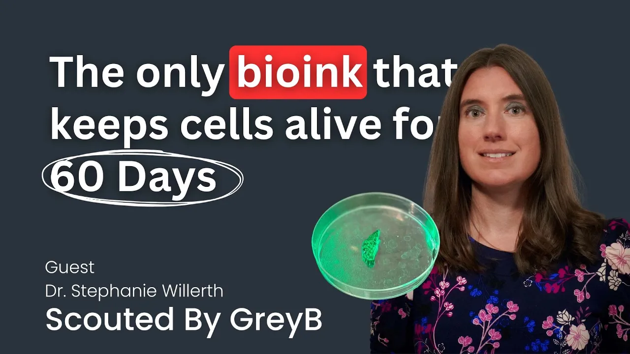
3D Bioprinting with Bioinks for Printing Neural and Cardiac Tissues | Axolotl Biosciences
193
Bioprinted tissue products follow distinct regulatory pathways based on intended use. Research-use products require end-user licenses, while clinical applications demand FDA approval and GMP manufacturing standards. Xeno-free bioinks present straightforward pathways toward human translation, with successful implantation already demonstrated in clinical settings. Future directions include expanding into skin grafts for burn patients and gut models for disease research. Strategic collaborations with established companies and access to appropriate manufacturing facilities are critical for advancing bioprinted tissues from research to clinical applications.
Click to watch →
3D-биопринтинг // Биоинженер Владимир Миронов на ПостНауке
9.6K
Translating bio-printing technology to clinical practice faces multiple barriers: the need for multidisciplinary teams combining expertise in computer science, chemistry, cell biology, and mechanical engineering; the absence of clinically certified bio-printers for medical use; and complex regulatory frameworks that create high barriers to entry. The regulatory environment, designed for traditional medical products, creates challenges for bio-printed tissues that may not fit existing categories. Additionally, the technology requires significant investment (5-10 years) that both academic research and industry may be reluctant to provide. Emerging applications include beta-cell replacement for diabetes treatment, where researchers have created insulin-producing cells responsive to glucose concentration that can be transplanted under the kidney capsule. Hair follicle regeneration represents another promising application, with stem cells grown in specialized bioreactors and implanted using robotic systems.
Click to watch →

3-D printing could one day help fix damaged cartilage in knees, noses and ears
2.2K
The research team discusses translating bioprinted cartilage technology into clinical applications for reconstructive surgery. Target applications include ear and nose reconstruction, spinal disc repair, and knee cartilage regeneration. The startup company Cell Inc developed a specialized printer priced at $5,000, selling approximately 30 units globally. Regulatory approval for surgical room use is pending, with plans for autologous tissue applications using patient-derived cells. The first clinical applications will focus on ear reconstruction, replacing current methods of silicone prostheses or rib cartilage carving with patient-specific printed cartilage structures.
Click to watch →
Deep Dive
The collaborative impact of open-source science in accelerating biomedical engineering breakthroughs and hardware development.

Raspberry Pi used in Biomedical Research
28.3K
Successful scientific instrumentation requires iterative engineering design, starting with functional prototypes and refining through multiple iterations to achieve robust, reliable systems. Professional engineering expertise combined with hands-on experimentation accelerates this process. Open source publication of complete hardware and software designs democratizes scientific research by enabling replication without expensive proprietary equipment. This approach increases scientific reproducibility, validates results through community scrutiny, and accelerates discovery by allowing researchers worldwide to build upon shared methodologies and innovations.
Click to watch →

TReND Open Labware Series (اردوينو): مقدمة عن الأجهزة العلمية مفتوحة المصدر
220
Open source hardware expands the developer base and reduces development costs. Products compete with commercial alternatives in quality, particularly in software. In scientific research, open source hardware facilitates experiment repetition and development by others in an active scientific community, especially in life sciences. This collaborative approach removes barriers to equipment development by allowing the largest number of interested parties to participate in the development process.
Click to watch →

How open-source hardware is changing our future | Amber Huffman | TEDxPortland
3.5K
Open source hardware applies software collaboration mechanisms directly to physical components since hardware is defined in software languages. The Calyptra security initiative demonstrates this power—fierce competitors like Google, Microsoft, AMD, and Nvidia collaborated despite competing in their respective markets. This collective approach enables solving problems no single entity could address alone. In medical research, open source hardware supports AI-driven breakthroughs like AlphaFold's protein structure database, accelerating cancer research and personalized medicine. Each technology era has been led by dominant companies, but the AI era offers an opportunity to enable broader community participation through open source hardware and standards.
Click to watch →

Integration, multi-disciplinary and Open Science in biomedical engineering
209
Effective biomedical engineering requires integrating multiple disciplines (engineering, biology, medicine) through collaborative projects that combine data analysis, computational modeling, and clinical translation, supported by open science practices that promote reproducibility and accelerate innovation in healthcare applications.
Click to watch →

Why Open Source Hardware is Important to Scientists
385
Open source hardware enables scientists to design, build, and share custom scientific equipment at significantly lower costs than commercial alternatives, allowing for rapid prototyping, customization to specific research needs, and accelerated scientific progress through collaborative innovation.
Click to watch →
Tissue Structure
Scalable Tissues
Tissue Structure
0:16- 1
Investigates how human tissue structure dictates function.
- 2
Demonstrates airway distension boosts blood mixing in lungs.
The Biological Complexity and Hype-Reality Gap in Bioprinting
While bioprinting vascular networks is a major engineering milestone, critics argue that the enthusiasm surrounding 3D-printed organs overlooks immense biological hurdles. Replicating the physical structure of blood vessels is vastly different from replicating the dynamic, metabolic, and endocrine functions of complex organs like the liver or kidney. Printed tissues often lack the cellular density, heterogeneous cell types, and mature extracellular matrix necessary for long-term viability. Furthermore, severe challenges remain regarding long-term immune integration and the potential for printed materials to trigger inflammatory responses or tumorigenesis. Consequently, many researchers argue that alternative approaches—such as xenotransplantation (using genetically modified animal organs) or decellularization (re-populating donor organ scaffolds with stem cells)—are far more realistic and closer to clinical viability than building highly complex organs from scratch using 3D printers.
Basic principles of additive manufacturing (3D printing), particularly layer-by-layer material deposition.
Fundamentals of tissue engineering and biomaterials, including the role of hydrogels and bioinks as cellular scaffolds.
The anatomy and physiology of vascular systems, specifically how blood vessels deliver nutrients and oxygen to sustain living tissue.
Basic cell biology concepts, such as cell viability, cell-extracellular matrix interactions, and stem cell differentiation.
The challenges of biocompatibility and host integration when transplanting bioprinted organs into living organisms (in vivo testing).
Advanced perfusion bioreactor technologies used to mature and sustain 3D-printed organs in laboratory settings.
Ethical, legal, and regulatory pathways (such as FDA approval processes) for translating bioprinted tissues to clinical human trials.
The collaborative impact of open-source science in accelerating biomedical engineering breakthroughs and hardware development.
73.4K views452likes2:45@riceuniversityOriginal Release: 2019-05-02
in this research we're asking what is the structure of human tissue and how does that structure underlie it function here what we're trying to do is figure out how to build living tissues made of soft gels and living cells in the lung we have the airway and its surrounding bloodstream what we are seeing here is that the distension of the airway is actually helping to boost the mixing of the blood in the vascular architecture that's flowing around the airway this is something that may happen inside of the human lung and it's something that we are exploring more to inform new designs for living tissue one of the challenges in our field is making large tissue blocks that have viable and functional cells and we can make these tissue blocks pretty easily but the challenge is actually maintaining the viability and functionality of cells inside them what we're really excited about is making scalable tissues that could one day be implanted into humans one of the biggest challenges in making living tissue is figuring out how to get nutrients and oxygen into the tissue and how to get waste products out the way that our body does this is with vascular networks but in fact it's been very difficult to get blood vessel architectures into soft materials in which cells can live this has been just a big challenge in the field for the past several decades and our lab has been adapting 3d printing technologies for growing soft tissues this project was really facilitated and enabled by open source projects like RepRap Ultima Sheen and Purusha and we're really excited about the ability to also contribute back to the open source community we believe that sine should be open when you think about your body your lungs your heart and other tissues are constantly in motion and so the fact that we were beginning to explore the 3d printing of these dynamic structures such as valves or dynamic lung like tissues is incredibly important for the field we are only at the beginning of our exploration of the architectures found in the human body we still have so much more to learn you [Music]
Up Next

21:22
Photobioreactor Design and Algae Cultivation: A DIY Turbidostat Build
@rafik.nassif
47K views•2023-11-09

12:54
Triumph of Orthodoxy Icon: Byzantine Art & History Explained
@BenCallan
2.1K views•2024-08-06

9:49
FastAPI vs Flask vs Django: Choosing the Right Python Web Framework
@TechWithTim
302.5K views•2024-05-26

1:42
Game of Thrones Opening Credits: A Cinematic Analysis
@gameofthrones
46.3M views•2011-04-18
Related Study Plans & Knowledge Roadmaps
Structured learning paths in General & Interdisciplinary Studies
general-studies
Elevator Engineering: History, Physics & Cities
01From Hoists to Safety Brakes: The Dawn of Vertical Lift
02Hydraulic vs. Traction: The Engineering Physics of Elevators
03Modern Speed, Safety, and Algorithms
+ 2 more learning modules
12h5 modules
Explore Roadmap
general-studies
Rubber: Material Science, History & Geopolitics
01Origins of Natural Rubber & Mesoamerican History
02The Chemistry of Natural Rubber & Vulcanization
03The Geopolitics of the Rubber Boom & Colonization
+ 2 more learning modules
12h5 modules
Explore Roadmap
general-studies
Containerization: Evolution, Logistics & Trade
01Ancient Trade & Bulk Cargo: Amphorae to Barrels
02The Industrial Revolution & Cardboard Packaging
03The Containerization Revolution: Malcom McLean
+ 2 more learning modules
12h5 modules
Explore Roadmap
general-studies
Textile Tech: Evolution, Labor & Global Impact
01Pre-Industrial Textiles: Handloom & Cottage Industry
02Mechanization: The Spinning Jenny to the Power Loom
03The Factory System & Global Labor Transformation
+ 2 more learning modules
10h5 modules
Explore Roadmap