



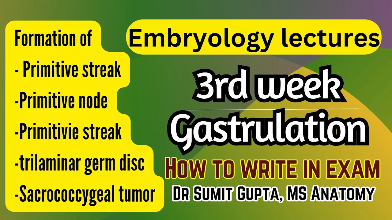


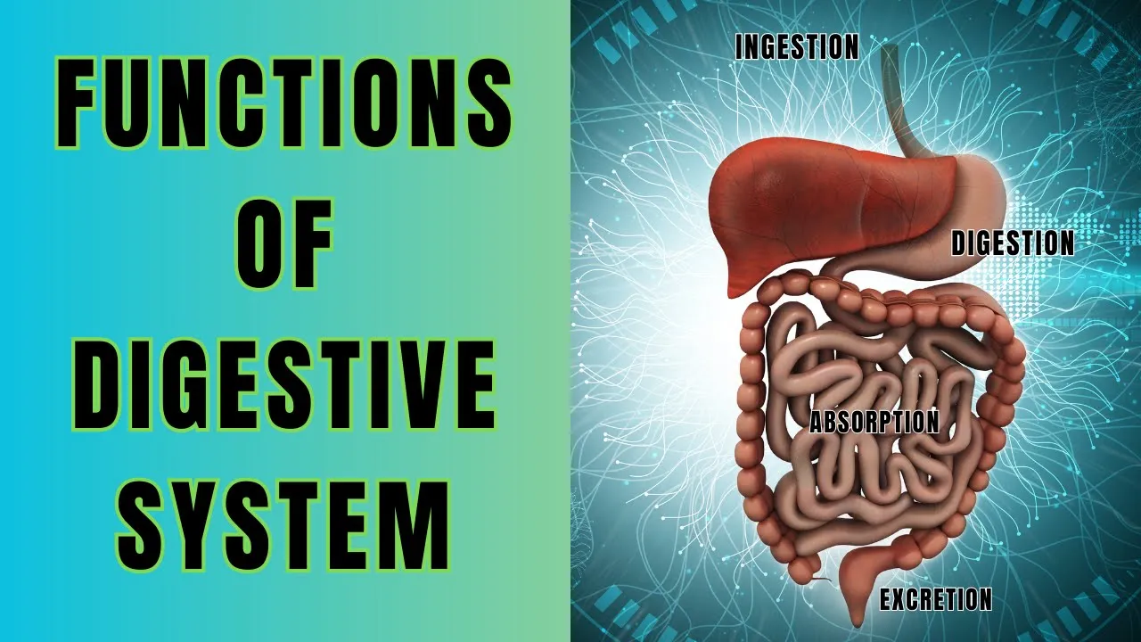
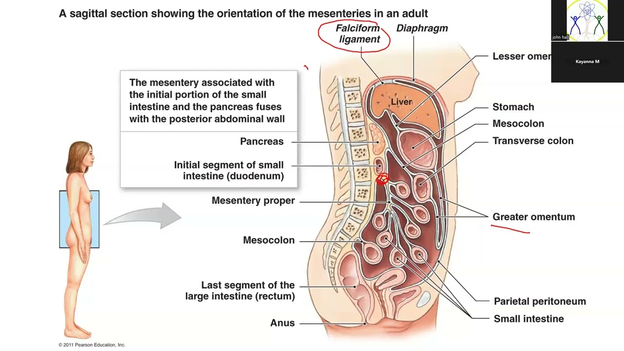

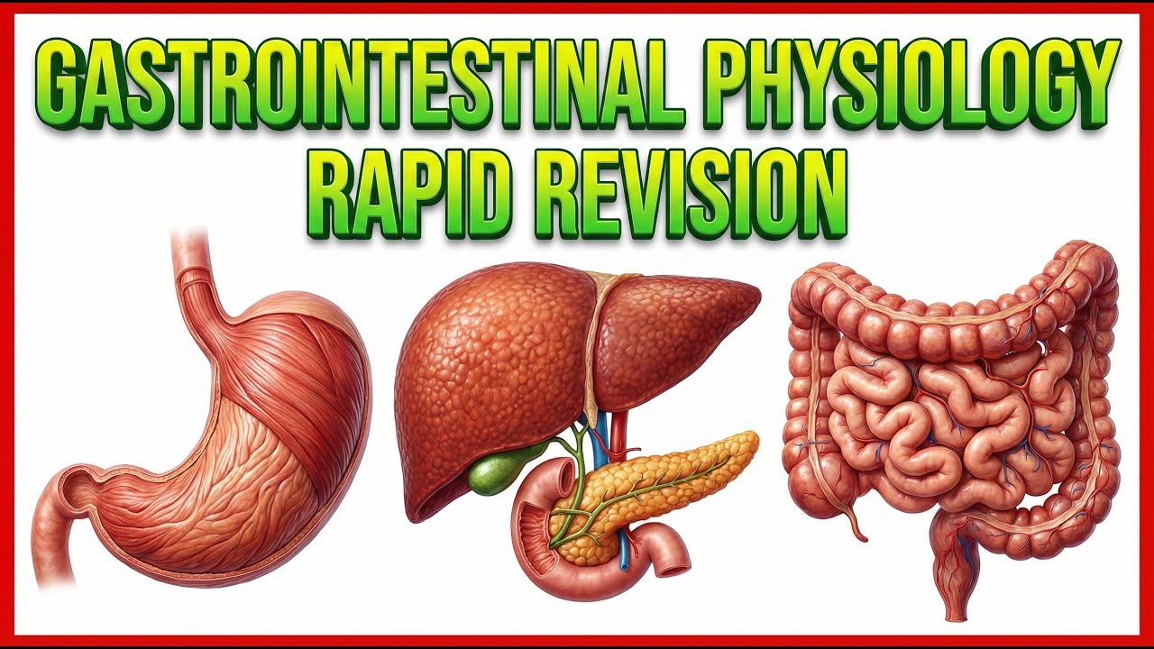





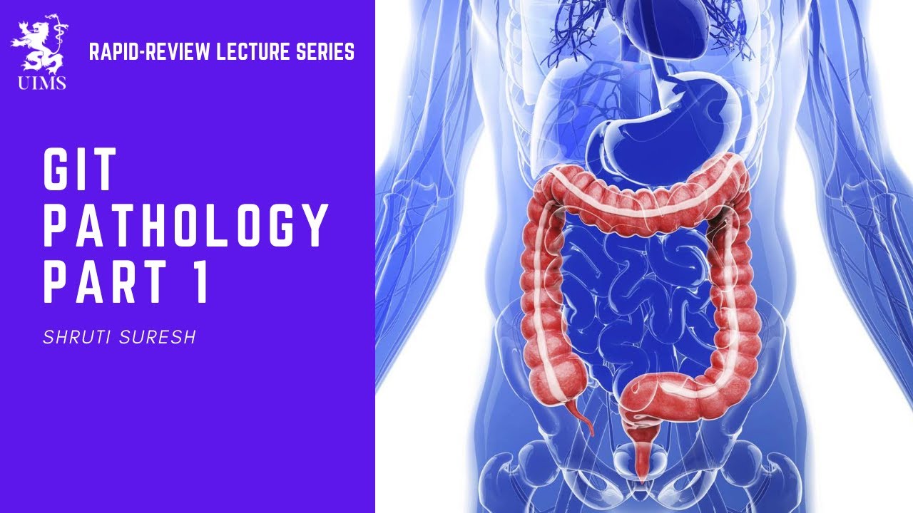






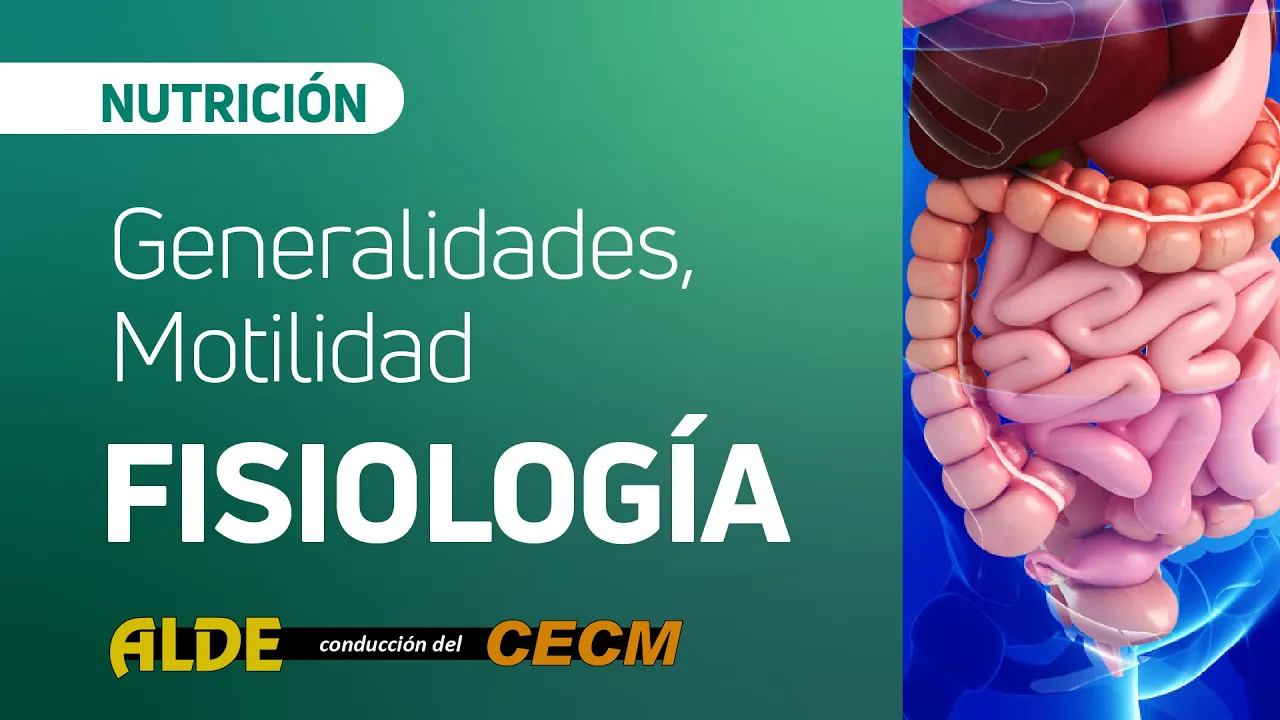
The gastrointestinal system is divided into three embryological regions: the foregut (esophagus, stomach, liver, gallbladder, pancreas, and proximal duodenum) supplied by the celiac trunk, the midgut (distal duodenum, jejunum, ileum, cecum, appendix, ascending colon, and 2/3 of transverse colon) supplied by the superior mesenteric artery, and the hindgut (last 1/3 of transverse colon, descending colon, sigmoid colon, and rectum) supplied by the inferior mesenteric artery. The gut tube develops from endoderm and lateral plate mesoderm, undergoes 270-degree rotation during embryonic development, and consists of four histological layers: mucosa (epithelium, lamina propria, muscularis mucosa), submucosa, muscularis externa (inner circular and outer longitudinal layers), and serosa/adventitia. The enteric nervous system includes the submucosal plexus (Meissner's plexus) controlling secretions and the myenteric plexus (Auerbach's plexus) controlling motility.
Anato-Bee 7: GI System Anatomy, Histology & Embryology (2025)
Added:hi everyone so just again if you could just keep yourselves muted as we go through this session that would be we would greatly appreciate it my name is Dr Hagerty I'm with Thomas Jefferson University and one of the co-founders of anabe I'm usually see Dr pasca introducing everyone tonight I will um be coming in and introducing this session so this is anatomy 7eventh tutoring session we're going to be looking at some GI and Anatomy histology and embryology and before we get started we like to do a couple of announcements so again I see we're still admitting people um please keep your camera and microphone off if you choose to not be recorded because we will post this on our YouTube channel um and we want to welcome you all back from Thanksgiving break so we hope you all had a wonderful holiday break you got some time to relax and we're very thankful to have you all here for us this uh with us on this Sunday um so again this is session number seven there are three more sessions after after this so you'll have two more sessions with our lovely medical students tutoring you and then you'll actually have a review session um with some of the uh faculty to go through an overall review before the competition our next session is going to be two weeks from now that's going to be session number 8 on reproductive and urinary system um and that'll be December 15th and then um just remember we're gearing up for the local competition so the competition itself is going to be on January 25th that's Saturday January AR 25th um and it is going to be based off of the local objectives so whenever you go in and look at our objectives online um and maybe I can I did have this up for everyone um so if we go into these can you guys see my screen okay so if you go online here's what the tutoring schedule looks like but you can go to our resources you can go to the learning objectives and when you click on any of these objectives I think I'm seeing the wrong screen I think I'm seeing a just like f files that you have all my files oh no okay well there we go I picked the wrong screen everybody I hope you got a good look so let's start again if you go to our website go to resources and our objectives and I know all of you are probably pretty familiar with this but just as a friendly reminder you click on one of the objectives it'll pull them up this is for hystology but remember we have anatomy hystology and embryology here you can see on the left column here those are the local objectives so these are the ones we we we will be testing you on in the local competition on um January 25th and that's Saturday um so basically the only other announcement I have is that if you have any friends that are interested in registering let them know last Daye register is going to be January January 13th at 12:00 a.m. eastern time so that's January 13th 12:00 a.m. eastern time um before we get started any other announcements all right amazing uh Dr Frank if you could kick it off for us okay welcome back everyone our first presenter this evening is uh kalista Mueller uh she's going to be presenting on the anatomy of of the digestive system uh kalista is from Nashville Tennessee and graduated from George Washington University start in 2023 with a major in public health and a minor in biology she is currently a second year at the Edward Bia College of osteopathic medicine and Auburn in her free time she enjoys going for walks spending time with friends and reading good books so far kalista's favorite part of anatomy lab has been learning nuro and cardiac Anatomy with her awesome nerve anatomy lab tablemates okay um I'm gonna share my screen and get the presentation going okay can everyone see that okay um uh like Dr Frank said my name's kalista and I'm really excited to talk to you guys today about um GI and digestive Anatomy so these are some of the learning objectives that we're going to cover today um we're going to talk just a little bit about the larynx and ferix we're going to focus most of our time talking about the for gut the mid gut the hind gut and we'll also talk about the major organs there and also their blood supplies and then we're going to talk about some of the accessory organs of the digestive system and then also the liver um let's see okay so to kick it off we're just going to talk basically about like what the overview of the digestive system does so um I kind of want you to think just like a thought exercise think back to the last thing you ate today and think about how those large pieces of food um are were broken down into nutrients that you know your body was able to process and use to function and move your body along so the digestive system is really amazing and it helps nourish our body with a constant supply of nutrients water electrolytes it's really important for the motion and movement of food through our body as well as things like absorption storage and even helping past waste um I think the best way to break down the digestive system is looking at both the elementary canal and then the accessory organs so your alementary Canal is where the actual are like the actual tupal structures that the food is going to be passing through so we have things like your esophagus and your stomach and the intestines whereas your accessory organs are going to be organs that are helping with digestive processes by releasing things like enzymes and um juices down your um food products but those don't directly have food pass through them um the major motion that we see for moving all this food along in the digestive system is something called peristalsis which is a slow wave contraction that's going to propel a food Bolis I like to think about it if you have a bit of toothpaste at the end of your toothpaste tube and you're squeezing it you're pushing out slight the muscles are Contracting at one point to move it a little bit along and then those muscles will relax and the next set of muscles kind of contract to move it along so it's like a wave of contraction that helps move the food um down through your Elementary canal and this is an involuntary motion um and then often your digestive system we talk about it like clinically in terms of the four quadrant system so up near your liver you have your right upper quadrant over here by your spleen you have your left upper quadrant and then you have your right lower quadrant and your left lower quadrant as well okay so this is kind of just a little table that we put together to talk about for gut mid gut and hind gut and in embryology I think they're going to talk a little bit more about where these terms kind of come from and how we get them but basically um what you need to know for the anatomy of it is for gut is the beginning part and then mid gut is the middle hind gut is the end so we're going to focus on the forut first which goes from your esophagus to the first part of your small intestine and the dadum um and then we'll talk a little bit more about what it does but main function here is we're thinking protein carbs and lipid breakdown and then also your forga includes some of your accessory organs like your gallbladder and liver that help with bile production okay so just like a quick overview of the laryn and fairx again someone will talk about this a little bit more in depth later but this is where your digestive and your respiratory systems intermingle so your fack has three portions there's this part up kind of in line with your nasal cavity that's called the naso ferx a part right below it called the orox and then your L lenio fenx um and the larynx is really important for connecting to your trachea and then the ferx um to help with air flow and to make sure so we want to make sure when we're swallowing that food doesn't go down the wrong pipe so we have this handy little flap of cartilage it's calls the epig glotus and it just covers up the trachea and it actually prevents any food from going down that way and directs the food instead down the esophagus okay so speaking of the esophagus we're going to start there um this is the first part uh contributing to your uh forut it starts at the distal end of your fings and it actually has some important anatomical things that we talk about quite often in lab and stuff so relationship wise it is posterior to both the larynx the trachea and also the heart so if you look at this image down here of the kadaver basically where you see this thoracic aorta if you follow that up and you just imagine the heart being right there it's actually in front of the esophagus um and the esophagus itself is basically just a large um circular muscle that uses peristalsis to move your food Bolis down into your stomach it has two sphincters there's both an upper esophageal sphincter right around here and then a lower esophageal sphincter closer to your stomach the upper esophageal and both of them just help prevent backflow of food and when those don't work you can get something like gird or gastric um reflux where you feel kind of that acidic burning in your mouth because the food is kind of came back up um and then some kind of a important anatomical relationship is we often talk about which level um different structures enter the abdominal cavity at so the esophagus enters the adominal cavity at the level of T10 and a fun little pneumonic to remember that is the I8 10 eggs at noon so I8 stands for the inferior vnea being at T8 the 10 eggs is we're talking about the T10 level for the esophagus and then at noon is for the aorta at T12 like I said we're focusing on the esophagus so that's going to be the 10x part um so yeah it's kind of fun little neonic and then the last part of the esophagus is the gastro esophageal Junction area and this is where you have that lower esophageal sphincter and that's what controls the food Bolis entry into your stomach okay so now we're in your stomach your stomach is part of your for gut and it has four main regions to it um the first right up near this uh lower esophageal sphincter area is the cardia the the cardia or the cardiac of the stomach next you have your fundus of the stomach then you have your body of the stomach and then your pyloris of the stomach which is the lower part down here by the pyloric sphincter the stomach also has two curvatures to it that are important because they have blood supplies that run along them so you have the Lesser curvature and the greater curvature um and the main function of your stomach is to store your food it's for motility and digestion so even like when food drops into your stomach there's a mo motion going on it pretty much all the time to help mix up that food so you're getting both a mechanical digestion with the mixing waves to create the Kim while you're also getting some chemical dig digestion by release of different gastric enzymes there's bunch of Bunches of specialized cells in your stomach that help with that and then also uh an important structural feature of your stomach to help with digestion is it has these sub mucosal folds that are called ruge um and these increase the surface area of your stomach and also help with the motion of digestion there and then at the bottom of our stomach as we transition into the first part of the small intestine the dadum we have another sphincter this one is called the pyloric sphincter and it just controls stomach emptying to make sure we don't have too much movement or too much at a time release into the stomach or into the small intestine okay and then kind of a little side from the our um Elementary Canal that we've been talking about we're now going to talk about an important accessory organ for the digestive tract which is the liver um so the liver's main job is it helps produce bile and bile helps emulsify fats which emulsifying the fats just means you're breaking down the fats into smaller globules that can more easily be digested by our body um because fats are kind of hard to digest on their own so the liver is really important it has a ton of blood supply that goes through it um and those vessels are huge when you see them like on a caver um and and you as well but that's kind of like from this picture down here you can see how big all three of those vessels are um and it acts like the body's filtration system so it's really important for removing toxins and substances um and it also helps absorb nutrients back into your bloodstream which is really important too um some bit about the anatomical relationship of the liver it's going to be right inferior to your diaphragm so if you were to palpate the liver you kind of go to the right upper quadrant and you can have the person breathe in and out and you can kind of feel the liver right there um and it's on the right side of your abdominal cavity so it's in your right upper quadrant and there's kind of two ways of breaking down the liver to look at it and study there's the four anatomical loes that we would like identify in lab these are things like the right lobe the left lobe this small Cotate lobe in the front and the quadrate lobe and then also there's a classification system called the conod I believe I'm saying that right classification system and that breaks the liver into functional loes which are important for you know clinicians especially like surgeons because each lobe of the liver actually has its own inflow outflow a vascular Supply and B bilary Supply so this is how you know they can remove a part of a person's liver and not have to take the entire liver out and you still have functioning liver left because they each have their own functional lobe um so moving on we're going to talk about the gall rather now which before you move on can we go back to that previous slide yes ma'am um can you just clarify what right upper quadrant means two things clarify that and then so that image on the left I really like it because I agree it shows all those big vessels going in but that's not how the liver sits in our body so if you could explain what the upper right upper quadrant um where that is and then um kind of explain a little bit how the the liver is oriented more so in that image on our right here versus how we're seeing it on the left yes okay so right upper quadrant is kind of what we were talking about earlier if you were to divide your stomach into four parts clinically you have the right side the left side and then you can split them into quadrants too so that's what the clinician is doing when they come to palpate your stomach to see where the pain is they'll palpate first in that right upper area and the main organ there is your liver um and then like Dr Hagerty was pointing out the liver isn't oriented in the way the image on the left shows it's on the right so if you see the image on the right if we imagine across the top right here you have the diaphragm and then you have the liver so if the liver is my fist the diaphragm sits right on top of it and then those um vessels kind of go through it like a straw almost goes through a cup and those then Pierce through the diaphragm was that good for clarification okay um so now we'll talk about the gallbladder and I think I mentioned this earlier but just to better clarify what um an accessory organ is is it's an organ that's really important for digestion and we couldn't do digestion without it but it doesn't actually have the food pass through it and you know help with like the actual mechanical digestion part with the food passing through it um so these just secrete digestive enzymes and the main function of the gallbladder is it's what holds um concentrates and releases your bile so I know it always looks green in pictures and people are like is it really green it's really green because your bile is green so it's kind of a fun fact um and it is going to be if we imagine our so this is our diaphragm this is our liver our gallbladder is going to sit just on the inferior surface of the liver down there and it's kind of like tucked up in it like a little it's pretty it's much tinier than the liver but it's tucked up in the bottom of the liver and it actually has three regions of it there's this area in the middle called the body you have the fundus at the top and then the neck which then inter goes and connects more towards the two ducks um so when you're eating something especially like a fatty meal your um gallbladder it's stimulated and hormones are secreted um to send stored bile from the gallad to go into the dadum to help with emulsification which is just that breakdown of the fats so they'll go they'll travel through the gallb but will get the stimulation to secrete bile the bile will go through the common hepatic duct into the common bile duct and then it will enter into the small intestine where it'll release um bile to help with that emulsification okay so now we talked about the um bio entering the small intestine we'll get into the small intestine so this is actually where we transition from being in the quote unquote for gut to the mid gut so the for gut is going to be at the it it ends at the first half of the dadum and then the mid gut is kind of the rest of the small intestine there into part of the large intestine and the main function with the small intestine is to just help finish up your digestion and absorption of nutrients and especially water absorption happens here as well um there's three parts of the small intestine you have your dadum which is the purple part up here the junam which is this greenish blue part and then the ilium which is this like pinkish red part so the dadum is an upper portion it actually runs right next to the liver if you can imagine the liver over here next to the stomach and um it wraps around the head of the pancreas which is another accessory organ we'll talk about in a little bit and there's the spot that we were just talking about where your bile enters into the duum to help with that digestion and the emulsification of fats that specific point is called the ampula of and that's where the pancreatic ducts and the common bile ducts come together they empty that um bile and other pancreatic juices into the um the dadum that way you can have the uh the mixing and the digestion multipication of fats um and then your Juna it's a middle portion it's mostly located in your left lower quadrant or left upper quadrant sorry and it also just helps finish digestion and absorption and then your ilum is actually the longest portion and it is what terminates into your seeum which becomes your large intestine um and then kind of just throughout all of your small intestine you have circular folds vill and microvilli that help increase absorption in surface area that way your small intestine is able to absorb all those nutrients in water okay so speaking of the pancreas and the deum wrapping around the pancreas we're just going to talk a little bit about the pancreas which is also an accessory organ and its main function is to produce the these things called pancreatic juices which are just fluid concentrated with digestive enzymes there's a bunch of different kinds um and you can get more into the specific weeds of that but the main portion you need to understand is that they empty into the dadum with the bile from the gallbladder and they help break down nutrients um and so the dadna or the pancreas has an in it kind of fits right along the dadum if you imagine the top of the dadum right there it kind of wraps right around the head and then the tail goes all the way out and kind of tapers right by the spleen um so it kind of stretches across both your right upper quadrant and your left upper quadrant and the pancreas the pancreas is a unique organ because it has both exocrine and endocrine function so the exocrine function is the secreting of the digestive enzymes and the endocrine is releasing hormones into the blood um which I think you guys will talk about endocrine stuff more in the future um and just down here I kind of outlined the pancreas portions on this cadaver image but your head is the part that's nestled into the dadum the tail is the part by the spleen over here there's the unate process which is also kind of closer to the dadum stomach area and then your body is what extends between okay so I have some whole questions the first one is um when you're talking about the GI track what part is the most important for nutrient breakdown in bile production okay I think I'm seeing a lot of answers and most of you guys are right it is the forut a um so that's really important because the forut is right where we said that your gallbladder is a part of that's producing your bile as well as part of your small intestine like the dadum that's uh all part of your for gut so good job there and then for the second question um um we have a patient presenting with reflux symptoms after they eat a big meal they kind of get heartburn and acidic taste in their mouth what sphincter do you think could be causing that problem okay I think most people have answered it now but great job again seeing most people um picked be the lower esophageal sphincter and like we said that makes a lot of sense since the lower esophageal sphincter is what opens up and is going to allow the stomach or the contents from your stomach to go into your esophagus um so that would definitely contribute to that kind of heartburn uh acid taste in the patient's mouth to be you move on it's it's Dr Peterson maybe just for those that don't understand what a sphincter is could you just describe it to them so they have a better understanding uh yes oops sorry um a sphincter is basically just a closure around it's a muscular kind of closure around part of the organs like in the esophagus and it opens and closes at certain times to allow things to flow through it I guess not things but food bis and so your sphincter and it's normally controlled by like kind of neural control or you know pressure and stuff like that and so the lower esophageal sphincter will open um and allow stuff to go down through into the stomach but if it stays open for too long and some of the stomach fluid goes pushes back up into the esophagus and then it closes that fluid has nowhere else to go back down so you'll get reflx I don't know if anyone else has something to add there to make it a little more clear good okay okay so now that we've kind of tackled the for gut we're going to move on to the mid gut and the hind gut so the mid gut is basically going to run from the distal part of your dadum all the way until the first two3 of your transverse colon whereas your hind gut is going to run from your transverse colon all the way down to your anus they both have different blood supplies we'll talk about those a little bit later and the function is kind of very similar to the forut because all these things are absorption but we have less actual breakdown going on here and more just absorption so in the midcut we're going to get absorption of nutrients like your vitamins your minerals and then in the hind gut it's going to be absorption of water and electrolytes and then also that process of storing and compacting for Waste okay um and so for the large intestine I thought it would be easier to break it down via chart so your large intestine is largely made up of the colon the rectum and the anus your colon is runs from your seeum all the way to your sigmoid colon and I have a picture of that later that'll show it a little bit more clear um and your colon is where all your gut flora and microbiomes are which is pretty cool it's important for Waste storage and it's kind of the last place that we get any sort of absorption um by the time you're moving into your rectum we're more talking about storing the waste in the fecal matter and you're not really absorbing anything anymore um and then your anus is mostly important for waste removal and your voluntary bowel emptying and you also have some sphincter there as well kind of like we talked about we talked about with your esophagus to help control your voluntary movement of a stool um oh okay so now we're going to talk about the specific regions of the large intestine so I kind of outlined them over here in a color so hopefully that kind of allows you guys to follow along with me as we talk about them but um like we said the main function of your large intestine is to absorb water and electrolytes and then also for storage of waste prior to going to the bathroom um so your four regions here are you have uh the a you have your that you start out with here then you move into your a ing colon you have a place called The hepatic flexure which hepatic is just another word for liver so it's right by your liver it's the corner and then you have the transverse colon so this is all mid gut and then about 2third into the transverse col we switch to the hind gut then we have this area up here which is called your splenic flexure which just means your it's by your spleen so you have that curvature then we move into your descending colon you have your sigmoid colon which is pretty uniquely looking um it kind of looks like an S if you look at it and then you move into your rectum right there so the large intestine doesn't have circular folds or bli like the small intestine um but it does have lots of mucus that helps with moving the waste products along and instead it has these kind of unique anatomical things you can see on it called HRA and tenia coli so the HRA are like tiny little out patchings of the colon so you imagine kind of like small little kind of like grape like out pouch hangings of it that's kind of what I think of when I think of a hastra and then the ten coli are just bands of smooth muscle um that go around the length of the colon and those are both just important for helping with movement as well and then now we're going to move on to talking about blood supply so for blood supply to the for gut mid gut and hind gut they're all coming off of branches of the aorta so I have this picture here and for the for gut I want you to think of the Celiac trunk that's kind of the main branch that we're going to talk about and then some surrounding branches for the mid gut I want you to think about the superior mesenteric artery and then for the hind gut we're going to think about the inferior mesenteric artery so I'll get a little bit into that um and so for the forut like I said our main blood supply is going to be the Celiac trunk and then off of the Celiac trunk as you can see over here we have three branches we have the splenic artery which runs kind of right along the pancreas the body of the pancreas down into the spleen and it um and it also has two branches and the splenic artery is going to supply your pancreas which makes sense because it's running right along the body your stomach it's got the posterior fundus and the left greater curvature and then your oops sorry I clicked that too early um your left gastric artery that's going to kind of run right along the area near the stomach there and it's going to supply your esophagus and your stomach along the Lesser curvature which if you guys remember that's kind of that uh C the semi semicircle I guess kind of round moon shape along the top of your stomach and the greater curvature is that one along the bottom of your stomach um and then your common hepatic artery is going to go to the right side over towards your liver and your gallbladder and so it's going to help Supply your liver gallbladder the right side of the Lesser and greater curvature of the stomach and then also the dadum and the head of the pancreas are over that away um so these small individual branches just kind of are named after the things that they're supplying so we have those branch are going to supply those areas so for example gastroduodenal is going to help Supply the um G the do oh c it's gonna help Supply the dadum and um also the Celiac trunk is the very first Branch off of the abdominal aorta okay so now moving into blood supply for the midgut um we're going to be thinking the superior mesenteric artery which is number eight over here on this image um and so it's going to have a bunch of branches the main ones um I think are important are you have your middle kolic artery which is going right here with number six and it's going right towards if you look at it the ascending col or the transver transverse and kind of this uh uh oh my God hepatic flexure area so that's what your middle colic artery is going to supply and then if you look right here at number four that's going to be your right colic artery and that's heading right towards your ascending colon so it's going to supply your ascending colon and then down here number 10 we have your aloc colic artery it's heading down towards that areum between your alium and your colon so it's going to app Supply your ilum your seeum and also there's an appendicular artery that runs to your appendix um and then you also have this inferior pancreatico doal artery which goes and um helps cover the pancreas and the dadum and forms an anastomosis with one of those arteries from the for gut that we had talked about and then you also have intestinal arteries so these are just branches that help go to both your junam and your ilium and help uh give them blood supply and then an important thing for the arteries when we're talking about the SMA and the inferior mesenteric artery is they help GP this uh Branch uh cardl marginal artery of druman and this creates an anastomosis to cover an area that doesn't get a lot of blood supply so if you look up here in the left area of the screen that's where your marginal artery is going to cover because your IMA covers all these branches down here but this area of your stomach it almost kind of creates an area that doesn't have enough blood supply so that's what that marginal artery of drumond helps cover helps cover um okay and so now when we're getting into the hind gut like we said the blood supply here we're thinking inferior mesenteric artery which is this one labeled IMA over here on the image and this does give branches that help uh form the marginal artery of drumond which kind of helps form that an asmosis to give that left upper area of your um poon blood supply and then it also has its own branches so there's the left cholic artery which is this LCA here that supplies is the descending colon then you have your sigmoid artery which is going right down here towards your sigmoid colon so that's going to supply part of the descending and the sigmoid colon and then you have the superior rectal artery which is going right down here and that is going to supply the proximal area of your rectum and again um IMA is also a branch of the abdominal aorta okay so I have a couple more questions to help nail in that blood supply because I know it can be confusing but starting with the first one clista can you read off that first question oh yes sorry brain fart there um so for this question we're talking about what two arteries help Supply the anastomosis that covers that marginal artery of drumond I'm trying to remember too if we've covered anastomosis in our previous sessions um but if we haven't and you guys were confused about that it just means that there's a communication between um two arteries uh creating uh some possibility for blood to come from multiple sources yeah sorry I should have talked about explain that a little bit better but to me anastomosis make the most sense when you think about there's an anastomosis right by your elbow and then also your knee and it's really helpful for when like your arm is bending if certain arteries may get compressed by that motion and so others can help fill its place so you still get blood flow okay so it looks like um a couple of us got that yes it's going to be our Superior mesenteric artery or inferior mesenteric artery there were some people that were thinking it was the Celiac artery um but let's just back up and look at that picture maybe one more time that first picture uh here sorry I have so many transitions that's making it go a little slower okay so your okay this one so your Celiac trunk uh if we remember that's the first Branch coming off of the abdominal aorta and then your Superior mesenteric is going to come off down here and the inferior mesenteric down here so Celiac trunk like we said is kind of doing more of this kind of area by your stomach your duad num we're not really reaching your Colton with the Celiac artery or Celiac trunk whereas your uh Superior mesenteric artery and your inferior mesenteric artery are the superior is this one up here a and then your inferior would be down here those are those two big arteries that are supplying that anastomosis um so if anyone has questions about that you can throw it in the chat and we'll try to answer and then we can move on to the second question which is kind of throwing back to some of the uh accessory or we talked about so a mass on the blank of the pancreas would cause compression of the dadum and then we just have all four sections of the pancreas all right and for the sake of the time I'll go ahead and close the poll in five four 3 2 1 sorry I kind of clicked the answer there at the end so some of you guys may seen but it's going to be your head of the pancreas so just for the sake of not having to back up through my entire presentation and try to see if I can go really quick uh just to show you that picture of the pancreas again but if we have a mass at so we're talking about a mass on the pancreas that's causing compression of the dadum I went too far of the dadum so if we look at our pancreas right here the part that's right next to our dadum is going to be the head of the pancreas so a mass on the head of the pancreas can actually sometimes present patients will have you know stomach pain sometimes they can have jaundice um problems with bioc concentration and stuff that because you're pressing right on that dadum and also that area that ampula of water um so yeah that's how that can present any questions I think that was the last side besides the references y great job kalista and thank you for that while people are thinking of questions um to either put out there or put in the chat one thing I wanted to put out there about thinking through how should you approach the arteries and how should you study that is uh don't think too hard about it uh some of the names can tell you where they're going like uh right uh colic for example is telling you that it's on the right side of the colon so if you know that the a sending is on the right side that's telling you um you know what what side that that's going to be on so the names can be helpful the other thing as far as um what they are supplying there's a saying that we have an anatomy is if it's nearby it will supply so um don't worry too much about sitting and memorizing everything that's being supplied I think it's better to just understand approximately where the arteries are and then you can sort of work them them out for there so I wouldn't sit and memorize um you know that that'd be way too much for you guys to sit down and do I see Sammy has a question for kalista um hey yes um two things uh to what you just said um I have a question so obviously I have a question but for if you do understand it can you still memorize or would just would that not be the smart idea to do so you can still memorize I don't think it's the the best tactic um moving forward I Know It Gets You Through high school and probably through some of your undergraduate classes but um I'll say working through if you can understand it I think that that's that would show that you've you've done that so um you you've made it through to that kind of proficiency and understanding so um sometimes you got to memorize to get to understanding so um but if you're to the understanding point then you're in a good place okay and my other question uh since I'm so big on cardio um I just recently learned about the um IMA and SMA and what so what problems would have to occur if because I know it's maybe I think it's AAA surgery that it happens with but what problems would it just be like a a bleed in there or um would it just be a bleeding them that would cause the need for surgery or just what um dealing with what have you had in uh in what have you learned about in medical school so far that you can kind of so when you're talking like about like a abdominal aortic aneurysm that's more what we learned as like a tear in the uh aorta so that isn't necessar a problem with your SMA or your IMA that's kind of the entire aorta tearing but some problems that could come from having like a blockage of blood flow with your like SMA or IMA is you could get a part of like uh we talked about something in class called like a um oh my gosh what's it called when the blood isn't flowing there uh iner like you can kind of get an infarction of parts of your bowel if the blood supply is cut off there and things that can cause that are uh it varies there can be kind of twisting of the gut in the wrong way like a volvulus or an interception or you can have just like a blockage um something like a mass growing that kind of help yes ma'am thank you okay I think that will finish our anatomy discussion uh we will move on to histology uh and we're looking especially at the hology of accessory organs of digestion as well as the GI track and uh presenting for us this evening is Hannah bash Hannah is from West Hartford Connecticut and graduated from Washington University in St Louis and 2022 with a baa in cognitive neuroscience and minors in Spanish and biology she is currently a second-year medical student at the Cindy Kimmel Medical College at Thomas Jefferson University with an interest in surgery Hannah enjoys running cooking spending time with friends and her dogs so with that Hannah is all yours sorry can you hear me okay perfect hi everyone once again I'm Hannah really excited to be here and be able to help with the hystology session for the GI system so we'll just go ahead and get started hey Hannah I'm sorry to interrupt really quick um do you mind changing your presentation mode I think we're seeing the presentation view is that better perfect thank you okay perfect okay awesome so our lecture objectives for today we're just going to describe and identify the four histological layers of the gut tube and then we'll describe the components of the enteric nervous system and explain what the what the function is of that nervous system and be able to see them on our histology slides so just to start off a really broad overview of the gut tube and what it is so the gut tube is part of our developing embryo which I'm sure Kyle will go much more into in his embryology presentation on the GI system um but it's developing in our embryo and it forms part of our digestive system and in our gut tube we have four layers A mucosa a sub mucosa the muscularis and then a Sosa or an adventia layer depending on where we are in our digestive system and we can see these layers in many organs such as the esophagus the stomach um the small intestine and many other organs in our digestive system in my presentation specifically I'll be showing you guys histology slides of the dadum just to keep it continuous throughout the presentation so that you can see this same slide showing all the different layers of the gut tube um but just keep in mind as I go through the presentation that these layers do exist in other organs not just the dadum of the small intestine um so just keep that in mind it's in all the organs of the digestive system so to start off we have the mucosa which is the innermost layer of our gut tube and if you look at the diagram on the right um our mucosa is what I highlighted in blue so that inner layer that surrounds the Lumen which is the opening of the gut to tube and that's where food particles and stuff like that would pass through and the mucosa is made of three layers so we have an epithelium which is the innermost layer that would be surrounding the Lumen and then a lamina propria which is made up of loose connective tissue and that's where we find blood vessels as well and then we have muscularis mucosa which is a smooth muscle layer um and that would be the deepest layer ofosa so to look at it a little bit on histology we'll do zoomed out pictures and then some more zoomed in pictures so you guys can see but we'll be looking at the dadum here and where my yellow line is highlighting is the mucosa layer itself and then if you look on the right a little bit further in we can get a more zoomed in image of what the mucosa would look like um so the Lumen would be all of the white surrounding this and then our mucosa is that big yellow line and we'll go to another slide so that we can see the differentiation of the layers okay perfect um so starting off we have the epithelium and that would be all around and this is for the example of the dadum a simple columnar epithelium so really simple columnar is just those tall skinny cells um and it's simple meaning just one layer versus stratified you could have multiple layers of these cells but simple we just have one layer usually they should be aligned very nicely as you can see in the more zoomed in image they should line up pretty close to each other and parallel and then we have our lamin propria which is on the one layer further in and this is made up of our loose connective tissue and this we can have blood vessels here as well so we can see some capillaries um and that will provide blood supply to the epithelial layer and then we have our muscularis mucosa further down and this would be our smooth muscle layer so a little bit smaller as you can see the lemon appropria is probably the biggest layer that you see and the epithelium can change too depending on where you are in the GI system this is just an example for the dadum but you can have different types of epithelium It won't always be simple columnar so just keep that in mind as well so Hannah it's Dr Peterson before you move on because I think this is a really nice slide um so kalista mentioned how the gut tube has increased surface area for mostly absorption and she talked about those folds and you can see a really nice picture here could you just maybe use your cursor and show everybody what we mean when we talk about the Villi those finger like projections that would be awesome of course yeah thank you yeah so here we are um as kalista said it's really important to have absorption in our GI system so these nice finger like projections if I just move my cursor around you can see that is a Villi and then we have the nice fold that it creates and then it continues on the other side so around if maybe if we go back you'll be able to see you can see all around multiple Villi creating these folds all around okay and then the sub mucosa is our layer just beneath the mucosa and this is composed of dense irregular connective tissue and then also blood vessels lymphatic lymphatic or lymph vessels and nerves and then a big part of the sub mucosa is glands depending on where we are in the digestive system the glands will have different names um but usually if you see glands that's a really big indication that you're in the sub mucosa so that's the best giveaway on histology to know where you what layer you're in so if we look at that a little bit further same picture we're just moving further down in our layers so underneath our mucosa layer will be the sub mucosa extending by that yellow line and if we zoom in more you can see the glands that I was talking about so these glands should glue you into the fact that you're in the sub mucosa so always keep that in mind if you see glands and then on the bottom here we can see our dense connective tissue if you can compare it to the loose connective tissue we saw in the lamin propria this dense connective tissue is usually more pink because there's more collagen there um lot looks a lot more put together than our loose connective tissue which is more like whirly kind of pillowy versus this is a lot more um organized in its nature and then if we go just a little bit further specifically to the dadum we would call these bruners glands um but that name is specific just to the dadum so if you see gland somewhere else it might not be called GL runers gland so just keep that in mind U but glands are always the giveaway of being in the submucosal layer and then as we move further down that dense irregular connective tissue layer um and just an example of some dense irregular connective tissue on the side over here and then just a quick practice question to make sure that we're keeping in all of our layers in check so this asks which layer of the gut tube is the innermost layer and then is further subdivided into three layers all right you guys are doing so great with participation on this one I'll close the poll in 5 4 3 2 1 awesome okay looks like most of the participants selected answer a which is the correct Choice the mucosa so our mucosa layer if we recall is the innermost layer surrounding the Lumen of the gut tube which is that opening of the gut tube and then it is further subdivided into three layers which are the epithelium the lamino propria and then the muscularis mucosa layer which is is our muscle layer so great job everyone and then as we go further we'll go into some of those other layers as well and then there's one more question um what layer of the gut tube would this be where my yellow line is and then we'll do the bonus question after and you guys could just type in the chat um if you have an answer okay awesome I see some people putting in yeah the sub mucosa layer so as I said before usually the glands are the dead giveaway that you're in the sub mucosa so always keep that in mind it's the easiest way to identify this layer on histology and then for a bonus question if anyone knows what these glands are called oh I see someone put it in already yeah the Bruner's glands yeah exactly so we're in the dadum here so these would be Bruner's glands great job that's a you guys are really smart perfect okay and then we'll move on to our next layer of the gut tube which is the muscularis layer um so this is composed of two muscle layers we have an inner circular layer and then an outer longitudinal layer really important here not to get confused between the muscularis mucosa which we saw in the mucosal layer um as one of those subdivisions versus this is just a muscularis layer some people call the muscularis externa um so just remembering not to get those layers confused used our muscularis mucosa here is within our mucosal layer versus this itself is a muscularis external layer and it has two um submuscular layers our circular and our longitudinal so inside our dadum we can see as well here just moving further down in our picture nice division between our two muscular layers here um so over here we have our circular layer and then our longitudinal layer on the outside so as we go further to a zoomed in picture we can just look at these again this is our inner circular layer um so in cross-section it would look like this picture on the left so our smooth muscle cells kind of bunched up versus our longitudinal section would look much more parallel which is how you see it in the picture here looks like a bunch of parallel lines with the cells um is usually the easiest way to identify that that's our outer longitudinal layer and then there our last layer of the gut tube is sosa and adventia this one gets a little tricky because depending on where you are in our GI system some organs will have Sosa depending on the part of the organ it will be Sosa versus adventitious so that can get a little tricky but to keep it as simplified as possible it's our outermost layer or most superficial layer of the gut tube so all the way out here and we Sosa is just a single layer of squamous epithelium found on our intra paratenial organs and then adventia is is usually a dense layer of collagen found on retrop peronal organs such as the esophagus so retrop peronal being organs that are more enclosed and behind other structures and then a nice little pneumonic um Sosa slides versus adventia adheres usually collagen you can think that that's adhering to something um so adventia adheres you'll see a collagen layer versus Sosa just our single also you could use SNS Sosa single layer of squamous epithelium um to remember that if you see just a single layer of squamous epithelium that would be Sosa versus adventia is when you see all of the collagen so we'll look at that in the next slide so back to our trusty dadum for the last layer that we see here is the adventia um lots of Pinks you would notice that this is all of the collagen making this advantia instead of Sosa if it were Sosa we would just see one line at the bottom of a nice squamous epithelia um but here we see lots of collagen so that would indicate that it's adventia and then just a quick practice question to make sure we're identifying the layers correctly um to know which one represents Sosa or adventia here all righty I'll close the poll in five four 3 two one okay awesome so this one definitely tripped people up a little bit more D is actually where our um adventia is over here this our outermost layer C is actually our muscularis externa so where we have the two layers an inner circular layer and then an outer longitudinal layer it can be confusing because they somewhat look similar but in our adventia with the collagen you won't notice cells as much so here in our muscularis layer we see all of these dots which are the nuclei and the cells um of the muscle so that can kind of clue you into the fact that you're in the muscularis external layer versus out here in our adventia this is just collagen and it's Dr Peterson again maybe with your cursor in the adventia you could just show van those rounded in cross-section blood vessels we don't have to know whether they're arteries or veins but that's another easy way to get a sense that you are in the Advent tissue when you see those larger blood vessels awesome yeah so if you guys can see my cursor right here we have some blood vessels definitely too far out to tell if they're veins arteries capillaries um but blood vessels can also be a nice clue to let you know that this is the adventitious so all of these right here different blood vessels that we have awesome and then to go into the anic nervous system just to round out the hystology of the GI system so our anic nervous system is uh the nervous system of our GI system so it has its own nervous system that will regulate the passage and absorption of food and it's further broken down into two plexuses as we would call the submucosal plexus which can also be called the misner plexus and then the mayic plexus so maybe based on the name you can guess where our first one is going to be um but as we go our misers and sub mucoso plexus however you want to call it um will be found in our mucosal layer so once again that's our second layer of the gut tube and this will control secretions so that's a really important function of this layer so controlling secretions and blood flow and then our myeric or also called the hour boox plexus so you know when you see it written either way this is found in our muscularis layer so that muscularis external layer where we have usually an inner circular layer and then an outer longitudinal layer this can be found in in between those muscular layers so as you see right here this nice nerve plexus coming in between our inner circular layer and then the outer longitudinal layer very nice nerve plexus there and this plexus is really important in controlling our smooth muscle contractions that will allow for motility of food in the gut tube so every time we have a smooth muscle contraction in the gut tube this will push the food Bolis further along in the process and then allow that food to move down throughout the GI system and then just closer up in this picture you can see our inner circular layer at the top the outer longit longitudinal layer in the bottom and then the nerve plexus running right between them so that would be our myenteric plexus or our boox plexus however you like to call it and then just a quick practice question about our plexuses e we'll go ahead and close this pole in five 4 3 2 1 okay awesome um so the right answer is a which you see about almost half of you got so our misner's plexus which can also be called the submucosal plexus to give it away um is found in our submucosa layer and it will regulate secretions and blood flow for the other answer choices I know I put some confusing ones in there um found within the muscular externa would be our myeric plexus or the hour boox plexus and then that will regulate motility so that would be answer B and then just switching up the layer on u c would be partially correct because our sub mucose plexus does regulate secretions in blood flow but it's not found in the mucosa layer so it would be in the sub mucosa layer and then D also partially correct this is found in the submucosal layer but it doesn't regulate motility that is the function of the myenteric plexus so just some quick answer choices there for you guys and then there's one more hystology practice question um so what plexus do we see in the image here so there's a more zoomed out picture on the left and then closer up on our right right side and you can just type in the chat sorry trying to get back to the chat if anyone has a guess and we labeled the layers to give a little bit more of a hint there on where we are looking at okay I see a few answers in the chat so I'll just jump in I see that people are writing sub mucoso plexus which is a good guess but unfortunately we're actually looking at the myeric plexus um so the adventia here on the outside is labeled to kind of help you guys the layer after the adventia closest into our gut tube is our muscularis external layer so we can see right here we have an outer longitudinal muscular layer and then closer up here our circular our inner circular muscular layer so in between our muscular external layers we would see a myenteric plexus which is what we're looking at our submucosal plexus would be closer up in this region where we have the mucosa on the outside is right here so that's um surrounding the Lumen closest and then underneath our mucosa would be the sub mucosa and if we saw a plexus in here it would be the sub mucosal or misner plexus they're actually usually very hard to find in histology images you mostly only see myenteric or hour boach plexuses um if that helps you in any way but usually it is really hard to find a submucosal plexus on Imaging so if you one it's probably a mayic um but make sure you're looking out the outside to see if you see Sosa or adventia that usually Clues you into the fact that you're in the muscularis external layer where you see that plexus and then that would be the myenteric plexus and then just references and any questions at all I know hystology can be really hard you guys are really amazing for doing this because I didn't even touch histology in high school had no idea what that was I don't even think we talked about it in college really until I got to med school so this is amazing that you guys are getting such a head start on it you'll be very prepared later on I was just goingon to say thank you Hannah for making me fall in love with histology again I think that was wonderful thank you okay I don't see any further questions so we will move on to our final presenter this evening um which is Kyle Yang uh Kyle is going to be covering embryology of the gastrointestinal system so a little bit about Kyle he is currently a seconde medical student at Drexel University College of Medicine he attended UCLA for undergraduate and majored in Biochemistry in his free time he enjoys watching movies playing games and sleeping he also enjoys traveling whenever he gets a longer break from school his specialty of interest is emergency medicine but he's open to exploring other Specialties as well so feel free to reach out if you have any questions with that Kyle the floor is yours all right good evening everybody uh thank you for being with me here today sharing my screen and slideshow see annotate and this see if I can do this here all right uh oops sorry give me one second screen's being weird not okay I'm just going to go here for now okay um move this off to the side here okay so I will be covering uh GI embryology today and for our lecture objectives uh we are going to be listing the organs that are derived from the for gut the mid gut and the high gut portions of the gut tube uh plus um a couple of extra things here and there along the way okay so just a quick review from last week embryology um so uh during week two after fertilization we have our B laminer disc which starts out with an epiblast and a hypoblast with a primitive streak and then by week three we have a traminer disc formed with the ectoderm the mism and the endoderm and this will be between weeks three and four and then folding really happens in week four and this is where we're really going to pick up our story um with g embryology and so between week 3 and week four Once the folding starts you have a longitudinal view here and then let's just imagine you sort of take a cross-section view of this longitudinal view right down the middle you would see something like this and so at week four you have your gut tube which is made primarily of endoderm and then you have the spanic layer of your gut tube which is primarily made of lateral plate mism and then uh in general your endoderm of your gut tube is responsible for the glands and the epithelium that we just covered in histology and then your uh plate mism your lateral plate mism is responsible for the smooth muscle and the connective tissue layers of your gut tube and with that quick review we just want to do one quick po question our first one of the day for embryology what germ layers does the gut tube derive from so we have ectoderm endoderm mism A and B and B and C oops sorry well uh I kind of gave away the answer so thank you Mary I'm just going to move on here for the time being so what germ layers does the gut tube derive from um those who picked B and C were correct so looking back at the image I showed on the first slide we sort of have the gut tube and which is sort of made up of the endoderm and the lateral plate musm combined um they will give you the four layers of your intestines as mentioned um in histology okay good job everyone okay moving on here so before we start thinking about GI embryo um just sort of giving you a quick sort of really simplified view of it you're just thinking about a tube that extends from your mouth to your rectum and that tube is mainly divided up into three parts you have your for gut your mid gut and your hind gut and then there are two really sort of major landmarks we use to distinguish where does the for gut end versus where the mid gut begins or where the mid gut ends and where the hind gut begins and that would be the major duano Pilla and the about 2/3 of the way through the transverse colon and we'll get to those in the further slides okay so focusing up a little bit more on the Primitive gut tube here uh we want to realize that Reg regionalization really begins at week five um around this point your gut tube is suspended from the dorsal body wall and so dorsal is sort of the backs side so the back side of the body wall you got tub is suspended uh from the back side um with a suspension membrane which will eventually develop into the major blood vessels and nerves um that will supply um sort of your GI system and then cell proliferation begins between weeks 5 through 8 uh which leads to a gut tube being too long and it's kind of shown here but we have a better depiction of it in the future cides uh but it ends up kind of being a little bit too long to fit inside the embryo's body and it sort of grows into the umbilical cord a little bit before sort of retracting back okay um and so more on this gut tube rotation so we have a picture here for you so in picture a we really see that the gut tube begins uh elongating cell proliferation happens and it sort of elongates into the umbilical core just imagine that right around here is the cut off where the umbilical cord is and the tube is actively growing into the umbilical cord in picture B1 here um this really cool thing happens where it actually rotates uh the cord actually rotates right because when we sort of look at um your GI system as a whole um it's sort of all curvy and it has all these Curves in it and it really gets that right here in uh embryology when it starts rotating and so B1 um it herniates into the umbilical cord and it rotates 90° um in B2 you're really thinking the inferior aspect of the tube really grows and becomes coiled and it's depicted here where it starts coiling and then in C um you see or yeah in C it sort of retracts from the umbilical cord and it sort of rotates another 180° eventually creating something like you see on the right here which um you know is more indicative of what you would see um in a sort of infant or as you grow up um what your digestive system would look like okay and then so the coil part of the gut tube is really centrally located and the straighter and larger tube surrounds the central intestines as the colon by week 12 post fertilization and there is a short GIF here that gif I'll repr pronounce it that I would like to show you all and so your fetus you're taking a cross-section here um you see your gut tube it really herniates into the umbilical cord and it starts rotating and it will rotate rotate rotate and then once it has rotated it starts retracting back into the embryo okay to form the GI system that um that is that you normally think of when you think of a fully developed GI system awesome slides show here okay moving on to the next slide we're have our second poll question here uh how many degrees does the elongated elongated gut tube rotate in total so we have 90 270 360 or 180 he we're still getting some answers in um I'll move on at 45 seconds here okay 45 seconds and it looks like the majority of you guys picked uh B which is correct it's 270 degrees so how do we get that number so like I as I was mentioning in the earlier picture here you have a 90° rotation in picture B1 so it's 90° when it is in the umbilical cord and then in picture C once it retracts it rotates another further additional 180° and so we get 180 Plus 90 you get a total rotation of 270° in um complete okay uh moving on to focus a little bit more on the for gut here I'm sure it was mentioned before already um but your main structures that come from your for gun are your esophagus esophagus sorry um depicted by the structure labeled one your stomach labeled three your liver with four pancreas with seven your proximal dandom of your small intestine uh with six uh and this is sort of everything before the major duano Pilla which is that Landmark that I mentioned earlier that separates the for gut from the mid gut okay uh moving a little bit moving on a little bit to the development of the for gut structures um I want to focus on sort of the bile duct uh which was mentioned earlier in anatomy um it sort of connects to your liver um and your pancreas and your gallbladder um and so your bile duct sort of grows from the Primitive gut tube right here right at the location of the major duano Pilla your gallbladder your liver and the vental part of your pancreas will develop from the bile duct meanwhile the dors or the uh sort of more uh backside part of your uh panc pancreas will develop on the other side um with the dorsal pancreatic bud and so and it grows directly off of the gut tube and then as the gut rotate as the gut tube rotates like we were discussing earlier it sort of rotates the vental pancreatic Bud um it sort of brings it in line with the dorsal pancreatic bud and then they fuse to become your complete pancreas and I think that's really cool here that it can rotate like that uh moving on to the structures of your mid gut so as I was mentioning earlier again um it your mid gut sort of spans everything from the major duop Pilla to about 2/3 of the transverse colon and so when we look at the picture of your colon here um it's really divided into four sections you have your ascending colon and then it curves at the hepatic flexture uh because your liver is right on this side to your transverse colon and then it curves again um this is called your splenic flexure because your spleen is right on this side and finally becomes your descending colon your sigmoid colon and your rectum and so right around where this dotted um black line is is sort of the cut off um between your mid gut and your hind gut structures and so it really only your mid gut is really only responsible for um as you can see about the first 2/3 of your transverse colon in addition to that it covers your distal duum because if you remember the for gut only really covers the proximal part of your Dum your mid gut does the distal Dum your rest of your small intestine which is made of the the junam and the ilium and then of course the colon like we were discussing um it covers the seeum the appendix the entire ascending colon and 2/3 of the transverse colon okay and that will bring us on to poll question number three so what structures would be missing if the embryonic bile duct failed to develop would it be your liver your spleen your gallbladder A and B or a and C okay get your answers in just like before I'll probably move on at 45 seconds be in five 4 3 2 1 okay awesome so we have a good spread of answers but the majority of you chose e which is correct um and I see there was a good there was a couple choices with A and C which are correct as well and so moving from the picture shown here um you see uh just just an earlier picture from an earlier slide we see our primitive embryonic bile duct and we said that your liver your gallbladder and the ventral part of your pancreas a pancreatic Bud will develop from your bile duct and and so liver and gallbladder is the correct answer um your spleen actually develops from a different part of your embryo okay finally moving on to the hind gut as we discuss as I mentioned already it's everything after the 2/3 transverse colon Mark so everything past this dotted line here um so it's the last last portion your last onethird of your transverse colon uh your descending colon your sigmoid colon and your rectum okay and then so sort of we have our summary here we have our for gut we our mid gut and our hind gut and we I want to sort of use this slide to bring a little bit more attention to the blood supply um blood supply to the GI system I think is a little bit less complicated than blood supply to many other parts of your body essentially your for gut um is entirely supplied by branches of your Celiac artery your mid gut is entirely um supplied by branches of your Superior mesenteric artery and then your structures of your hind gut are completely supplied by blood supply uh from your inferior mesenteric artery okay and sort of this is a sort of like a very typical question that I remember being asked um when I was learning about the GI system and it was essentially just asking what major blood vessel supplies blood to the transverse colon and so we have our answers here we have our inferior mesenteric artery Superior mesenteric artery Celiac artery and it depends and so okay once again we'll probably stop at around 45 so in about 5 Seconds here two one okay and so a majority of you chose a um which isn't 100% wrong um it is correct um but as I was mentioning earlier uh um so your mid gut um does do a good port a good part of your colon including the ascending colon and 2/3 of your transverse colon but that little onethird in the back there is technically a structure of your hind gut right and um as I mentioned all the structures of your mid gut come from your Superior mesenteric artery um and then all structures of your hind gut um um derive get their blood supply from the inferior mesic artery and so we have to realize and this is very important here that your transverse colon is um actually derived from both sections your mid gut and your hind gut which have two different blood supplies okay and so which ones um it would be your inferior mesenteric artery and your Superior mes mesenteric artery as I discussed earlier okay and these are my references and are there any questions is just a very um short intro into Embry GI embryology here and it most of my work was done for me by the two awesome previous presenters um they really laid the foundations of everything else um question yeah I know you're like of course he has a question but um what are the most um gastrointestinal bleedings and um how do you see them in like a presentation um that's actually a good question I actually haven't seen any GI bleeds um yet I know we've done our anatomy lab um but you know there's no active bleeding going on there so I might actually have to pass this question up on to maybe Dr Peterson or um one of the um faculty I haven't seen him either yet that's I was like man somebody it so Sammy I just didn't hear your question can you just repeat it yes ma'am yes ma'am um what of the most gastrointestinal bleedings in um how do they differ in presentation I can kind of jump in with just a really vague answer to your question because it's it really depends um one thing that's kind of interesting would be where the blood is coming from right so if someone is vomiting up blood or coughing up blood right it's likely to be from like the upper GI tract from the stomach or something like that but then if you had blood and your stool right it'd be more likely to be below that pyloric sphincter down more into the intestines um that the bleed would be coming from but gosh there's so many different reasons why your your GI traps can bleed um but that would be kind of one thing that if someone showed up you know in the ER and they saw blood in the toilet right that might be the the instigating reason why someone would then kind of undergo an investigation of of where that bleed is coming from and why it's happening so that would cause um hesis or um uh hemo typis yes okay okay good question Sammy um are there any other questions um about sort of embryology of the GI tract I think we had one good one in the chat um that was uh from Angel if the liver is so huge during development does it get less ambitious after birth why does it shrink in proportion to our body as we grow um Dr Frank had an answer in there um Kyle if you'd like to read it off or if you'd like to put your two cents in there but just for the people that are attending this afterwards that can't read our chat um actually I can't read the chat either um actually can't see the chat um but actually can't see the question either let me see if I can um Dr Frank do you mind reading it off I I really can't see it or I don't know how to it to show up on screen no problem if if the liver is so huge during development does it get quote less ambitious after birth why does it shrink in proportion to the body as we grow um I don't think the liver shrinks right it just it it grows and it's a massive part of your stomach cavity I think in prop they're maybe talking in proportion to the size of the infant um the liver is quite large at Birth comparatively and as the body grows the liver doesn't keep up at the same Pace as the body and becomes Less in proportion as as the body grows it can also shrink with age and there are other pathologies U one in particular would be curosis of the liver um that can also shrink uh the uh uh liver uh remember that the liver is one of the organs that can regrow right I'll just it just related to embryology Dr Frank and I know you know you have presented this to our drexal students um more than a few times but the liver also Angel and this is something that's really you're not going to be tested on but it's just a cool fun fact that after um the embryo gets Beyond about when it has a a fully developed liver by week eight it will take over the function of what early on in embryonic development were those blood islands that we saw on the surface of the the Yol Sac remember those were making red blood cells and white blood cells well after about week eight when the again liver is at least in some rudimentary way completely formed it will it will take over that function for the developing fetus up until about month seven so it has to be relatively large because as the embryo grows it needs more and more of those blood cells so that's a very that's a very cool fun fact thank you Dr Peterson and I noticed Sammy I think you had your hand raised again um hey yes so this is about the studying um so I've been uh studying everything for our upcoming local test and I was wondering is it going to be everything that we've learned from one through um everything we finished or so we need to be looking at all of that um everything that we have learned from um should we be studying all that material over and over yeah um it will be all the content um for the specifically the local learning objectives so if you look at that um sheet that Dr pulled up uh for the learning objectives um there's some of the regional objectives that I think we've kind of tickled on a little bit and kind of go a little bit more in in depth at times but um the the most concise answer I can give you is that if you can answer the learning objectives in there then you're in good shape okay also Dr Peterson has something to add as someone who also okay okay and you so last year we did the uh patient um you remember the patient thing with yeah so are we going to do that again so I can be a little more okay yeah the we'll give we we're hoping to get more details for the regional competition to you guys soon and we'll we'll make those widely available to everyone on our website when we lock in again so yeah yeah and and just to say Sammy we appreciate all your questions they're they're very inquisitive and so don't don't be afraid to ask questions we're glad that you're here thank you they help me during rounds so y'all help me a lot y'all help me a lot so thank you good good and then I think another one in the chat Kyle um another really good question with angel that I think is just really thinking outside the box here is if bile starts forming before birth but digestion isn't active yet is the gallbladder like a storage closet that babies don't even need well they need it at some point um so I mean you wouldn't be wrong but uh you know we grow all of the stuff that we have basically in birth right so we we do need it at some point but yeah you I think you'd be right and it's part of the reason why um the ve the baby's very first poop Angel is kind of really thick and uh kind of has the consistency of tar and some of that dark color comes from the the bile that has been sitting in the gallbladder for so long so that's when people talk about the moonium that's the first baby's poop and that's why it's so dark dark and tar likee in color then there's one question that I think we can help answer from the anatomy standpoint is is if if there was a tutoring session for the standardized patient diseases uh one thing Dr hagy mentioned at the start of this was that we'll do a review session for the local objectives you guys will get familiar that that's going to be us just uh basically the same way that we've been doing interactive questions during the lecture we're going to have some that are um uh kind of targeted in that way and it's going to be a very Interactive session the whole entire way through um when we get to the regional session where you actually would be responsible for those um standardized patient cases uh we will go through an example of those and then as well as when we put that information on the website we'll put another example on the website as well so you'll get it minimum two examples to work through for those uh standardized patient cases and then um a really good question here from Elum is why do some people end up with two gallbladders is it like having a backup hard drive or does it make digestion more confusing I like these analogies that's so great yeah so you know like occasionally during birth um you know something fails to divide um and another analogy I can think of is um for your urinary system right sometimes you can actually have two uror because um the structure that it derives from fails to divide and so um you can think of like a second gallbladder almost like just not like something major went wrong um during development and I wouldn't even really call it going wrong because you can function normally right you just sort of have a backup gallbladder um whether or not it sort of impacts your functioning um really varies on a case-by Case basis sometimes I think you could just have an extra gallbladder that's non-functioning and you'll be fine and then maybe other times they can develop into a functioning gallbladder um so yeah it it really depends but it is completely a possibility uh but we really only need one so good question okay um are there any other questions Mary I think that's it I think we might be all done um I got one more in here we'll we'll do this last one and then um we'll let you guys go early uh I know I'm still digesting all my things giving food and needs some more rest so maybe you guys are in the same boat uh this question is if the gallbladder is just a bio storage tank why does it even need its own unique developmental pathway could the liver do fine without it um so you can survive without your gallbladder right people have had their gallbladders removed um as to why we have it I'm not entirely sure besides the fact that it just is a storage for bile um I think it's just another step to Ure that um you have adequate secretion of bile and I'll just jump in and add but more than just a storage tank it's also concentrating the bile and so that's really important as well and people that don't have a gallbladder um they um typically and that's surgically that's something that you know they agree to let a physician do typically so their bile is much more dilute and it's constantly being produced by the liver and it just is draining or trickling down through that ampula of Vader or Vader pronounce it either way so it's constantly in the dadum rather than only when you have a really heavy lipid uh Laden Meal which is when it gets signals um from the stomach to actually secrete that uh uh bile into the dadum so yeah okay all right so thank you guys yeah thank you so much Kyle um final question here is all the content taught in the tutoring session good for the naab local competition is there anything else inabe that is not taught here everything should be covered uh through um the learning objectives that we have posted at the beginning so um as far as the local competition I will say there's many times where we go beyond what you guys need to learn so again if you can concisely um understand and and sort of work your way around those learning objectives then you'll you'll be in good shape um so with that um I see we actually had one more question come in I'm not sure Dr Pon you got time for one more right we got uh the foret rotates during development to put everything in its right place what would happen if the rotation got stuck would we be walking around with livers on the wrong side or up down let Kyle give that a shot and then I'll jump in well I think it it depends on um the degree of stuck right because um if it's just a little bit and it's mostly done I think you could still completely function your things would everything inside would just be maybe a little bit misaligned just a just a little bit but I feel like if you you know had you know really early on it just didn't rotate I just don't believe it would be compatible with Life but I could be completely wrong and we've talked about this before good answer Kyle um there are different scenarios um congenitally where everything is rotated to the opposite side so early on in embryology um there's some signaling factors that have to be made at the appropriate time in the appropriate concentrations so that all of the organs that are not not paired so that they end up on unilaterally on the right side of the body that they're supposed to and again if the if things go wrong and that those signaling factors aren't there at the appropriate time you can get this reversal of organs and Dr Frank just put some information about it in the chat so yay that's a very cool idea to end to end on so it was great to see all of you and for all of you out there watching the video again keep studying and we're just really grateful as Dr presura said for the time you're willing to spend with us and again the time that the medical students are willing to share with all of you so thank you and have a good evening can we remind them where they can get the uh videos for these sessions again yeah a couple people asked so if you just go to our website uh anatomy.org then we post post all of the videos within 24 hours of the session um you can go to our tutoring Tab and scroll down and there there'll be a link to the most recent uploaded video and that's how you can get to our YouTube channel conversely if you're on YouTube if you just Google or search in the YouTube search bar Anatomy then we should come up there as well um and you can see all the sessions I I will say make sure that you're looking at this year's tutoring sessions because we've slightly modified some of the learning objectives from the previous year and we want to make sure that you're studying all the right content for the 2025 competition so um just everything ditto to what Dr Peterson said thank you for reminding us to say that Dr Frank and we will see you guys again on December 15th so everyone have a great evening and we'll see you next time
Up Next

Gut Microbiome & Mental Health: The Gut-Brain Axis Explained
@DocSnipes
34.8K views•2023-02-19

Bacterial Communication: Quorum Sensing and Biofilm Formation Explained
@JHUAAP
12.6K views•2011-06-28

Enteric Nervous System Explained: The Gut's Brain | Neurobiology Lecture
@alumniu6029
438 views•2018-09-12

Bacteriophages: Earth's Deadliest Killers and Future Antibiotics
@kurzgesagt
34.6M views•2018-05-13
Related Study Plans & Knowledge Roadmaps
Structured learning paths in Biology