

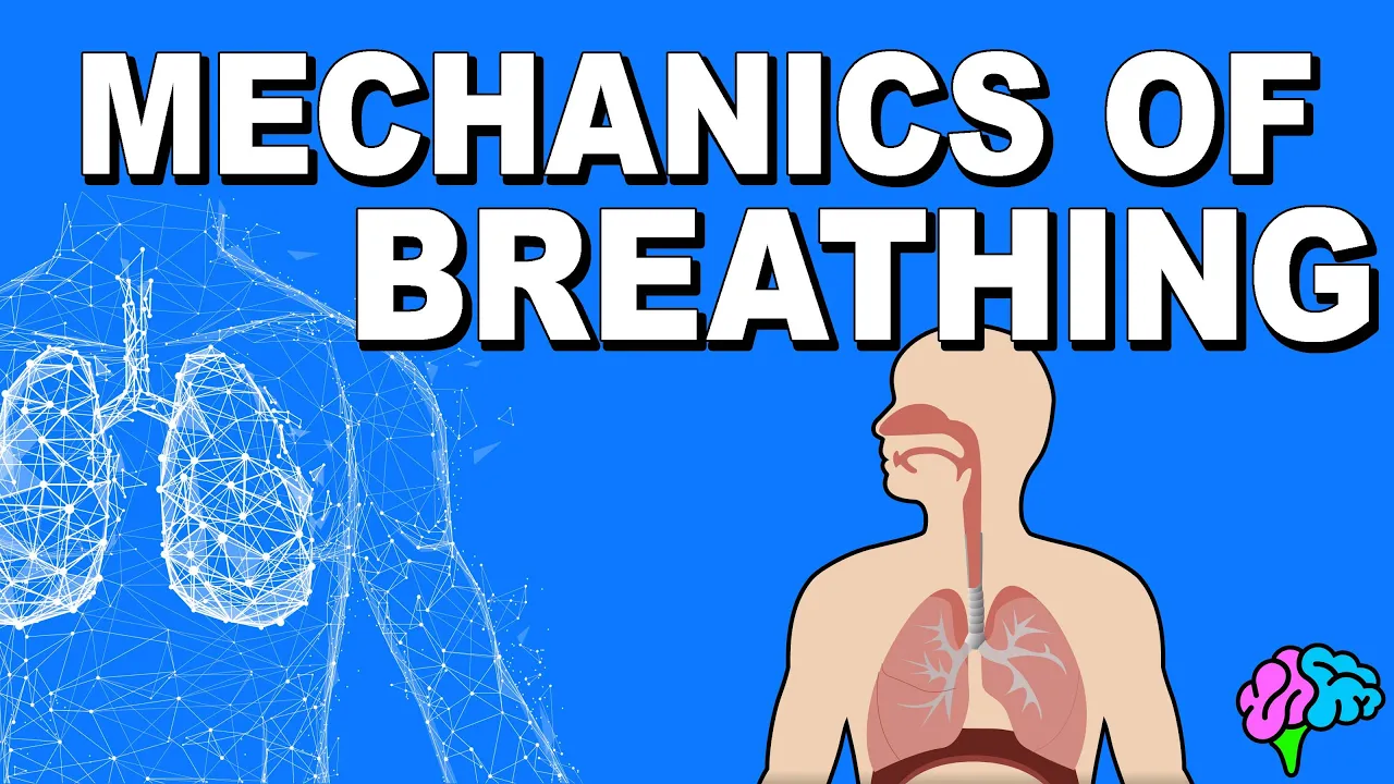






![Mechanics of Respiration [Thorax Anatomy 3 of 16]](https://i.ytimg.com/vi_webp/yalN1suRCas/maxresdefault.webp)












Negative pressure ventilation is the body's normal breathing mechanism where diaphragm contraction creates negative intra-thoracic pressure that passively draws air into the lungs; positive pressure ventilation uses a ventilator to actively push air into the lungs by creating positive pressure within the airways, typically requiring paralysis of the patient's respiratory muscles.
Negative vs Positive Pressure Ventilation | Respiratory Physiology
Added:Basic anatomy of the respiratory system, including the lungs, pleural cavity, and diaphragm.
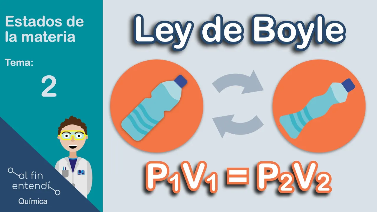
Boyle's Law and its application to gas flow, specifically how volume changes alter pressure.
Normal physiological mechanics of quiet breathing, including active inspiration and passive expiration.
The concept of pressure gradients (atmospheric, alveolar, and intrapleural pressures) and how they drive airflow.
Advanced modes of positive pressure ventilation, such as Volume Control, Pressure Control, and Assist-Control.
The physiological and hemodynamic complications of positive pressure ventilation, including barotrauma and reduced venous return.
Non-invasive positive pressure ventilation (NIPPV) techniques, such as CPAP and BiPAP therapy.
The clinical management of Acute Respiratory Distress Syndrome (ARDS) using lung-protective ventilation strategies.
226.9K views2.8Klikes18:12Original Release: 2014-11-01
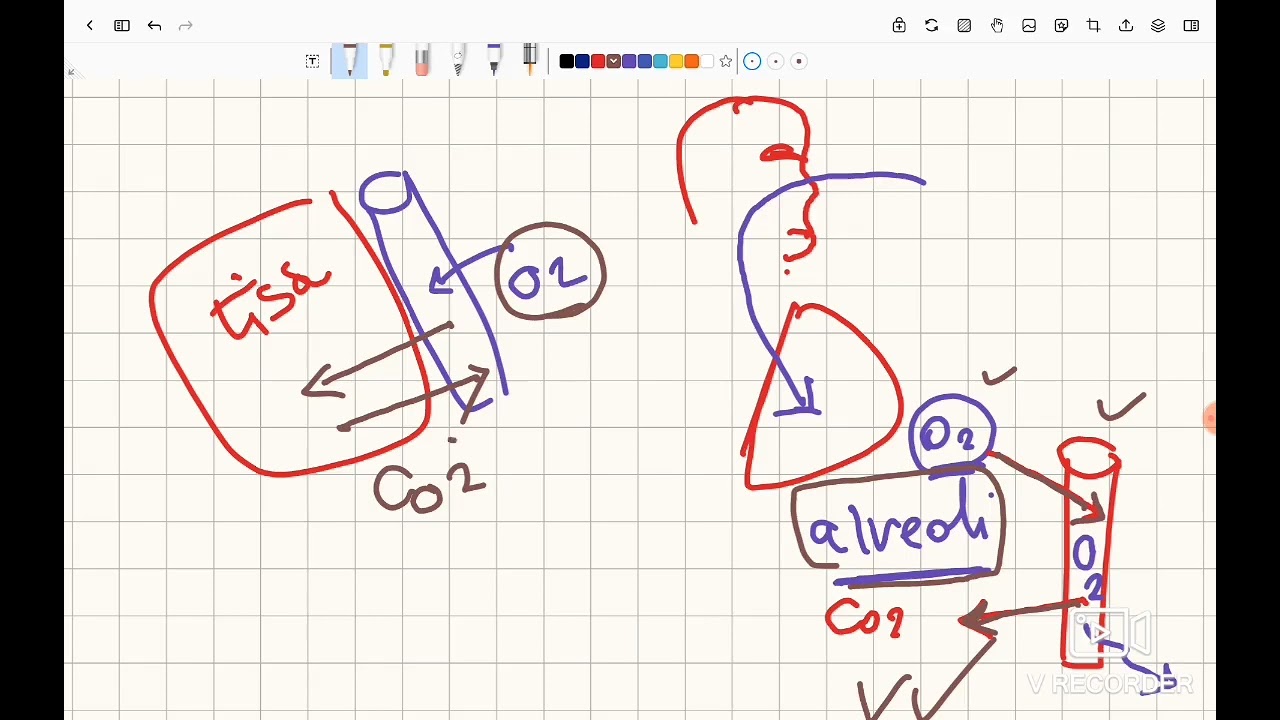
welcome everybody my name is Tyler Foster and in this video we'll be touching on the basics of the differences between negative pressure and positive pressure ventilation so negative pressure ventilation is going to be our body's normal way of breathing positive pressure ventilation is US introducing the patient to a ventilator and we're going to ventilate the ventilate the patient using positive pressure or nonphysiologic ways of breathing so let's always start off with the normal way to breathe um we have lungs here we have our Apex we have our base of the lung let's draw the other one in here we have our trachea so just to kind of Orient you excuse me here's our trachea we have our uh primary bronchioles we have uh kind of our collection of alvioli within our lungs we have a whole bunch of connecting Airway in between up here we have our our trachea it's going to connect to our larynx which will go uh into our Oro FNX it'll go to the Nars and then to the mouth I'm not going to draw that in it's just too much however just know that the trachea is going to have the glotus which can close so our vocal cords can go together to close or they can open up to pretty much allow our lungs to see the atmosphere so those are our two scenarios we're going to have uh our vocal cords which can close that's kind of how we cough our glotus or vocal cords are going to be closed while we build up some pressure in our lungs eventually it'll open up giving us a cough however that has nothing to do with ventilation so let's get back on topic here we're going to have to draw in some other structures here uh in the lungs we're going to have our chest wall so our thoracic cavity I'm going to draw it in in a circle and I do that for a reason it's because our our chest wall is not going to have let's let's put that a different way our lungs are not going to be exposed to the atmosphere except for our breathing passageway so unless we if we close off our glotus here our lungs are not going to be exposed to the atmosphere within our chest wall cavity so that's why I kind of Drew this blue chest wall over our trachea then also one other structure I need to put in here is our diaphragm it's going to be our maj Maj muscle sitting just below the lungs and that's going to be our major muscle for inhalation now why do I do all of this well let's take a look at a normal breath for a normal breath physiologic breath our diaphragm is going to contract our diaphragm is going to contract which brings it downwards now what that does is it's going to create a negative pressure within our closed system okay so let's put put that again this is going to be a closed system our lungs are not exposed to the atmosphere our diaphragm is going to contract making this cavity bigger when we have a bigger cavity it creates a vacuum and that's how that's how an air compressor works so I'm just going to draw this off on the side here we're going to have a piston this is going to be our piston we're going to have a cylinder that that piston is in and then here's our air compressor out here uh tubing etc etc our piston is going to push push push push compacting all of this air all that air is going to go through our tubing into a big collection and then when we when we squeeze a trigger we're going to have a nice uh stream of air or if we want a water uh gun we would have a stream of water however if we were to pull the cylinder back we're going to create a vacuum in this space kind of like when you have a syringe so let's say this is going to be our syringe it kind of looks like a syringe when you push the plunger forward you're going to eject all of the fluid however if you pull this the the lever back you're creating a vacuum which sucks up some fluid so let's say you're attaching this uh syringe to an IV you're going to withdraw some of the blood so you're getting a Venus sample for example um and that's because the Venus blood will go up into the syringe because you have a negative pressure vacuum same thing with the lungs our diaphragm is going to pull kind of like we're pulling the little stopper here we're going to pull creating a negative pressure within our lung space what that's going to do is it's going to expand our lungs now let's introduce another topic here we're going to have a pink layer enveloping our lungs and I'm just going to draw it for half and then we also have a pink layer that's going to envelop our chest wall now it's normally not this large of a space we have something called the plura the plura we're going to have two types of plura we're going to have our parietal plura parietal versus our visceral that's an S so visceral and parietal plura now kind of fancy words our parietal plura is going to envelop our chest wall while our visceral plura is going to envelop our lungs notice how I drew it in a very continuous fashion our plura is going to be continuous there are not going to be holes in our plura so have you ever heard of a plural effusion plural effusion if you heard that fancy term an effusion is going to be fluid buildup in our plural space so a plural effusion is where somehow we get water built up between these two layers normally our parietal and our visceral plura are touching one each other they have a nice little kind of wet slippery surface but they're touching each other if we get water between the layers we have something called a plural affusion and that's bad because we lose well I guess for a lot of different reasons which I won't get into however normally our parietal and our visceral plura are touching and the reason why it's called visceral plura is because the lungs are an organ viscera means organ so any layer that uh lines our organs can be called the viscera or the visceral layer of our of our plura so any of this plura plur plur plur plura the plural layer is going to be our visceral plur all right so if I say hey can you take out the body's viscera I'm really just saying take out the body's organs so that's a little terminology in there um normally our plura is touching so when I say that we're going to move our diaphragm downwards we're creating a negative intra plural pressure oh man that's a big negative intra plural pressure man that is a lot to consider we have a negative pressure here since we have a negative pressure on the inside it's going to pull our visceral plura with it so our lungs are going to expand that is how we take a breath when our diaphragm contracts we're going to create a negative intra plural pressure and what that's going to do is it's going to pull on the lungs and create a direction of force like this if you can see it we're creating a outward Force which is going to expand our lungs and when we expand our lungs it's kind of creating vacuum just like here we're creating more space as we pull the plunger back which is creating a negative space in our volume which our volume is going to be our lung Airways and what that's going to do is it's going to take this pressure that's in the atmosphere and it's going to pull it into the syringe now same situation here we're going to take the atmosphere so atmosphere is going to be our big pink ball here it's just going to be air our glotus is going to open as our lung expands due to this negative int plural pressure we're going to draw air and create a negative Force inside our lungs a negative atmospheric pressure that air is going to passively go into the lungs so that is going to be negative pressure ventilation it's where we we have a diaphragm that's going to contract and it's going to pull downwards that pulling downwards is going to pull on the pl the parietal plur that pull on the parietal plura is going to create a negative intra plural pressure so negative in here a lot of negative signs that negative pressure is going to pull on the visceral plura which is attached to the lung which is going to expand that expansion is creating a space a negative pressure space which is going to pull in the atmospheric air so this negative pressure space is going to be at a pressure that's below our atmospheric pressure and when we have a pressure that's below atmospheric pressure our air is going to passively come in so we're actively pulling down to passively pull air into our our lungs now that is going to be our negative pressure system when our lungs are fully expanded and we're ready for exhalation our diaphragm stops Contracting and it's actually going to return upwards it's also going to ex we're going to Exhale because our thoracic cavity doesn't like being expanded all the way so what it's going to do is it's going to have natural elastic recoil which is going to push on the lungs which is going to create a positive pressure in here so we're pushing which is going to collapse the lung as we collapse the lung we're losing volume so we take our cylinder here and we're going to push our plunger what that's going to do is it's going to compact all the air that's currently in here creating a more positive pressure which is going to expel the contents outwards so we have a positive pressure in the lungs it's going to override the atmospheric pressure and when this pressure becomes more than atmospheric pressure our air air is going to return to the environment into the atmosphere that is going to be our negative pressure ventilation normal body physiology so what happens when we put somebody on a ventilator so we're going to introduce our patient to a ventilator let me uh let me get different color here I'm going to have to draw the lungs again but I'm going to draw it a little further down now because uh we're going to have an altered uh kind of scenario here so again here's a terribly drawn lung here's our trachea going to draw it a little smaller this time we're still going to be in our closed system don't get me wrong here it's just going to become a little less important so I'll bring in the same factors that we just discussed and I'll try and keep everything the same color for clarity sake so we have our diaphragm we have our thoracic wall we're going to also have that plural space um at etc etc the body is still the same however this time what we're going to do is we're going to introduce a breathing tube we're going to introduce an endot tracheal tube uh into the system or you can do a non-invasive mask uh for positive pressure ventilation as well um that's where you put a nice tight fitting mask around the mouth so air cannot cannot leak out the sides so really it's kind of like inserting an endot tral tube just less invasive um so let's say we stick an endot tral tube down the mouth into the Oro ferin we thread it up um up into the glotus and then inflate a cuff right around the trachea so we're sealing off off the lungs from the environment except now we have this nice uh Hollow endot tracheal tube that Hollow endot tracheal tube what we're going to do is we're going to hook it up to our tubing we're going to take our tubing here it'll be connected to a machine and in our machine we're going to have some components that we need to discuss we're going to have a cylinder now didn't we just talk about cylinders yeah it's the exact same thing with a ventilator you can have a Bellow within the cylinder um so let's let's draw a little piston here same thing that we just dealt with so here's our plunger here's our piston here's our little cylinder at the end of the cylinder we're going to have a little output that output is going to hook up eventually to our our endot tral tube tubing that tubing the circuit system is going to attach to our endot tral tube which is going to go directly into our lungs thus creating a closed system so again we're not exposed to the environment in this situation we're hooked up to a uh to a cylinder which is going to create pressure so here in positive postive pressure ventilation our diaphragm is not going to be contracting downwards if we paralyze the patient completely with a with a paralytic agent we give sual choline the patient is not going to be able to breathe on their own because we paralyze the diaphragm we paralyze the chest wall uh at least the active motion of the chest wall so muscles can't contract all right now let's talk about how positive pressure ventilation differs in this scenario we're going to take our cylinder and we're going to push on the plunger so we're going to compact the air that's in here that compaction of air is going to create a nice high pressure that high pressure is going to push through the tubing we're going to meet a little bit of resistance in the tubing however most of our pressure that we created is going to enter the trachea it's going to go down onto our bronchioles it's going to go into the terminal Airways and eventually reach our alvas our Alvis then gets pushed open because we're creating a positive pressure through our ventilator so this is going to be our ventilator if if I haven't made that clear we're creating a positive pressure through kind of like a like a compression uh like a air compressor for example if if you have an air compressor at home in your garage that's the same thing we're doing we're creating a positive pressure it's going to go through our tubing it's going to push open our Alvis so our Alvis we pushing air in there instead of being driven by a negative pressure which is going to pull on the walls of the Alvis on the outside we're pushing air from the inside to push this Alvis open that's the difference in positive pressure ventilation we're pushing from the inside of the Airways to open up the Alvis our diaphragm is not Contracting so our lungs expand because we're are pushing air from the inside out instead of pulling from the outside to create a negative space we have a positive pressure inside which pushes that Alvis open so our lungs are going to be fully expanded how are we going to get the air out well I said our thoracic wall muscles are paralyzed however we're going to have natural recoil our chest wall still doesn't like to be expanded we've expanded all these Alvi our lungs are now completely taking up our thoracic space pushing our chest well outwards we're getting our chest well movement now as we exhale we're pushing air out so our ventilator stops feeding all this nice positive pressure into our system allowing the Alvis to collapse why does it collapse it's because our chest well pushes back it's it's resisting the force of our lungs being expanded thus creating a negative or a positive pressure in our in plural space that positive pressure in our inter plural space is going to push the lungs collapsed again thus exhaling so let's say we need another breath we're going to create some pressure in our ventilator some positive pressure goes through here positive pressure goes through our circuit down into our endot tral tube that positive pressure is going to enter our lungs it's going to eventually enter into our terminal uh Airway which is going to be our Alvis it's going to have that positive pressure on the inside pushing open the Alvis again pushing the lungs open pushing the thoracic wall outwards and then we're going to stop then then the ventilator says we need to Exhale so it's going to stop with this positive pressure our thoracic wall is going to push inwards again thus uh thus completing our exhalation so really in a nutshell that's our difference between negative and positive pressure ventilation we have different modes of positive pressure ventilation which is going to be my next video that I'll make hope you enjoyed if you have any comments let me know if you'd like a little more detailed explanation of any subject here be sure to ask have a good day
Up Next

17:43
Non-Invasive Positive Pressure Ventilation (CPAP & BiPAP) | Lecture 6
@StrongMed
274.4K views•2011-11-21

48:47
Graphic Medicine: Comics for Collaborative Healthcare Communication
@nationalpatientadvocate
189 views•2023-12-04

10:28
Neuroanatomy: Central and Peripheral Nervous System Divisions Explained
@AKLECTURES
136.2K views•2014-09-20

3:05
Stages of Labor and Vaginal Birth | Childbirth Animation
@nucleusmedicalmedia
52.1M views•2017-08-18
Related Study Plans & Knowledge Roadmaps
Structured learning paths in Medicine
medicine
Abnormal LFTs: Diagnostic Workup & Pathology
01Liver Anatomy and Basic Physiology
02Introduction to Liver Function Tests (LFTs)
03Hepatocellular vs. Cholestatic Patterns
+ 3 more learning modules
12h6 modules
Explore Roadmap
medicine
Analyzing Blood Smears: RBC, WBC & Platelets
01Introduction to Blood Components & Smear Preparation
02Normal Morphology of Blood Cells
03Erythrocyte Pathologies & Morphological Variants
+ 2 more learning modules
12h5 modules
Explore Roadmap
medicine
Mechanical Ventilation: Waveforms & Alarms
01Anatomy and Physiology of Breathing
02Introduction to Mechanical Ventilation & Core Modes
03Ventilator Settings and Waveform Interpretation
+ 2 more learning modules
12h5 modules
Explore Roadmap
medicine
Neuro Exam: Localization & Stroke Syndromes
01Foundations of Neuroanatomy and Vascular Supply
02The Neurological Examination: Step-by-Step
03Principles of Neurological Localization
+ 3 more learning modules
18h6 modules
Explore Roadmap