






















Electron microscopes overcome the 200nm resolution limit of light microscopes by using electron beams instead of visible light, enabling visualization of structures as small as 0.1nm; Scanning Electron Microscopes (SEM) produce 3D surface images with easier sample preparation, while Transmission Electron Microscopes (TEM) generate 2D internal images with higher resolution but require complex sample preparation.
Electron Microscopes: SEM vs TEM Explained
Added:Basic optical microscopy principles, including how lenses focus light and the physical limits of resolution (Abbe's limit).
Wave-particle duality and the de Broglie wavelength concept, explaining why accelerated electrons have much smaller wavelengths than visible light.
Fundamentals of electromagnetism, specifically how electromagnetic lenses are used to focus and manipulate charged electron beams.
Basic atomic structure and electron-matter interactions, including the concepts of elastic and inelastic electron scattering.
Advanced specimen preparation methods, such as sputter coating for SEM and ultra-microtomy, grid preparation, or plunge-freezing for TEM.
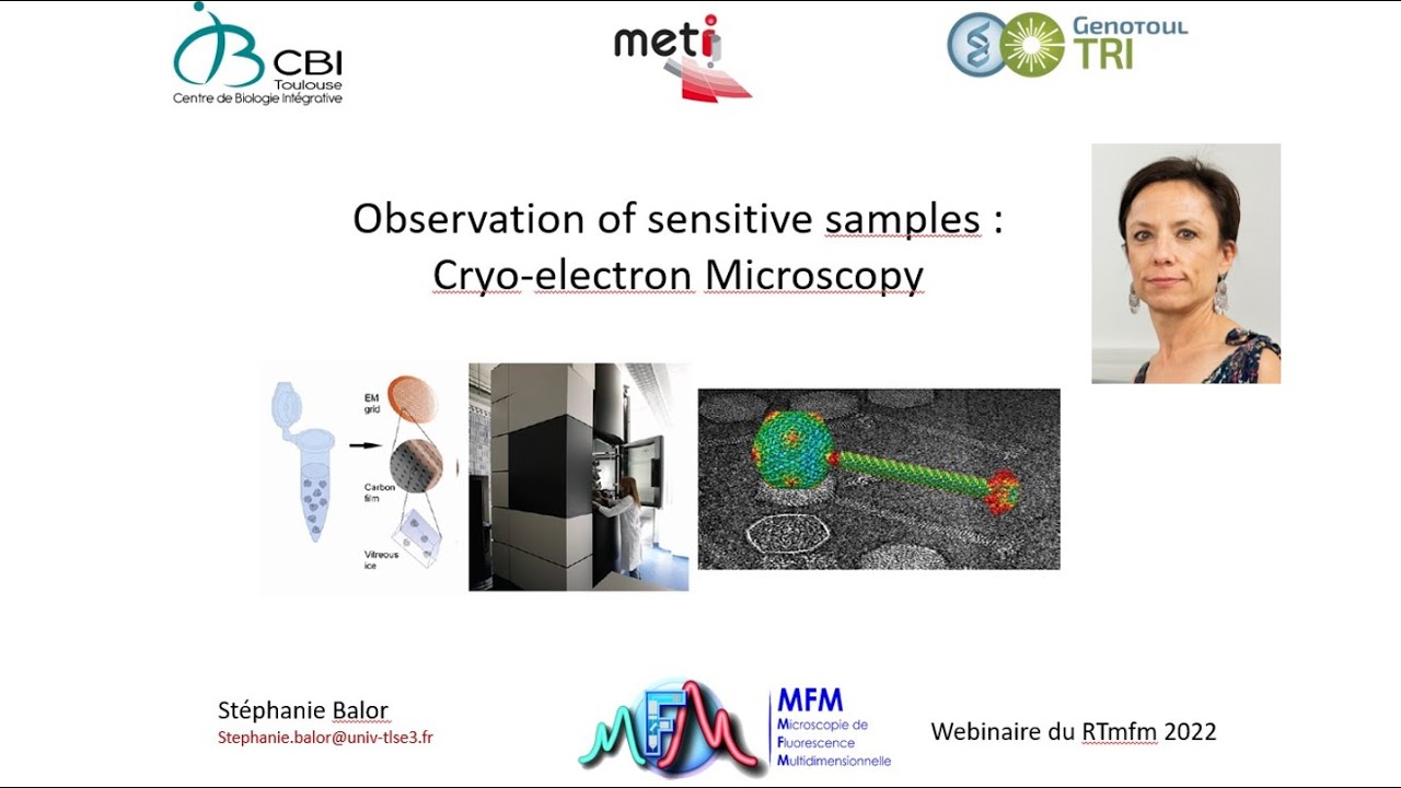
Cryo-Electron Microscopy (Cryo-EM) and its application in structural biology to image biomolecules in their native state.
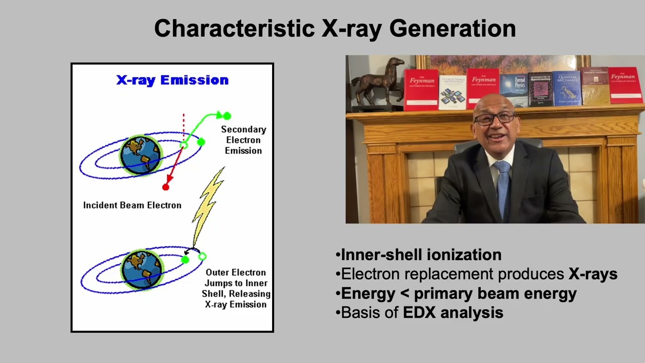
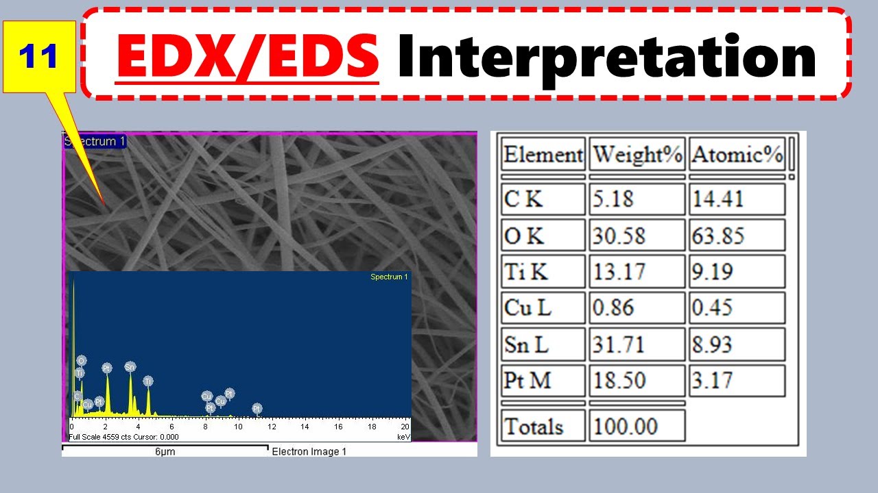
Energy-Dispersive X-ray Spectroscopy (EDS/EDX) and Electron Energy Loss Spectroscopy (EELS) for elemental and chemical composition analysis.
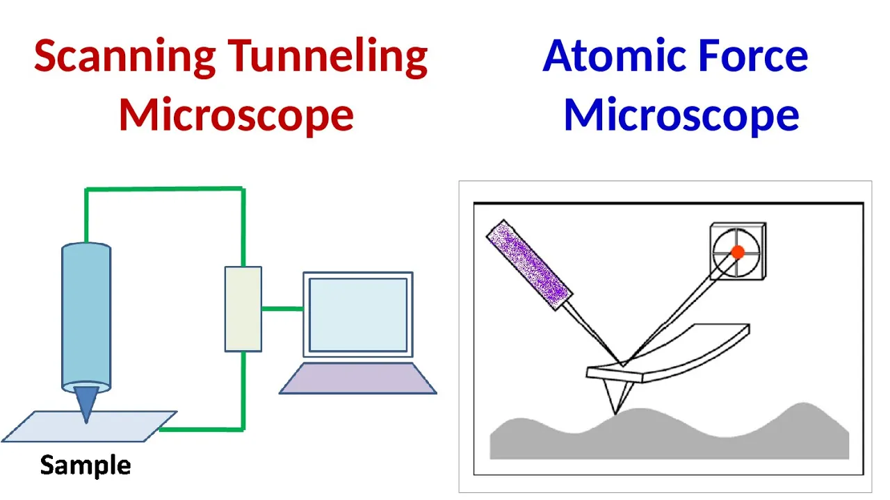
Alternative high-resolution imaging modalities, such as Atomic Force Microscopy (AFM) and Scanning Tunneling Microscopy (STM).
910 views17likes4:37Original Release: 2024-11-29
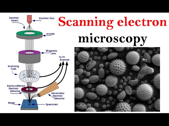
hello students welcome again for another learning bite before that please subscribe my channel to support my effort thanks in advance today we will learn about electron microscopes scanning and transmission electron microscopes let's begin let us see first why electron microscope what are the limitations of light microscopes light microscope has resolution of around 200 nanom or 0.2 microm this means they can't distinguish two points that are closer than 200 nanom apart the Practical magnification limit of a light microscope is 1,000 X in transmission mode the thickness of the sample is limited to tens of micrometers light microscopes cannot provide in-depth information on the three-dimensional structure of specimen because of these limitations electron microscope is invented following are the features of electron microscope electron microscopes can resolve features that are more than 1 million times smaller than Optical microscopes some electron microscopes can detect objects as small as 1/ 12th of a nanometer instead of visible light electron microscop use a beam of electrons to illuminate the sample electron microscopes use electromagnetic coils to focus Electron Beam instead of glass lenses electron microscopes can be used for defraction contrast Imaging phase contrast Imaging high resolution imaging chemical analysis and electron defraction there are different types of electron microscopes scanning electron microscopes sem and transmission electron microscopes t let us see where exactly electron microscope is needed studying the ultr structure of cells microorganisms and large molecules to diagnose gastrointestinal tumors detect microorganisms in the gastrointestinal tract and analyze the effects of treatments on patients testing new vaccinations and medicines comparing Blood and Tissue samples and identifying diseases and viruses researching Metals polymers batteries and geological samples they can also be used to perform quality control and failure analysis anal ing gunshot residue comparing bullet markings and identifying Firearms so we can see electron microscopy is a blessing in research let's now study the scanning and transmission electron microscopes one by one first the internal structure of scanning electron microscope it consists of an electron gun which generates an electron beam which passes through anode lens first then it passes through two condenser and one objective lenses making it more columnated this columnated Electron Beam Falls on on the sample kept on a motorized stage which moves sample up down or sideways to scan the Electron Beam reflects back as three signals back scattered electrons X-rays and secondary electrons which are received by respective detectors and sent through amplifiers to computer to generate graphical image of the scan shown here is the actual image generated by Sam let's now study the transmission electron microscope it also starts with electron gun which emits electrons which are then passed through anode to get accelerated after passing through condenser lens the columnated Electron Beam falls on Sample transmitting through the sample electron being passes through few more lenses and at the end through projector lens which projects the electron on the floor ENT screen fluorescent screen shows the inside image of the sample T also has three signals to be processed to create actual image scattered electrons energy loss electrons and characteristic x-rays processing which the actual image is obtained as shown in the example so which one is better sem or t let's see the difference between sem and T sem samples are easily produced sem gives 3D image same sample ranges from 1 cm to 10^ minus 30 nano m s is used to study surface morphology while temp sample requires complex preparations temp produces 2D images with range down to 0.1 nano m is used to study the insights of the sample however broadly comparing the two sem is Affordable and easier technology that's all for today I hope you enjoyed learning see you in next video
Up Next

12:24
XRD Characterization Technique for Nanomaterials | X-Ray Diffraction Explained
@hydeduhub3341
6.9K views•2024-12-16

48:00
Clean Water, Green Concrete & Nickel from Plants: Sustainable Inventions Explained
@business
94.3K views•2025-11-06

54:16
Polymer Environmental Degradation: Mechanisms & Stabilization
@iit
1.8K views•2012-07-10

33:39
How a Student's Question Saved a NYC Skyscraper from Collapse
@veritasium
22.8M views•2025-04-26
Related Study Plans & Knowledge Roadmaps
Structured learning paths in Engineering
engineering
Building Acoustic Levitators: Physics & Arduino
01The Physics of Sound and Standing Waves
02Piezoelectric Transducers and H-Bridge Drivers
03Arduino Programming: Generating 40 kHz Signals
+ 2 more learning modules
14h5 modules
Explore Roadmap
engineering
Building Peltier Cloud Chambers: Particle Tracks
01Cosmic Rays & Cloud Chamber Physics
02Thermoelectric Cooling (TEC) Fundamentals
03Custom Liquid-Cooling Loop Design
+ 2 more learning modules
18h5 modules
Explore Roadmap
engineering
Yagi-Uda Antennas: Modeling, Build & VNA Test
01Introduction to RF and Antenna Fundamentals
02Yagi-Uda Antenna Design Principles
03Antenna Modeling and Simulation in 4NEC2
+ 2 more learning modules
18h5 modules
Explore Roadmap
engineering
ECG Monitor Design: Op-Amps, Filters & Noise
01Electronics & Breadboard Basics
02Op-Amps and Instrumentation Amplifiers
03Bioelectricity and ECG Fundamentals
+ 2 more learning modules
18h5 modules
Explore Roadmap