In vitro transcription (IVT) is a laboratory technique for synthesizing mRNA by first linearizing plasmid DNA with restriction enzymes, then using RNA polymerase to transcribe the DNA template into mRNA; the resulting mRNA is stabilized through capping and polyadenylation before being used in therapeutic or vaccine development.
mRNA Synthesis via IVT: A Step-by-Step Guide
Added:Deep Dive
The Central Dogma of molecular biology, specifically the biological mechanisms of DNA transcription and mRNA translation.
Grade 10 SCIENCE | Quarter 3 Module 4B | Replication, Transcription and Translation
245K
The central dogma describes genetic information flow from DNA to mRNA to protein. Proteins are made of amino acids, the building blocks of cellular function. RNA and DNA differ in nitrogenous bases: DNA uses adenine-thymine pairing while RNA uses adenine-uracil pairing. Three RNA types serve distinct functions: mRNA carries genetic codes from nucleus to ribosomes, rRNA (80% of total RNA) forms ribosome structure, and tRNA transfers amino acids. DNA replication produces two identical molecules through a semi-conservative process using four key enzymes: helicase (unzips DNA), primase (provides starting point), DNA polymerase (builds new strands), and ligase (joins fragments). Base pairing follows strict rules: cytosine pairs with guanine, thymine pairs with adenine.
Click to watch →
Genetics - Central Dogma of Life - Lesson 17 | Don't Memorise
695.9K
Transcription is the process by which genetic information flows from DNA to mRNA. A gene is a DNA segment encoding a specific protein. During transcription, the DNA double helix opens at specific points, enzymes assemble, and nucleotides gather to form mRNA. The resulting mRNA is single-stranded and differs from DNA by containing uracil instead of thymine. This process is essential because without transcription, mRNA cannot be synthesized, and proteins cannot be made. The mRNA serves as an exact copy of the DNA sequence coding for the protein, ready to be transported to the cytoplasm for translation.
Click to watch →

المراجعة المركزة - الفصل الخامس - المسائل الوراثية
252.2K
DNA to mRNA transcription: To transcribe DNA to mRNA: (1) Replace thymine (T) with uracil (U) in the DNA template, (2) Replace adenine (A) with uracil (U) in the mRNA, (3) Replace guanine (G) with cytosine (C) in the mRNA, (4) Replace cytosine (C) with guanine (G) in the mRNA. The DNA template strand is read in the 3' to 5' direction, and the mRNA is synthesized in the 5' to 3' direction. DNA template to tRNA translation: When translating DNA template to tRNA: (1) First transcribe DNA to mRNA, (2) Then translate mRNA to tRNA, (3) The DNA template is read in the 3' to 5' direction, (4) The tRNA anticodon is read in the 3' to 5' direction. Each codon in mRNA is read in the 5' to 3' direction, and each codon pairs with its complementary anticodon in tRNA.
Click to watch →

الخبر الوراثي live
171.2K
Transcription converts DNA to mRNA using base pairing rules: A→U, C→G, G→C, T→A. If DNA is transcribed, copy directly; if not, reverse first. Codons (groups of 3 nucleotides) code for specific amino acids. This process produces mRNA that will be translated into protein.
Click to watch →
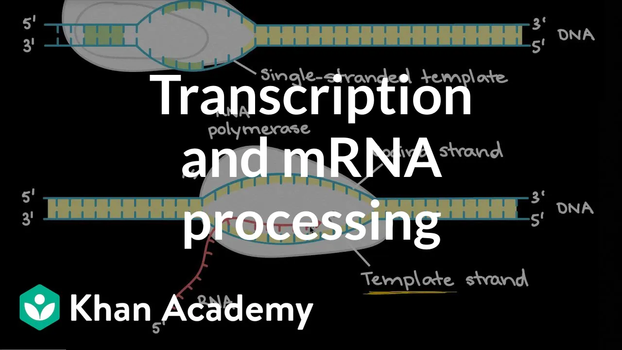
Transcription and mRNA Processing: DNA to mRNA in Eukaryotes & Prokaryotes
1.8M
Transcription is the cellular process of copying genetic information from DNA to messenger RNA (mRNA), serving as the first step in gene expression. In this process, the DNA double helix unwinds, and RNA polymerase reads one strand (the template strand) to synthesize a complementary RNA strand. The resulting mRNA carries the genetic code from DNA to ribosomes for protein synthesis. This fundamental mechanism enables cells to translate genetic instructions into functional proteins, forming the basis of molecular biology and genetic inheritance.
Click to watch →
Deep Dive
Structure and function of plasmid vectors, including restriction site mapping and the concept of plasmid linearization.

Recombinant DNA Technology - Animated Video
93.6K
Common cloning vectors like E. coli plasmids (such as pUC) contain three functional regions: an origin of replication enabling independent plasmid replication outside the bacterial cell cycle, a drug resistance gene (such as ampicillin resistance), and a multiple cloning site (polylinker) containing numerous restriction sites. The polylinker region allows insertion of foreign DNA at specific locations determined by the restriction enzyme recognition sequences.
Click to watch →

Asosiy fan olimpiadasi 10-sinf viloyat bosqichi Biologiya yozma ish topshirig'i
322
Restriction mapping determines the locations of restriction enzyme cut sites on plasmid DNA. A plasmid is circular DNA. When cut with a single restriction enzyme, it produces linear fragments. Fragment sizes indicate distances between cut sites. For example, a 1000 bp fragment indicates a single cut site. Multiple cut sites produce multiple fragments whose sizes sum to the total plasmid length. When enzymes are combined, fragment sizes change, indicating overlapping cut sites. Unchanged fragments indicate sites not affected by the additional enzyme.
Click to watch →

AP Biology Plasmids and Restriction Mapping
36.1K
Plasmids are circular DNA molecules found in bacteria that can be genetically engineered with specific genes and restriction sites; restriction enzymes cut DNA at precise recognition sequences, enabling scientists to create restriction maps by analyzing fragment sizes from single or double enzyme digestions, which allows them to determine the relative positions and distances between restriction sites on a plasmid or linear DNA molecule.
Click to watch →

Plasmid design (bacterial expression vector)
33.5K
Plasmids are circular DNA molecules naturally found in bacteria that serve as essential cloning vectors in genetic manipulation. They function by moving DNA between different species, similar to how mosquitoes transmit malaria parasites. Bacteria contain both chromosomes with essential genes and plasmids with optional genes like antibiotic resistance. Plasmids are self-replicating and range from 1,000-20,000 base pairs, making them ideal for inserting typical genes of 2,000-4,000 base pairs. Three fundamental components are required for functional plasmid vectors: (1) Origin of replication (ori) - a specific DNA sequence enabling DNA replication machinery to recognize and copy the plasmid, controlling copy number per cell (2-3 to hundreds); (2) Selectable marker - provides visible phenotype (like antibiotic resistance) to identify cells that have taken up the plasmid, since transformation efficiency is very low (~1 in 10,000 cells); (3) Multiple cloning site (MCS) - contains multiple restriction enzyme recognition sites for inserting genes of interest. The standard cloning workflow involves: (1) amplifying the gene of interest via PCR; (2) digesting both plasmid and gene with restriction enzymes; (3) purifying and ligating the fragments using DNA ligase; (4) transforming the recombinant plasmid into host cells (typically E. coli).
Click to watch →

pBR322 Gene Cloning Vector Explained | Biotechnology @biologyexams4u
197.6K
pBR322 is the first widely used plasmid vector in genetic engineering, discovered by polymer antrotric in the 1970s. It contains three essential elements: (1) an origin of replication (ori) derived from E. coli that enables vector replication in host cells, (2) at least 12 unique restriction sites (such as BamH1, Sal1, Pst1, Pvu1) within the selectable marker regions that allow insertion of genes of interest, and (3) two selectable markers—ampicillin resistance and tetracycline resistance genes. The vector is 4361 base pairs in length. The selectable markers enable selection of recombinant colonies through insertional inactivation: when a gene is inserted into a restriction site within a resistance gene, that gene becomes non-functional, allowing researchers to distinguish recombinant colonies (which grow only on ampicillin medium) from non-recombinant colonies (which grow on both antibiotics).
Click to watch →
Deep Dive
Eukaryotic mRNA anatomy, including the physiological functions of the 5' cap and the 3' poly(A) tail in transcript stability and translation.

La maturation de l'ARN pré-méssager
739
The 5' cap and poly-A tail are essential modifications for eukaryotic mRNA stability and function. The 5' cap consists of N7-methylguanosine linked via 5' to 5' triphosphate, formed co-transcriptionally by RNA triphosphatase, guanylyltransferase, and methyltransferase. It protects mRNA from ribonucleases, facilitates nuclear export, and recruits ribosomes for translation. The poly-A tail is a sequence of adenosines added by poly(A) polymerase using ATP, triggered by the AAUAAA motif recognized by CPSF. The tail length typically exceeds 200 nucleotides and serves multiple functions: protection against degradation, nuclear export facilitation, and translation promotion through recruitment of poly(A)-binding proteins.
Click to watch →

CUET PG 2026 | Molecular Biology Part 2| Zoology, Botany, Life Sciences & Applied Microbiology
3.3K
Eukaryotic mRNA has 5' cap and 3' poly(A) tail. Cap-binding protein (CBP) interacts with poly(A)-binding protein (PABP) via eIF4G, circularizing the mRNA. eIF4A helicase unwinds secondary structures. The small subunit binds to the circular mRNA, and the large subunit joins after dissociating initiation factors. eIF2 delivers methionyl-tRNA to the P site, guided by the Kozak sequence near the start codon.
Click to watch →

Control Of Eukaryotic Gene Expression 04 Translational Control
1.6K
The 5' cap (7-methylguanosine) prevents 5' exonuclease degradation by being unrecognizable as a substrate. The 3' poly(A) tail shields mRNA from 3' exonucleases by being degraded first, protecting the coding sequence. Both structures determine mRNA stability and half-life; longer tails mean longer mRNA persistence in the cytoplasm, enabling more translation rounds.
Click to watch →

Kevin Ahern's Bite-Sized Biochemistry #46 - Transcription II (RNA Synthesis)
9.2K
Eukaryotic mRNAs undergo extensive processing including 5' capping and 3' poly(A) tail addition. The 5' cap (7-methylguanosine) protects mRNAs from degradation and aids translation. The poly(A) tail (200+ adenylates) signals nuclear export and regulates mRNA stability—the longer the tail, the more stable the mRNA. These modifications distinguish eukaryotic mRNAs from prokaryotic transcripts and contribute to their longer half-lives (24 hours vs. ~10 minutes).
Click to watch →
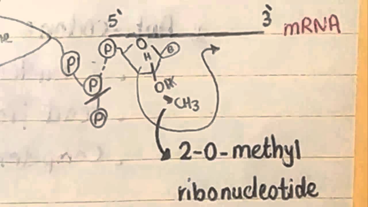
8 CBR, mRNA & tRNA
5.6K
The 5' cap consists of a modified guanine nucleotide (7-methylguanosine) that is linked to the first nucleotide of the mRNA. The 3' poly-A tail consists of a sequence of adenine nucleotides (typically 100-250 in length) added to the 3' end of the mRNA. The 5' cap serves as a recognition site for the ribosome during translation initiation and helps protect mRNA from degradation. The 3' poly-A tail interacts with the 5' cap through poly-A binding proteins, creating a circular structure that enhances translation efficiency. Together, these modifications protect mRNA from degradation and facilitate its export from the nucleus to the cytoplasm.
Click to watch →
Deep Dive
Basic laboratory concepts of enzymatic reactions, specifically the requirements of bacteriophage RNA polymerases (e.g., T7, T3, or SP6).

IYCN Science Café: Chemical Synthesis of RNA and DNA
114
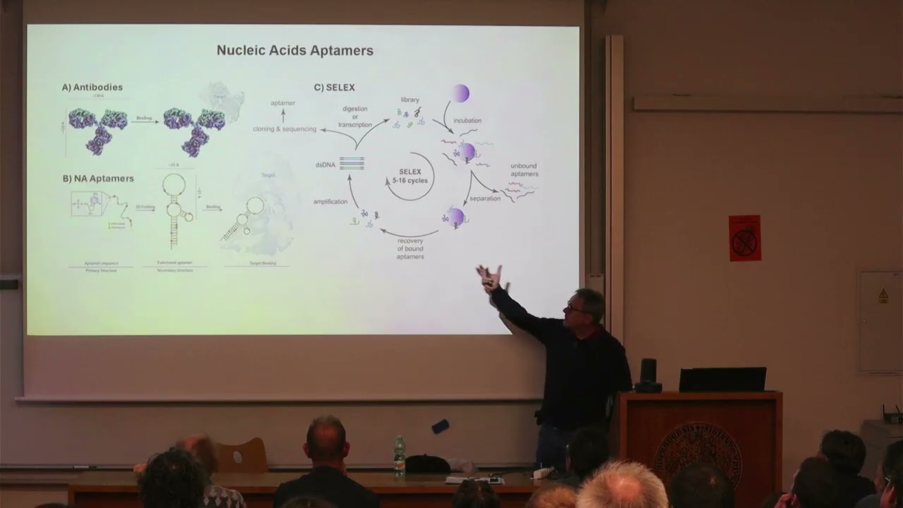
In vitro transcription is a molecular biology technique that recapitulates the transcription process outside of a living system. The process requires three main components: (1) Bacteriophage RNA polymerase (T7, T3, or SP6), with T7 being most commonly used; (2) Template DNA containing a promoter sequence and the gene of interest; (3) Ribonucleotide triphosphates (ATP, GTP, UTP, CTP) as building blocks. The polymerase binds to the promoter, reads the DNA template, and synthesizes complementary RNA. Linear DNA templates are used to define transcription endpoints. This technology allows production of large quantities of RNA needed for clinical applications, with the process being cost-effective compared to chemical synthesis.
Click to watch →
Biologické čtvrtky ve Viničné; M. Hocek: Maškarní ples nukleových kyselin; přednáška
423
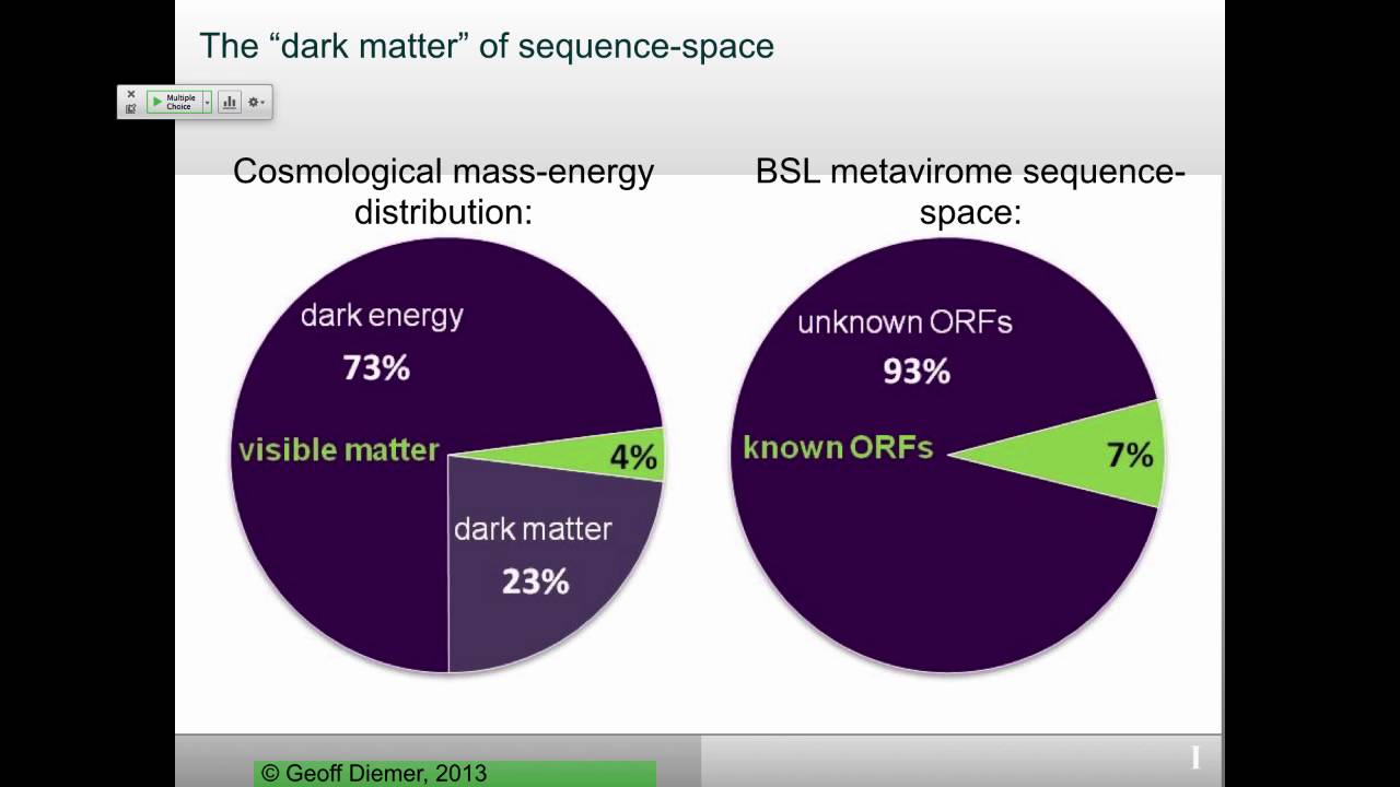
DNA polymerases require templates and primers, cannot initiate from nothing, and extend primers by adding nucleotides through phosphodiester bond formation. Family B DNA polymerases are most significant for eukaryotic replication. RNA polymerases use the same bond formation mechanism but require double-stranded DNA templates and can initiate from zero. Eukaryotic transcription initiation is extremely complex, requiring many transcription factors. T7 RNA polymerase from bacteriophage is commonly used for in vitro applications as it requires only a promoter sequence.
Click to watch →
VIr_S16_Review1
125
T7 bacteriophage encodes its own DNA-dependent RNA polymerase, which transcribes all viral genes from the injected DNA template. This viral polymerase is about 2.5 times faster than the cellular DNA-dependent RNA polymerase. The polymerase binds to specific promoters on the viral genome to initiate transcription, producing viral proteins needed for replication and assembly.
Click to watch →

Lecture 3 T7 bacteriophage
315
T7 bacteriophage is a midsize DNA virus infecting E. coli with double-stranded DNA containing equal A, T, G, C bases and no unusual bases. The 39,936 bp genome has 160 bp terminal redundancy and contains 50 closely packed genes plus 5 overlapping genes. The genome demonstrates extreme economy: leaders/spacers are only a few nucleotides, and for half the genes, the Shine-Dalgarno sequence lies within the preceding gene's coding region. The life cycle follows a precise timeline: attachment at T=0, slow DNA injection (~10 minutes total), host transcription shutdown at 4 minutes, phage DNA synthesis at 8-9 minutes, first phage appearance at 15 minutes, and lysis at 25 minutes. The first gene product inhibits E. coli restriction endonuclease. Transcription is temporally regulated in two stages: early stage uses host E. coli RNA polymerase to transcribe genes 3-1.3, while late stage uses newly synthesized T7 RNA polymerase. Three classes of transcripts (1, 2, 3) are synthesized from the right strand. Class 1 transcripts use three promoters to increase synthesis rate 3-fold and are cleaved by host RNase III into five mRNA molecules encoding protein kinase and T7 RNA polymerase. Protein kinase phosphorylates E. coli RNA polymerase, inactivating it and reducing host transcription. Class 2 and 3 require T7 RNA polymerase but are made sequentially due to slow DNA injection. Class 2 transcripts code for a second inhibitor of E. coli RNA polymerase that completely shuts down host transcription. By 8 minutes post-infection, no E. coli RNA synthesis occurs. Class 2 also contains DNA replication enzymes and structural protein genes. T7 DNA replication begins bidirectionally from one end, forming concatamers that are cut to release individual progeny genomes. The product of gene 3.5 binds to T7 RNA polymerase and is responsible for overall transcription inhibition and cell lysis.
Click to watch →
Vax-Hub & CPI Process Development Webinar - mRNA Vaccine Manufacture
3K
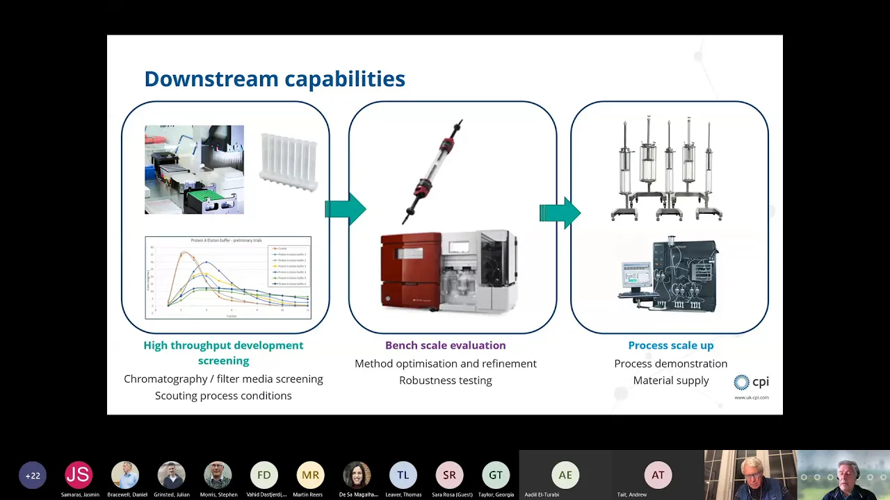
mRNA is synthesized enzymatically through in vitro transcription using linearized DNA templates. The T7 RNA polymerase (~100 kDa) is the smallest RNA polymerase requiring no cofactors. It binds promoters, separates DNA strands creating a ~14-15 base transcription bubble, incorporates capping molecules, and polymerizes RNA nucleotides through a Brownian ratchet mechanism taking ~20 microseconds per base pair. Reactions at ~37°C yield ~5g/L mRNA. Critical components include pyrophosphatase to consume pyrophosphate byproducts, all four nucleotides, capping reagents, and RNase inhibitors to prevent degradation.
Click to watch →
Prerequisite Knowledge
- Concept 01The Central Dogma of molecular biology, specifically the biological mechanisms of DNA transcription and mRNA translation.
- Concept 02Structure and function of plasmid vectors, including restriction site mapping and the concept of plasmid linearization.
- Concept 03Eukaryotic mRNA anatomy, including the physiological functions of the 5' cap and the 3' poly(A) tail in transcript stability and translation.
- Concept 04Basic laboratory concepts of enzymatic reactions, specifically the requirements of bacteriophage RNA polymerases (e.g., T7, T3, or SP6).
Subsequent Learning
- Step 01Downstream purification processes for mRNA, such as chromatography (HPLC/FPLC) and tangential flow filtration (TFF) to remove template DNA and double-stranded RNA.
- Step 02Formulation of mRNA into delivery vehicles, particularly Lipid Nanoparticles (LNPs), to facilitate cellular uptake and prevent in vivo degradation.
- Step 03Quality control assays for therapeutic mRNA, including cap-index assays, fragment analysis for integrity, and cell-free translation assays.
- Step 04Regulatory standards and scaling challenges associated with transitioning from laboratory-scale IVT to industrial GMP (Good Manufacturing Practice) vaccine production.
mRNA synthesis
mRNA synthesis
0:00Playing Section
- 1
Process starts with linearizing circular plasmid DNA using restriction enzymes.
- 2
RNA polymerase transcribes the linear DNA template into an mRNA strand.
- 3
mRNA is capped and polyadenylated to improve stability for therapeutic use.
Circular RNA (circRNA) as an Alternative to Linear IVT mRNA
While in vitro transcription (IVT) of linear mRNA is currently the standard for RNA therapeutics, circular RNA (circRNA) has emerged as a major technological alternative. Unlike linear mRNA, circRNA forms a covalently closed continuous loop. This unique structure inherently protects it from exonuclease-mediated degradation, drastically increasing its stability and functional half-life within cells without requiring the complex and costly 5' capping and 3' polyadenylation steps involved in standard IVT. Consequently, circRNA can provide more sustained therapeutic protein expression at lower doses, addressing the transient nature and high-dose requirements of traditional linear mRNA vaccines and therapies.
Deep Dive
Downstream purification processes for mRNA, such as chromatography (HPLC/FPLC) and tangential flow filtration (TFF) to remove template DNA and double-stranded RNA.

Getting Started with mRNA - Increasing Your Probability for Success
311
mRNA purification removes residual nucleotides, unincorporated cap analogs, enzymes, and DNA templates through multiple techniques: precipitation, silica membrane purification, oligo(dT) affinity chromatography (capturing poly(A) tails to improve integrity), and preparative reverse-phase HPLC for clearing double-stranded RNA. Tangential flow filtration serves dual purposes: initial purification based on molecular weight cut-offs and buffer exchange/concentration adjustment. Strategic workflow sequencing minimizes processing steps while maintaining product quality. Final formulation requires 0.2-micron filtration to prevent particulate contamination. Each purification step addresses specific impurity classes, with method selection dependent on scale, purity requirements, and downstream application compatibility.
Click to watch →
Gene-based Vaccine and Drug Development; mRNA Vaccines
295
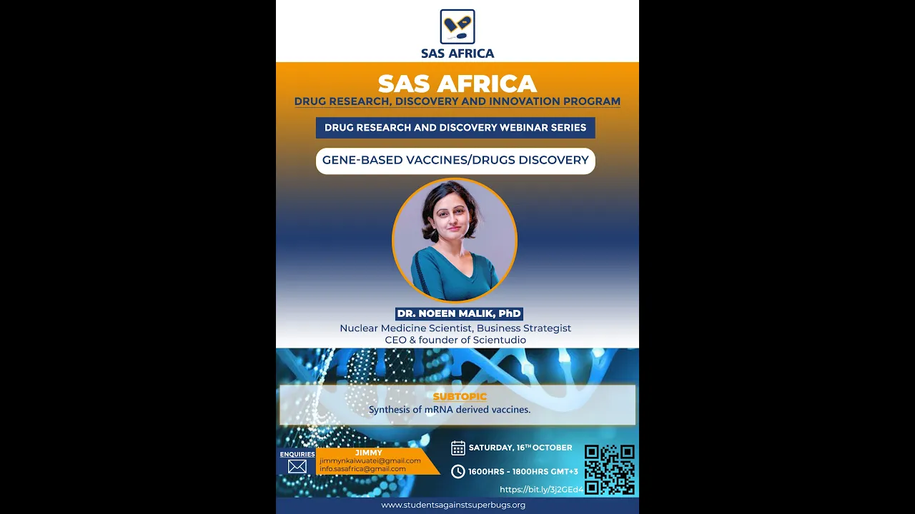
mRNA purification leverages its negatively charged phosphodiester backbone. Techniques adapted from DNA purification include size exclusion chromatography, reverse phase chromatography, ionic exchange chromatography, hydrophobic interaction chromatography, and thiophilic adsorption chromatography. For routine pre-clinical work, RNA can be precipitated using lithium chloride in combination with ice-cold ethanol, which neutralizes the backbone charges and decreases solubility. However, this technique is non-GMP. Iron exchange affinity chromatography combined with size exclusion chromatography is used for GMP-grade purification. Tangential flow filtration (TFF) is used for larger mRNA molecules (10,000+ bases) to remove lower molecular weight species using appropriate molecular weight cut-off membranes.
Click to watch →

Katalin Karikó: Developing mRNA for Therapy
4.7K
The speaker discovered that double-stranded RNA contamination was present in all their in vitro transcribed RNA preparations. This contamination was problematic because double-stranded RNA is highly immunogenic. After two years of work, they developed an HPLC (high-performance liquid chromatography) purification procedure that successfully removed the double-stranded RNA. This purification was essential for producing high-quality, non-immunogenic mRNA suitable for therapeutic applications.
Click to watch →
Exploring the Use of Multimodal Chromatography for the Purification of mRNA Vaccines
206
Multimodal chromatography using strong anion exchange resins with hydrophobic interactions offers superior selectivity for separating mRNA from challenging impurities like dsRNA compared to traditional affinity chromatography, enabling high-purity mRNA purification (>88%) with over 80% recovery in a single chromatography step by exploiting differences in hydrophobicity and charge between single-stranded mRNA and double-stranded RNA impurities.
Click to watch →
Chromatography in mRNA Production From Analytics to Purification
1.9K
mRNA purification leverages its unique properties through specialized chromatographic approaches: oligo(dT) affinity chromatography exploits the poly-A tail for selective binding; multimodal ligands (PrimaS) combine anion exchange and hydrogen bonding properties for non-polyadenylated RNAs; reverse phase chromatography separates double-stranded RNA based on hydrophobicity. Critical findings include flow rate dependence in oligo(dT) chromatography and enhanced mRNA stability from chromatographic purification compared to precipitation methods. Advanced polishing techniques ensure final quality: reverse phase chromatography using styrene-divinyl benzene monoliths separates dsRNA from ssRNA, scaling to 8-liter columns purifying 10 grams per batch with fragment analysis confirming purity profiles.
Click to watch →
Deep Dive
Formulation of mRNA into delivery vehicles, particularly Lipid Nanoparticles (LNPs), to facilitate cellular uptake and prevent in vivo degradation.

Lipid nanoparticles for mRNA delivery
17.5K
mRNA therapeutics enable protein production in vivo by delivering genetic instructions, offering solutions when direct protein administration fails due to degradation, poor targeting, or improper function. However, mRNA delivery faces fundamental barriers: mRNA is a long, negatively charged polyelectrolyte that cannot easily cross the negatively charged cell membrane; the body naturally degrades foreign mRNA; and finding biocompatible vehicles remains challenging. Lipid nanoparticles (LNPs) solve these challenges through four key components: cationic ionizable lipids (e.g., MC3DMA) that complex with mRNA and facilitate endosomal escape; helper lipids like DSPC that stabilize structure; cholesterol (~38.5%) that modulates membrane properties; and PEGylated lipids for steric stabilization. The cationic lipid has an apparent pKa of 6-7, becoming positively charged at physiological pH but neutralized at lower endosomal pH to disrupt membranes. LNPs are prepared using microfluidic mixers combining ethanol-soluble lipids with aqueous mRNA in 1:3 volume ratios, with standard compositions including ~50% cationic lipid, ~10% DSPC, ~38.5% cholesterol, and ~1.5% PEG lipid.
Click to watch →

mRNAs as Medicines
40.2K
mRNA medicines require a delivery vehicle, some sort of packaging material that serves three purposes: to protect it from degradation, to direct it to the desired tissue and cell type, and to help it gain entry into the cell cytoplasm. Although many different types of mRNA delivery vehicles are being tested, the most commonly used to date are lipid nanoparticles or LNPs.
Click to watch →
Lipid‐nanoparticle Formulations for mRNA Delivery: A Focus on Cellular Uptake and Trafficking...
12.4K
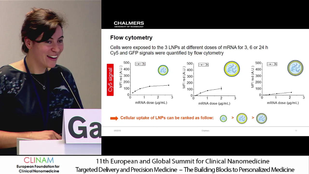
mRNA therapeutics offer transformative potential for genetic diseases through high target selectivity, transient genomic effects, and cytoplasmic delivery requirements. However, mRNA's negative charge renders it vulnerable to extracellular RNase degradation, necessitating sophisticated delivery vehicles. The Formula X initiative addresses these challenges through collaborative academic-industry partnerships focused on lipid nanoparticle development inspired by natural extracellular vesicles. Microfluidic technology enables precise formulation of LNPs with controlled inner cores (mRNA trapped with MC3 and cholesterol), DSPC-enriched middle layers, and PEGylated outer coatings. Systematic studies reveal that nanoparticle size and surface composition critically influence cellular uptake kinetics: smaller particles internalize more rapidly initially, while larger particles demonstrate superior uptake efficiency at later time points. Surface composition modifications consistently reduce overall uptake efficiency. These findings establish fundamental principles guiding the rational design of mRNA delivery systems for therapeutic applications.
Click to watch →
The technology behind the new COVID-19 mRNA vaccines
204.7K
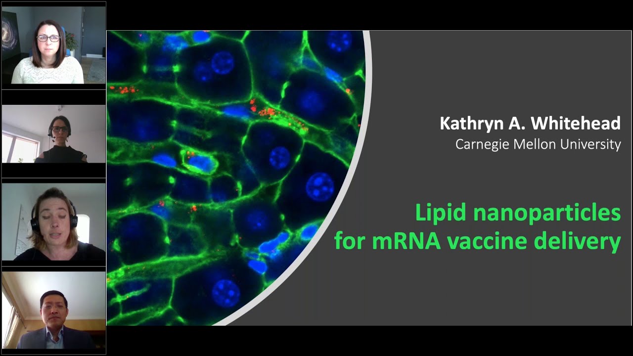
Lipid nanoparticles serve as effective delivery vehicles for mRNA vaccines by overcoming the natural barriers that prevent mRNA from entering cells, with the particles consisting of ionizable lipids that become positively charged under acidic cellular conditions to facilitate endosomal escape, cholesterol for structural stability, helper lipids for proper packing, and PEG for stability and immune shielding, enabling the mRNA to reach the cytoplasm where it can direct cells to produce viral spike proteins that train the immune system to recognize and fight infections.
Click to watch →
Getting Started with mRNA - Increasing Your Probability for Success
311
Lipid nanoparticles (LNPs) represent the predominant delivery vehicle for mRNA therapeutics, comprising cationic or ionizable lipids, helper lipids, PEGylated lipids, and cholesterol at approximately 100 nanometer diameter. LNPs facilitate cellular uptake through endocytotic mechanisms and protect mRNA from RNase degradation in the bloodstream. Upon cellular entry, LNPs bind to endosomal membranes, and pH changes cause lipid dissociation, releasing mRNA into the cytoplasm for translation. The innate immune system recognizes exogenous mRNA through pattern recognition receptors including TLRs (detecting ssRNA/dsRNA) and cytosolic receptors like RIG-I, MDA5, and PKR (detecting dsRNA and improperly capped RNA). While controlled stimulation benefits vaccines, excessive activation proves detrimental for other therapeutic applications.
Click to watch →
Deep Dive
Quality control assays for therapeutic mRNA, including cap-index assays, fragment analysis for integrity, and cell-free translation assays.

The potential of therapeutic mRNA — and how to measure it
394
Therapeutic mRNA vaccines, such as those developed by BioNTech for cancer and infectious diseases, require rigorous quality control testing including assessment of identity, concentration, integrity, and impurities; nanopore technology offers a promising platform method for these QC tests by enabling sequence-independent RNA analysis, modification detection, and integrity measurement without requiring prior sequence knowledge, though further improvements in base calling accuracy are needed for industrial implementation.
Click to watch →
mRNA Manufacturing
46.3K
mRNA quality control encompasses capping efficiency, dsRNA content, poly(A) length, residual proteins, residual DNA, RNA concentration, identity, purity, endotoxin testing, and sterility. Some USP assays require more RNA than small patient cohorts need, requiring careful QC planning. Despite common perception, RNA is chemically stable without metal ions or RNases, withstanding multiple freeze-thaw cycles and room temperature storage. Stability studies should be planned early with accelerated testing followed by long-term studies. Balancing metabolic stability and translation efficiency remains challenging—wild-type RNA shows optimal translation but may be too immunogenic, while modifications reduce innate stimulation but may decrease translational potential. m6A caps increase cellular stability, and poly(A) tail length affects stability.
Click to watch →
Streamlined mRNA Synthesis for Increased Protein Expression-TIDES Digital Week
975
mRNA quality attributes measured include: IVT reaction yields (mg/mL), full-length mRNA integrity (measured by IP-RP-HPLC analysis), capping efficiency, purity (DNA, protein residuals), and double-stranded RNA byproducts (measured by J2 immunoblot). ARCA manufacturing resulted in lowest integrity (78.6%) and highest double-stranded RNA levels, while enzymatic and CleanCap samples maintained high integrity (93-94%) and lower dsRNA levels. Both cap 1 methods achieved 99% and 96% capping efficiency respectively.
Click to watch →
Getting Started with mRNA - Increasing Your Probability for Success
311
Comprehensive mRNA characterization requires orthogonal assays addressing multiple quality dimensions: integrity (gel electrophoresis, ion-pair RP-HPLC), identity, content, purity, safety, and functionality. Different analytical methods serve distinct purposes with specific capabilities and limitations, necessitating strategic assay selection based on development stage. Manufacturing process variations significantly impact quality attributes: ARCA cap analog protocols with limited GTP produce higher double-stranded RNA levels and lower capping efficiency compared to optimized IVT with cap one analogs. These differences translate directly to functional outcomes, with optimized processes demonstrating significantly higher in vivo protein expression in luciferase reporter assays. Systematic comparison of cap analog structures reveals significant impacts on therapeutic efficacy: Clean cap AG (m7GpppAG) serves as baseline, while Clean cap AG with 3'-methyl modification shows intermediate improvement, and Clean cap AG with N6-methyladenosine modification demonstrates highest protein expression. Remarkably, Clean cap N6 at one-third the dose of standard Clean cap AG maintains equivalent protein expression levels, offering substantial manufacturing cost advantages and potentially improved pharmacokinetic profiles.
Click to watch →

Lecture 5 - From DNA to Protein Transcription (Chapter 7, Part 1)
9.3K
In eukaryotic cells, transcription occurs in the nucleus while translation occurs in the cytoplasm, enabling quality control. Newly transcribed mRNAs receive three critical modifications: a 5' cap (methylated guanine added in unusual 5'-to-5' linkage), a poly(A) tail (string of adenines added after cleavage), and splicing to remove introns. These modifications certify mRNA integrity—only capped, tailed, and properly spliced mRNAs can exit the nucleus through nuclear pore complexes. The 5' cap and poly(A) tail also interact to circularize the mRNA, enhancing translation efficiency. This multi-layered quality control ensures only properly processed mRNAs reach the translation machinery.
Click to watch →
Deep Dive
Regulatory standards and scaling challenges associated with transitioning from laboratory-scale IVT to industrial GMP (Good Manufacturing Practice) vaccine production.

Юлия Латынина / Код Доступа / 05.09.2020 / LatyninaTV /
249.1K
This segment explains the critical difference between laboratory vaccine production and industrial-scale manufacturing. The speaker describes the vaccine production process: (1) cells are grown in bioreactors, (2) the virus is introduced, (3) parameters (temperature, environment, time) must be carefully calibrated, (4) the process must be scaled up from small bioreactors (1.5 liters) to larger ones. The speaker warns that during scaling, 'anything can happen' - the virus may mutate, die, or not reach sufficient titer. The speaker notes that the vaccine was administered to oligarchs and officials using 'hand assembly' rather than industrial production, raising concerns about quality and consistency.
Click to watch →
(CAR)NK 세포 치료제를 개발하는 연구자를 위한 밀테니바이오텍의 솔루션
495
Transitioning from lab-scale to GMP manufacturing requires careful protocol validation. Regulatory agencies evaluate whether lab-scale protocols can be equally reflected in GMP facilities. Many researchers discover that commercial products used in research lack GMP-grade versions, requiring protocol re-optimization. For companies planning commercialization or technology transfer, selecting GMP-compatible products from the research phase is essential to avoid delays.
Click to watch →

Five tips on scaling up IVT for mRNA production
652
Successful scaling of in vitro transcription (IVT) for mRNA production requires five key measures: using high-quality GMP-grade raw materials to minimize testing requirements, optimizing reaction parameters such as time, temperature, buffer composition, and nucleotide concentrations for maximum yield and purity, ensuring a sufficient and steady supply of DNA templates through GMP-grade plasmid DNA or synthetic options, implementing a modular approach with scalable technology from microliter to liter volumes and adding automation to reduce staffing strain and increase productivity, and partnering with experienced technical teams who can provide end-to-end workflow solutions and support throughout process development and scale-up stages.
Click to watch →

We Asked The World’s Leading Vaccine Expert About COVID-19 Vaccine | STAY CURIOUS #17
531.9K
Vaccine manufacturing requires scaling up from small laboratory quantities to industrial production. This involves moving from tabletop experiments to large stainless steel reactors capable of producing millions or billions of virus particles per milliliter. Companies must develop manufacturing processes that maintain quality while producing sufficient quantities for global distribution.
Click to watch →

Debate-'Las vacunas sintéticas de ADN frente al SarsCov-2'
17.8K
For vaccines to be used in humans or animals, they must comply with Good Manufacturing Practice (GMP) standards. This includes establishing master and working cell banks to ensure batch-to-batch consistency, characterizing starting materials, establishing manufacturing processes with standard operating procedures, characterizing the final product, validating each process step, and qualifying the facilities and equipment where production occurs.
Click to watch →
mRNA synthesis
mRNA synthesis
0:00- 1
Process starts with linearizing circular plasmid DNA using restriction enzymes.
- 2
RNA polymerase transcribes the linear DNA template into an mRNA strand.
- 3
mRNA is capped and polyadenylated to improve stability for therapeutic use.
Circular RNA (circRNA) as an Alternative to Linear IVT mRNA
While in vitro transcription (IVT) of linear mRNA is currently the standard for RNA therapeutics, circular RNA (circRNA) has emerged as a major technological alternative. Unlike linear mRNA, circRNA forms a covalently closed continuous loop. This unique structure inherently protects it from exonuclease-mediated degradation, drastically increasing its stability and functional half-life within cells without requiring the complex and costly 5' capping and 3' polyadenylation steps involved in standard IVT. Consequently, circRNA can provide more sustained therapeutic protein expression at lower doses, addressing the transient nature and high-dose requirements of traditional linear mRNA vaccines and therapies.
The Central Dogma of molecular biology, specifically the biological mechanisms of DNA transcription and mRNA translation.
Structure and function of plasmid vectors, including restriction site mapping and the concept of plasmid linearization.
Eukaryotic mRNA anatomy, including the physiological functions of the 5' cap and the 3' poly(A) tail in transcript stability and translation.
Basic laboratory concepts of enzymatic reactions, specifically the requirements of bacteriophage RNA polymerases (e.g., T7, T3, or SP6).
Downstream purification processes for mRNA, such as chromatography (HPLC/FPLC) and tangential flow filtration (TFF) to remove template DNA and double-stranded RNA.
Formulation of mRNA into delivery vehicles, particularly Lipid Nanoparticles (LNPs), to facilitate cellular uptake and prevent in vivo degradation.
Quality control assays for therapeutic mRNA, including cap-index assays, fragment analysis for integrity, and cell-free translation assays.
Regulatory standards and scaling challenges associated with transitioning from laboratory-scale IVT to industrial GMP (Good Manufacturing Practice) vaccine production.
119.3K views136likes1:34@thermofisherOriginal Release: 2022-11-02
mRNA synthesis in this video we will describe the process of invitro transcription and show how therapure GMP products have been used in synthesizing mRNA on a global scale for vaccine and therapeutic development the process begins with the linearization of circular double stranded plasma DNA using restriction enzymes following linearization of the plasmid mRNA is synthesized using RNA polymerase along with nucleo Ides in the appropriate buffer conditions invitro transcription is initiated by RNA polymerase binding to the linearized DNA template the enzyme then uses nucleotides from the solution to assemble an mRNA strand following transcription the MRNA is modified by adding a chemical cap to stabilize the MRNA polyadenylation also improves the stability of the MRNA strand this is done by mixing the MRNA strand with polya polymerase and a TP to add a suitable polya tail following capping and tailing the MRNA is ready for further purification analysis and incorporation into an mRNA therapeutic or vaccine to learn more about therapure GMP products please visit thermofisher.com therapure
Up Next
3:28
mRNA Capping Methods and CleanCap® Technology Benefits
@Trilinkbiotech
364 views•2023-12-18
14:12
Quantitative Real-Time PCR (qPCR): Principle, Method & Data Analysis
@animatedbiologywitharpan
129.2K views•2023-09-06
23:33
Microbial Degradation of Plastics: Biodegradation Pathways & Sustainability
@majeedhammad
2.9K views•2021-04-11
16:04
CRISPR and Genetic Engineering: How Gene Editing Works and Why It Matters
@kurzgesagt
30.5M views•2016-08-10
Related Study Plans & Knowledge Roadmaps
Structured learning paths in Biotechnology
biotechnology
AAV Production: Transfection, Prep & qPCR
01Introduction to Gene Therapy and AAV Biology
02Mammalian Cell Culture: Suspension HEK293 Cells
03Upstream Bioprocessing: Triple-Transfection Optimization
+ 2 more learning modules
18h5 modules
Explore Roadmap
biotechnology
Building Gut-on-a-Chip: PDMS, Cells & TEER
01Foundations of Microfluidics and Gut Anatomy
02Cleanroom Fabrication & PDMS Soft Lithography
03Epithelial Cell Culture and On-Chip Seeding
+ 2 more learning modules
26h5 modules
Explore Roadmap
biotechnology
Optimizing CFPS: E. coli Extract & Yield
01Fundamentals of Molecular Biology and Translation
02Introduction to Cell-Free Protein Synthesis (CFPS)
03Preparing E. coli Crude Lysate (The Extract)
+ 2 more learning modules
19h5 modules
Explore Roadmap
biotechnology
Haematococcus PBR: Design, Stress & Astaxanthin
01Biology and Life Cycle of Haematococcus pluvialis
02Bench-Scale Photobioreactor (PBR) Design and Engineering
03Culture Media Formulation and Inoculation
+ 2 more learning modules
15h5 modules
Explore Roadmap