














![[Neurociencias] Procesamiento del Lenguaje (Area de Broca y Wernicke)](https://i.ytimg.com/vi/PdhFGq96eg8/maxresdefault.jpg)



The auditory brain processes sound through complex pathways where information from both ears travels to both hemispheres (ipsilateral and contralateral), with primary pathways crossing to the opposite hemisphere and secondary pathways remaining on the same side; the primary auditory cortex (A1) in the temporal lobe is tonotopically organized, containing neurons tuned to specific frequencies, and like the visual system, it uses separate 'what' and 'where' pathways that integrate with visual processing in shared brain regions.
Auditory Brain: Pathways & Processing | Perception Lecture 10.1
Added:Basic physics of sound waves, including concepts of frequency (pitch) and amplitude (loudness).
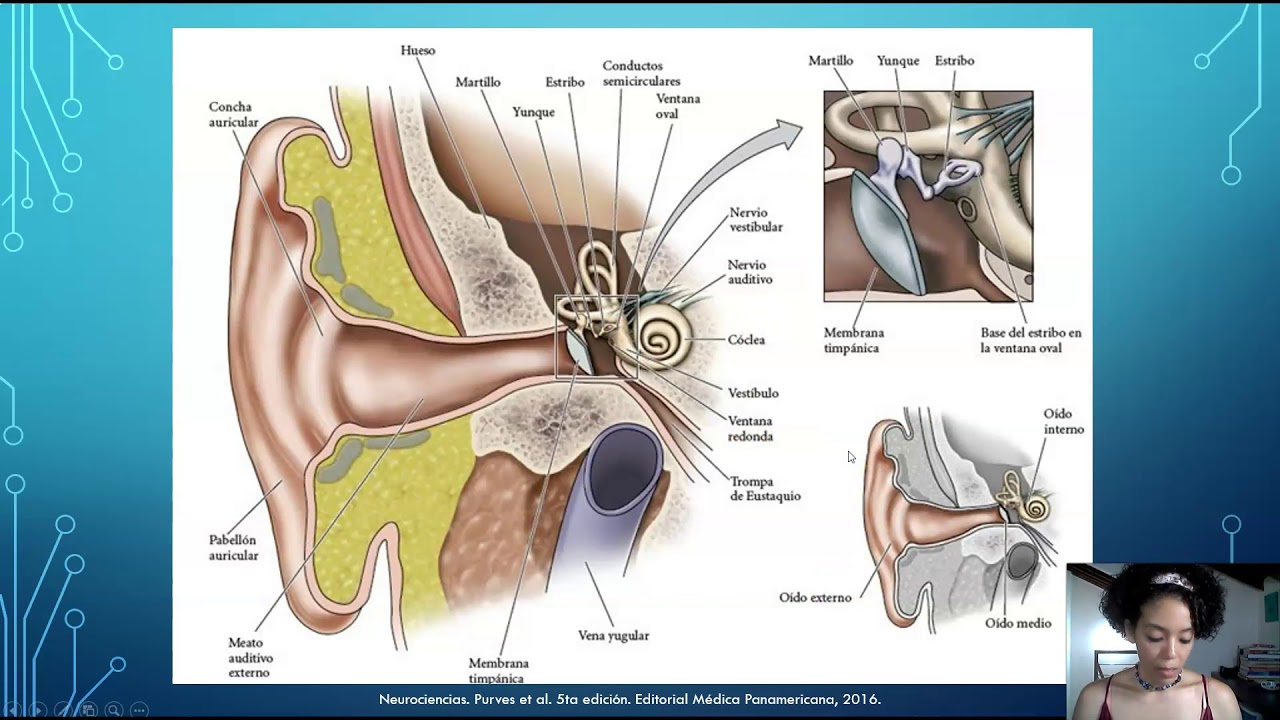
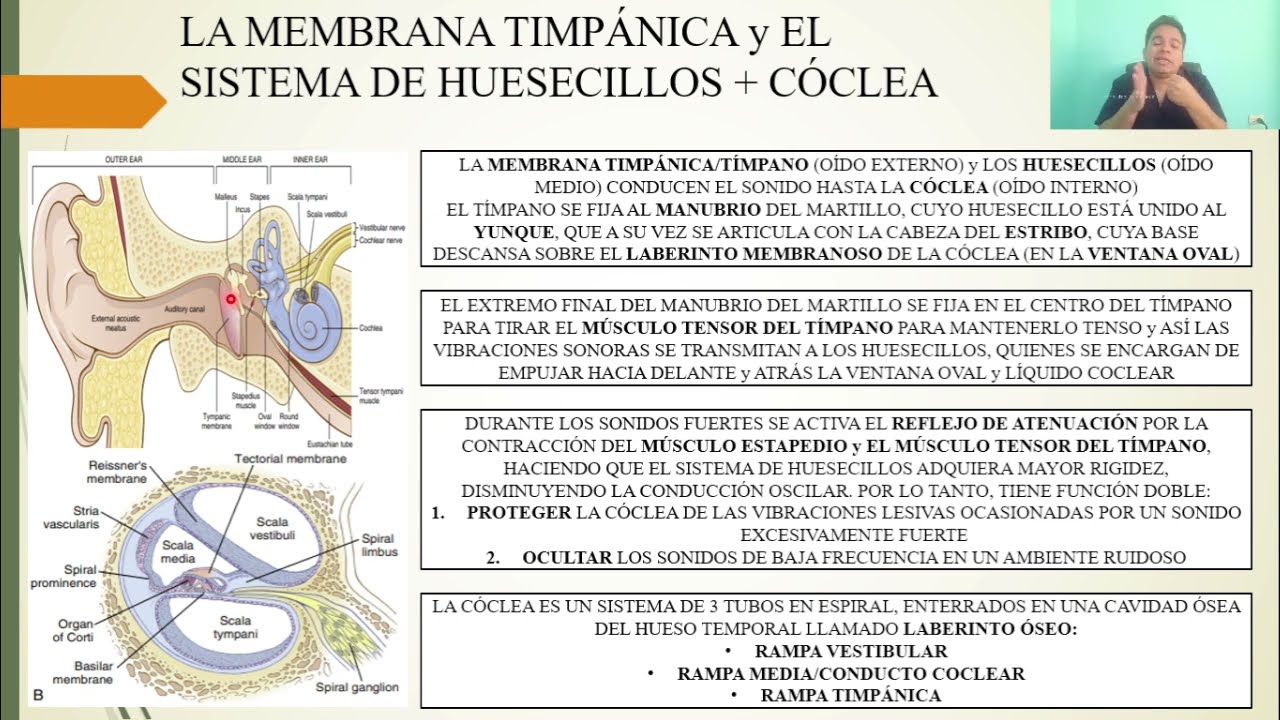
The anatomy of the peripheral auditory system (outer, middle, and inner ear) and the process of mechanoreception by hair cells in the cochlea.
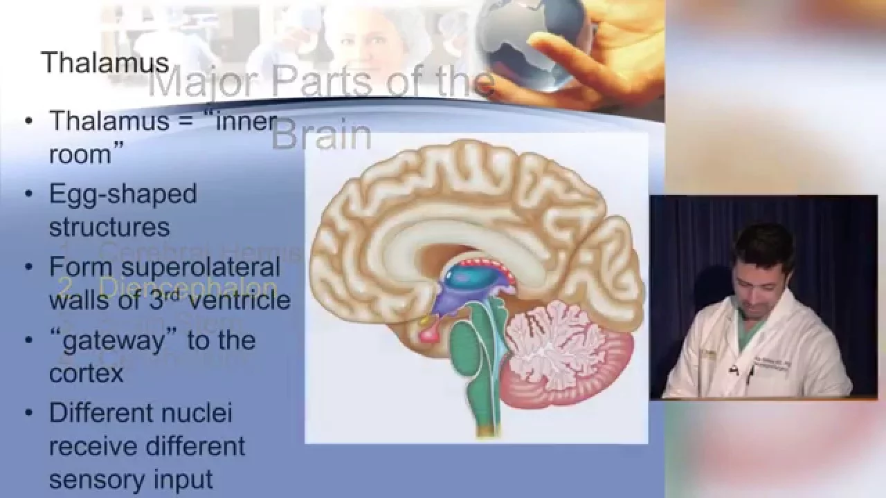
Fundamental neuroanatomy, specifically the structure of the temporal lobe where the primary auditory cortex is located, and the role of the thalamus as a sensory relay station.
General principles of sensory transduction and how neural action potentials transmit sensory information to the brain.
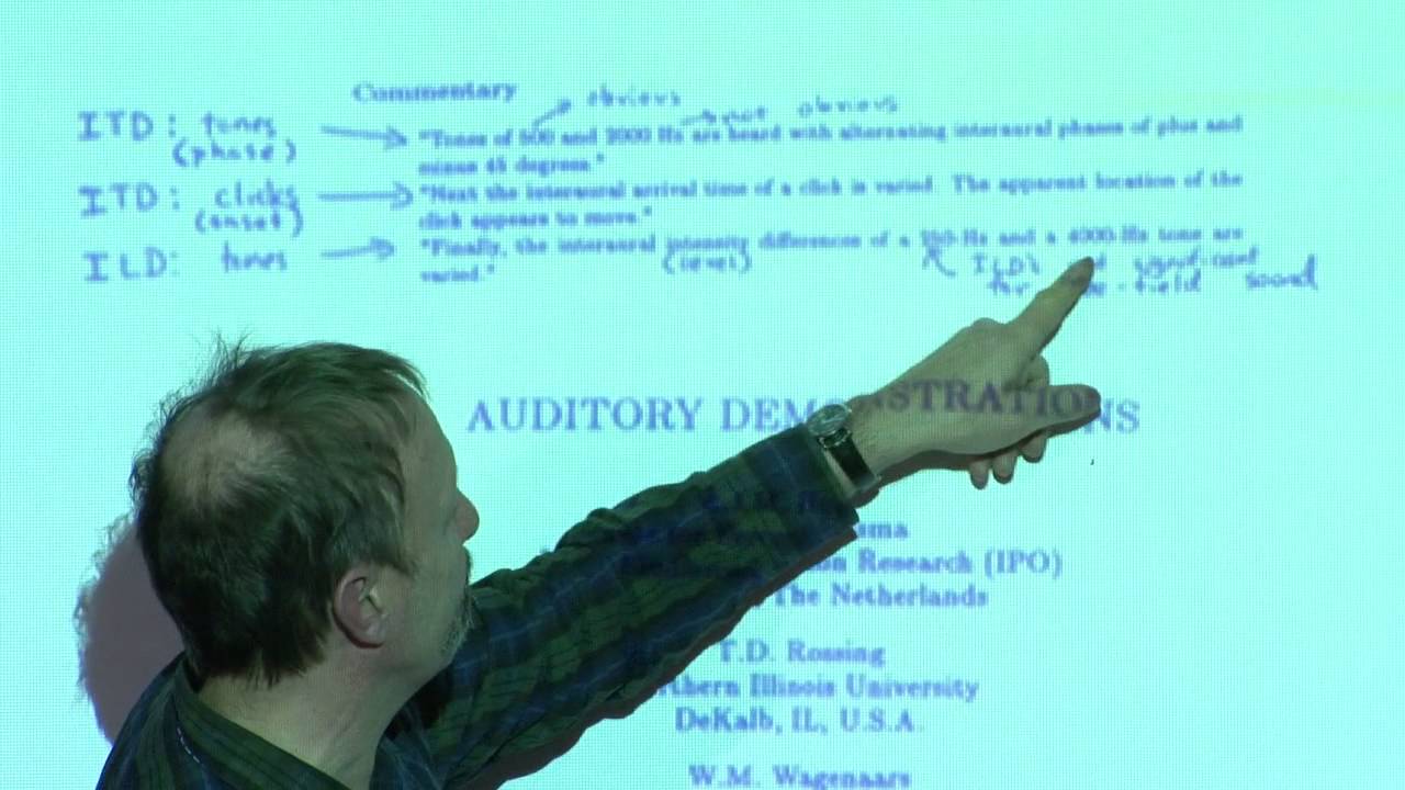
Specific mechanisms of sound localization, including Interaural Time Differences (ITD) and Interaural Level Differences (ILD) processed in the brainstem.
Auditory Scene Analysis (ASA) and the 'cocktail party problem'—how the brain segregates complex, overlapping acoustic environments into distinct auditory objects.
Higher-order language and music processing, including the functions of Wernicke's area and the neural pathways underlying speech perception.
Clinical pathologies of the central auditory system, such as auditory agnosia, word deafness, and the neural basis of tinnitus.
40.4K views634likes15:45Original Release: 2016-11-11
Kristen Asherson here and we're talking about chapter 10, the auditory brain and perceiving auditory scenes. And this is our first lecture and we're talking about the auditory brain. So auditory pathways are pretty complex um as were visual comp um pathways as well um but we don't know quite as much about the auditory pathways um as we do the visual. um partially due to their complexity, partially due to um kind of the differences in how things were studied and the ease in which things were studied. Um so you'll notice that this is a lot more truncated ver compared to the discussions we had about the visual pathways. We'll spend a lot less time on it. Um again, partially that's um to do with um the complexity of it and partially it's to do with they know a lot less about it. So most of this information crosses over just like we have in the visual pathways where we'll see um you know that visual pathway from the different uh the left visual field versus the right visual field was split and it would go to one side or the other. Um we're going to see that most of that is happening too with the auditory pathways but we actually see that the auditory pathways still send information to um the same side. So ipssolateral is same side. So we'll see that information from the left ear will go both to the left hemisphere and to the contrlateral side, the right hemisphere. Um but primarily we're going to see most of that information is going to that contrlateral side, that opposite side, but we will still have some going to the ipsilateral side. So again, sounds from both the left and the right sides of the environment aren't really separate. um and as they are in terms of visual information. So visual information we take the left side and goes to one side and we take the right side and goes to the other side. Um we don't necessarily have this because um of that we won't see that when there's damage to one hemisphere we won't see that it's going to have a major effect on hearing because hearing is happening um it's sending information to both hemispheres. So when we have unilateral brain damage, it's not going to have those same massive effects um that we saw in vision. Um so we're not going to have that. So let's talk about these pathways. And there is a also a website link um in Canvas where it lets you kind of um play with these things a little bit more and see them a little bit more in a little bit more detail. Um so structures in the auditory pathway, they come in pairs. Okay, so we've got two of everything. Um we've got one for um the left side and we've got one for the right side. Uh we so one for the left hemisphere, one for the right hemisphere, one for the left side of the brain stem, one for the right side, one for the left ear, one for the right ear.
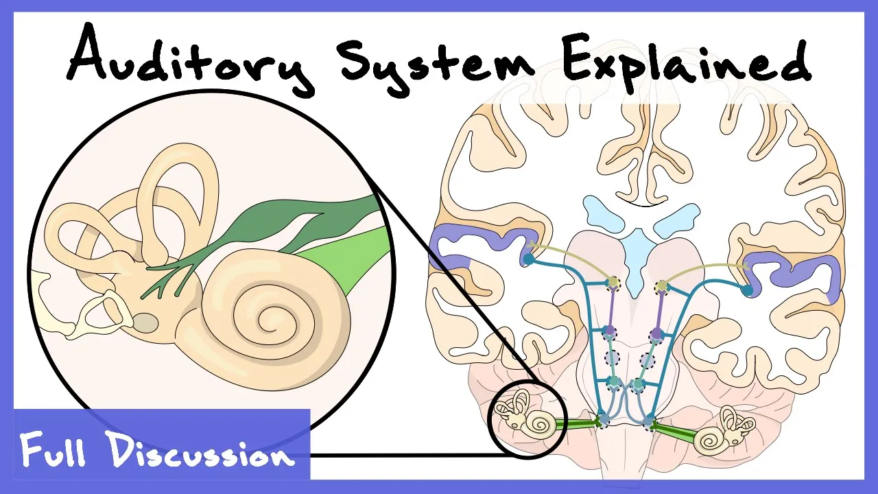
Um and what we where it starts is these type one auditory nerve fibers that are coming out of the cookia um are going to the ipssolateral cclear nucleus. Now that sounds big and scary, but remember ipspsilateral just means it's the same side. So, it's going to go to the colear nucleus that's in the same side um of the brain stem as that ear as that cookia is.
Okay. So, again, here's another way to show you that exact same thing. First, it goes into that ips ipssolateral uh cookar nucleus straight out of the cookia into the auditory nerve. Um and and again, it's going to stay on that ipssilateral side at the start. From here, the klear nuc nucleus is where it starts to split. Okay? and it will split into the main pathways um which will be contrateral. So this is where it's going to say okay most of that information is going to cross over and cross over to the contrlateral side. So the primary pathway is going to cross that dotted line um and it's going to go to the opposite side from where it was hurt.
That being said, there are secondary pathways um which you can see in this image are the thinner lines um and so some information is still maintained on that ipssolateral that same side. Now remember this is really beneficial to us because if we do have brain damage if there are injuries um we're going to see um very little impact. Um this is also has to do with some of the ways that we process sounds um the kinds of sounds in our environment. Um speech is very very complex and speech is one of the main things um that we have evolved to listen to. Um and because it's so complex we can't really have it in just one side of the brain or the other. Um and so information has to go to both sides of the brain um to be able to process this really complex information. So let's talk about some more of the areas that it goes to. um the information. Um so we talked about it going into the cular nucle nucleus um and then either splitting down to the primary um pathways or the secondary pathways. Um we'll talk about each of these where it one of the places it ends up is the medial geniculate body. Um and this is in the phalamus. Um and again there's one on each side of the brain.
So there's a pair of them. Um so if the information is coming from the right cclear um cookia and the right cular um nucleus it will on the primary pathway go to the left hemisphere it'll go contrateral right um and this is the um kind of one of the the last stops um on on its pathway the kind of the end of the pathway um and these are ascending pathways so we talk about them going up you see all these arrows are pointing up um so this is your book discusses as ascending pathways because it's going um kind of that bottom up um kind of thing. The inferior caliculus um is the structure. It's in the midbrain um and it's again a step on that ascending um pathway and it also goes the information after it goes um to from the nucleus um one of the primary places it goes is to the superior olivary complex. Um and this is in the brain stem st still too and again there's two of them. Um and what's really interesting is that they will receive information from both cclear nuclei. Um so the left superior olive complex will receive information from both the right and the left both the ipsilateral and the contrlateral cular nucleus. Um so they're getting information from both of those things.
Um and this is again one of those steps on that ascending pathway.
And this is the primary pathway that we're going to talk about. So again, um you can your book does a really great job of lining that out. We've talked about it some here. And again, there's that website that's linked on Canvas um that shows you that as well. Now, there are also descending signals. And this is kind of the opposite. Um this is kind of going to be from the brain back down. Um and your book talks about it vaguely.
We're not going to really cover it mostly because it's again not well understood. Um, also one of the reasons that we're not going to really cover it much um other than that know that they exist is that um um the the kind of pieces that we do know about them are information that we um didn't cover in this class um really you know to kind of maintain a better level of understanding. So it's important to know that they descending singles do exist.
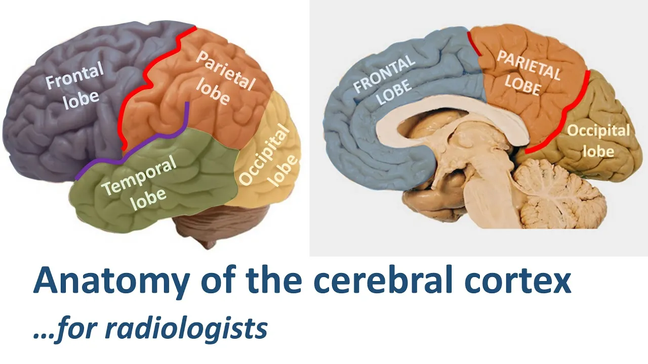
How they work where they go um what functions they're doing none of that's important. um just to know that there is kind of a top down feature going on as well um that we're calling descending signals. So once it gets to the brain the the main place just like we had a visual cortex, we have an auditory cortex um and our auditory cortex um is tucked into that temporal lobe. Okay? So remember when I said you put your hands on your ears um you've got your hand on your temporal loes. Again, we know that our ear, our temporal lobe is going to do hearing because it's right by our ears. This is very, very helpful for us.
Um, again, um, here in the picture, that's going to be this pink area. Um, when it says it's under the lateral sulcus, that's just that fancy way to say like a big kind of like fissure kind of thing. You can see this line, this kind of dotted line they have cut through. Um, that would be the lateral sulcus. And when they kind of cut or take you give you a cross-section across that lateral sulcus, um that's where we're going to be able to start to see um kind of um that this is really happening on both sides of the brain. Um that we've got an auditory cortex on the left and the right um temporal lobe. Um and within that, just like we had a primary visual cortex, we have a primary auditory cortex. Um, and that's the part of the auditory cortex and the auditory core region that I most want you to know about is the auditory cortex, the primary auditory cortex. The other pieces not as interested in. So, this primary auditory cortex, which we call A1, just like we had V1, A1, um, is part of the auditory core region in the auditory cortex. Um, and this this is those main structures that I want you to know that the kind of the the pieces of that aren't important um for your understanding of the material in this class. That's for subsequent classes if you're really interested in this stuff for higher level advanced versions. So the auditory cortex um this primary auditory cortex that I said that I'm interested in you knowing about um A1 is organized tonotopically.
So when we talked about renotopic mapping and we're saying okay well the way the retina maps maps onto the visual cortex this is saying the exact same thing um that the auditory cortex is going to be um tonotopically organized.
There's a map in there um about frequencies. Okay. Now, how exactly it does this, I don't need you to know that it goes you you can see that there's differences in there um and kind of the different patterns where we see low frequencies in certain places and high frequencies in certain places. Um all that's important to know is that it is organized tonotopically.
um that again we'll see kind of these frequency gradients um playing out just like we see in the Basler membrane these frequency gradients we're seeing these frequency gradients playing out in the auditory the primary auditory cortex in the brain. So just as we had in the visual system, this renotopic map that played out in the brain um where the ret the mapping on the retina corresponded to the mapping in the brain, we have the same thing going on with the auditory cortex where the mapping um from the basler membrane is indicative of the mapping that we're getting um in the auditory cortex. the auditory cortex neurons just as the visual neurons were tuned um to certain kinds of things. They were um they had certain receptive fields. They um preferred certain kinds of stimuli um just the same way that the auditory cortex does the same. The neurons do the exact same thing. Um and we can have neurons that are really narrowly tuned um and they only really like a small band of frequency. Um, so this one it would be do best kind of it looks like at about oh somewhere less than 3200 hertz. Um, and it's pretty narrow. And so this where it's its point is is its characteristic frequency. Um, that's where it's happiest. That's where it's firing the best. Um, that's where it's really tuned into that frequency. But we also have broadly tuned neurons as well.
And this is can have still have a characteristic frequency that it's going to fire for, but as you can see, it's much much wider um that we're going to have um it it still fires best um for this one characteristic frequency, but it has a bigger band of frequencies surrounding it um that it also produces this response for. For both of these however um as the amplitude increases which is what you see going up from 0 to 80 um on the y ais as that amplitude increases the bands become much broader.
And this happened for the narrowly tuned neuron just as it's happening for the broadly tuned neuron. Obviously it's gotten a lot broader here um than it did on the one before. Um but as again as this amplitude increases um the frequencies that it's responding to become broader. So the what and the where pathways we have them in the auditory brain too. So all that hard work you did on the visual pathways is paying off. Um so um just like this visual system, the auditory system has two pathways and we can still call them what and where. The what pathway is important in determining the item that's emitting sound. So if I'm trying to figure out what is making that sound, I hear a banging sound or a clicking sound, that information is going to the what pathway. If I'm trying to figure out where this sound is coming from, where that item is that's the sound is coming from, that's going to be on that where pathway. Um, and that's where it it's where it's gonna go. Um so again um just as we had in the visual system how we had these auditory pathways split up into what and where we have the same thing going on the visual pathways. What's really interesting is a lot of this information is going to the same places. Um so you see here in this graph um the auditory what and where pathways are the red lines um in this image and the per the dark dark purple almost black lines are the visual pathways. Okay. So those visual pathways that we talked about before, those are here. Um the auditory pathways are here as well. What's interesting um is these really like kind of light blue tealish areas of the brain. Um those are areas that process both auditory and visual information. Um so these are areas that are telling us where something is. These are areas that are telling us what something is regardless of whether it's an auditory stimuli or a visual stimuli.
What's most important is these things are being integrated in these areas. Um so we talk about these senses as they're completely separate. They're not. Um you're using both your auditory the stream of information from your auditory cortex and the stream of information from your visual cortex. You incorporate that information together. Um and we can see this happening um in the same regions of the brain. Um that the auditory what um the auditory what pathways and the visual what pathways end up in the same place. Um and um or very similar places very adjacent places that they can be connecting. Um the same thing with the um the wear. We're ending up in the same place. Um they may have some specific areas themselves. You can see some areas that are purple that are just auditory processing. You can see some areas that are green that are just visual processing. But there's also a lot of areas that are this teal color that's processing both auditory and visual information. So again, it's really important to remember that we talk about these senses as if they're discreet and which there's not overlap, but they really there really is and there's a lot of overlap. Um and we can see this even at the neural level, even at the structure, the module of the brain level. So this ends our discussion about the auditory brain. Thanks.
Up Next
51:11
Audition Brain Pathway & Hearing Loss Psychology Lecture
@thecognitivepsychologist
699 views•2023-03-13

21:55
Deciphering Neural Circuits Controlling Anorexia in Mice | Richard Palmiter
@AllenInstitute
2.3K views•2011-10-10

4:10
Vagus Nerve (CN X): Anatomy, Nuclei & Functions Explained
@Alilamedicalmedia
305.2K views•2022-10-31

13:03
How Exercise Benefits Your Brain: Science Explained
@TED
11.4M views•2018-03-21
Related Study Plans & Knowledge Roadmaps
Structured learning paths in Neuroscience
neuroscience
Appetite Regulation: Brain Circuits & Hormones
01Foundations of Endocrinology and Brain Anatomy
02Introduction to Energy Homeostasis
03Peripheral Signals: Leptin, Ghrelin, and Insulin
+ 2 more learning modules
14h5 modules
Explore Roadmap
neuroscience
Neuroeconomics: Value, Risk & Brain Mechanisms
01Foundations of Neuroscience and Brain Anatomy
02Introduction to Neuroeconomics and Subjective Value
03Dopaminergic Prediction Errors & Reinforcement Learning
+ 3 more learning modules
18h6 modules
Explore Roadmap
neuroscience
Spatial Navigation: Cells, Grids & Brain Maps
01Foundations of Brain Anatomy and Memory
02Place Cells and Head-Direction Cells
03Grid Cells: The Brain's Coordinate System
+ 2 more learning modules
12h5 modules
Explore Roadmap
neuroscience
BCIs: Neural Decoding & Prosthetic Control
01Neuroscience Foundations: How the Brain Signals Intent
02Signal Acquisition: Recording the Brain's Activity
03Neural Decoding: Translating Brain Waves into Data
+ 2 more learning modules
25h5 modules
Explore Roadmap