




























Single crystal X-ray diffraction determines three-dimensional molecular structures by exploiting the wave nature of X-rays; when X-rays interact with a crystal's ordered atomic lattice, they undergo elastic scattering that creates constructive interference at specific angles (described by Bragg's Law), allowing scientists to calculate atomic positions and understand molecular arrangements in materials ranging from minerals to proteins.
Single Crystal X-ray Diffraction: Bragg's Law Explained
Added:Understanding of wave mechanics, specifically wave interference (constructive and destructive) and diffraction principles.
Basic concepts of crystallography, including crystal lattices, unit cells, and the significance of Miller indices (hkl planes).
The nature of X-rays, including their position on the electromagnetic spectrum and why their wavelengths match atomic distances.
Fundamental trigonometric relationships to comprehend the geometric derivation of Bragg's Law equation.
The 'Phase Problem' in crystallography and the mathematical methods used to solve it, such as direct methods and molecular replacement.
Practical structure determination, including data collection, intensity integration, and structure refinement using specialized software (e.g., SHELX).
Macromolecular crystallography (MX) and its application in determining complex protein and nucleic acid structures for rational drug design.
Comparative study of related techniques, such as Powder X-ray Diffraction (PXRD), neutron diffraction, and electron diffraction (MicroED).
165.1K views3.1Klikes4:44Original Release: 2020-11-19
In a single crystal X-ray diffraction experiment, a crystal is placed in the center of the instrument.
The crystal is constantly rotated in a beam of X-rays, and a 2-dimensional detector records the signals from the sample. From the position and the intensity of these signals, the three-dimensional structure of the sample can be unambiguously determined. Many minerals such as diamond and salt, organic molecules such as sugar and drugs, such as salicylic acid appear as small crystals. In these crystals, atoms, ions or molecules are arranged in a highly ordered structure, forming a crystal lattice that extends in all three dimensions.
This crystal lattice acts as a diffraction grating for X-rays.
It is at this scale that the story of single crystal X-ray diffraction begins.
X-rays are high-energy light with a repeating period called a wavelength or lambda.
Since the wavelength of X-ray light is similar to the distance between atoms in a crystal, X-ray diffraction can be used to determine the distance between atoms in the crystal.
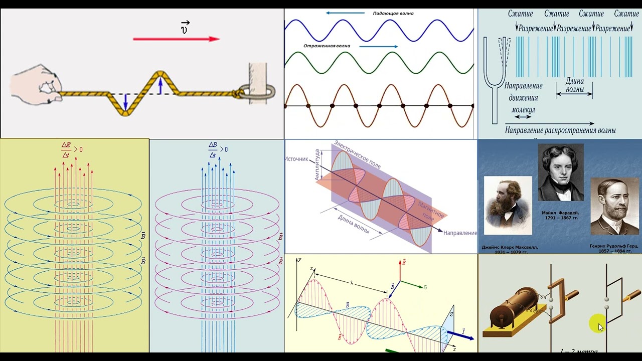
To understand X-ray diffraction, we first have to look into wave properties called interference.
Very similar to throwing two rocks into the water, the circular ripples that result from the two stones seem to pass through one another as they propagate out from where the stones entered.
This is known as interference. If the waves are in alignment, the signal is amplified; this is called constructive interference. If the waves are out of alignment, the signal is destroyed; this is called destructive interference. When an X-ray encounters an atom, its energy is absorbed by the atom's electrons. Electrons occupy special energy states around an atom.
Since the absorbed energy is not large enough to release the electron, the energy must be re-emitted in the form of a new X-ray, with the same energy as the original.
This process is called elastic scattering. In a crystal, the repeating arrangement of atoms, ions or molecules forms a lattice with well-defined repeating distances. When planes in these lattices are exposed to an X-ray beam, X-rays are scattered by this regular arrangement.
Strong amplification of the emitted signal occurs at very specific angles where the scattered waves constructively interfere. This effect is called diffraction. The angle between the incident and scattered beams is called 2-theta. For constructive interference to occur, the scattered waves must be in alignment, meaning the second wave must travel a whole number of wavelengths.
In this case, one half of a wavelength is traveled on the incident side, and one half of a wavelength is traveled on the scattered side, resulting in one additional wavelength.
In the case of the next X-ray, one wavelength is traveled on both the incident and the scattered sides, resulting in two wavelengths. This reinforcement occurs throughout the crystal.
The exact angle at which diffraction occurs can be determined from the red triangle.
The angle at the top is theta, half the angle between the incident and scattered beams.
The long side is the distance between the atomic planes, and the short side is one half of a wavelength.
The relationship between the diffraction angle and the spacing between the atoms can be determined by applying the sine function. Rearranging this equation yields an equation commonly known as Bragg's law, named after Sir William Henry and William Lawrence Bragg, the father-son team who won the Nobel prize in 1915 for their work analyzing crystal structures with X-ray diffraction.
Today, the technique of single crystal X-ray diffraction is used to determine the three-dimensional structure from a wide variety of crystals. Single crystal X-ray diffraction helps scientists to identify the products resulting from chemical reactions, and to understand the mechanisms behind these reactions. Single crystal X-ray diffraction is also applied to determine the structure of proteins, which play a key role in all organisms, including humans.
For example, our ability to see light depends on a protein called rhodopsin, located in the retina of our eye.
Single crystal X-ray diffraction help to understand the mechanisms behind seeing.
Similarly, the detailed knowledge of protein structures is essential to develop new life-saving pharmaceuticals.
Single crystal X-ray diffraction provides important information to finally understand the interaction of new drug candidates with the proteins in our body.
As scientists push their ability to engineer materials on the atomic level, single crystal X-ray diffraction becomes an increasingly important tool in their toolbox. Advances in equipment design have made single crystal X-ray diffraction easier to use and more powerful than ever.
Up Next

48:09
X-ray Diffraction Techniques II | Solid-State Chemistry
@mitocw
37.8K views•2020-12-07

83:56
Biocatalysis and Catalysis in Islatravir Synthesis | Merck Webinar
@SCIwheresciencemeetsbusiness
675 views•2020-10-08
10:06
1H NMR: Determining Number of Peaks from Structure
@MSJChem
59.2K views•2017-04-06

17:31
Edible Water Bottles: A DIY Guide to Sodium Alginate Spherification
@ryan
10.5M views•2019-06-21
Related Study Plans & Knowledge Roadmaps
Structured learning paths in Chemistry
chemistry
Nuclear Chemistry: Decay, Meds & Fuel Cycle
01Introduction to Nuclear Structure and Radioactivity
02Decay Dynamics and Kinetics
03Radiation Interaction and Detection
+ 2 more learning modules
24h5 modules
Explore Roadmap
chemistry
Supramolecular Chemistry: Forces & Machines
01Foundations of Chemical Bonding and Intermolecular Forces
02Introduction to Host-Guest Chemistry
03Thermodynamics and Dynamics of Self-Assembly
+ 2 more learning modules
16h5 modules
Explore Roadmap
chemistry
Photochemistry: Excited States & Reactions
01Foundations of Light-Matter Interactions
02Molecular Orbitals and Electronic States
03Photophysical Processes and Jablonski Diagrams
+ 2 more learning modules
18h5 modules
Explore Roadmap
chemistry
Coordination Chemistry: Bonding & Properties
01Introduction to Transition Metals and Ligands
02Crystal Field Theory (CFT) and Geometry
03Magnetic Behavior and Spin States
+ 2 more learning modules
14h5 modules
Explore Roadmap