Histones are proteins that package DNA and regulate gene expression; HDAC enzymes remove acetyl groups from histones, causing them to bind more tightly to DNA and silence genes. In cancer cells, abnormally high HDAC levels cause excessive deacetylation, silencing critical tumor suppressor genes like p-53 and promoting uncontrolled cell growth. HDAC inhibitors, such as Zalinska (SAHA), block HDAC activity by docking into the enzyme's active site, preventing excessive deacetylation and allowing DNA to remain accessible for normal transcription.
Histone Deacetylation Explained: HDAC Enzymes and Cancer
Added:Deep Dive
Understanding basic chromatin structure, including nucleosomes, histones, and the packaging of DNA within the nucleus.
LECTURE 8.18 Cell-The Unit Of Life
124
Chromatin is the thread-like material inside the nucleus that condenses to form chromosomes. The basic structural unit of chromatin is the nucleosome. Each nucleosome consists of DNA wrapped around histone proteins (H2A, H2B, H3, and H4 - eight histones forming an octamer). This nucleosome model explains how DNA is packaged and organized within the nucleus.
Click to watch →

BIOLOGÍA MAÑANA 04/01
308
Chromatin is the uncondensed form of DNA and proteins found in the nucleus during most of the cell cycle, while chromosomes are the condensed form that appears during cell division. Histones are proteins that help package DNA into chromosomes. In eukaryotic cells, DNA wraps around histone octamers (containing eight histone molecules) to form nucleosomes, the basic units of chromatin. This packaging allows the long DNA molecule to fit inside the cell nucleus while remaining accessible for gene expression.
Click to watch →

03:00 PM - CSIR UGC NET 2020 | Life Science by Priyanka Ma'am | Chromatin Structure and Packaging
20.8K
Chromatin is DNA plus histone proteins that packages DNA within the nucleus; it consists of nucleosomes (histone octamers of H2A, H2B, H3, H4 with 146 bp DNA wrapped around) plus H1 histone and linker DNA (54 bp between nucleosomes), with the complete nucleosome including H1 binding approximately 200 bp of DNA; during S phase chromatin is loosely packaged for replication, while during mitotic phase it becomes highly compact to prevent transcription.
Click to watch →
Histones, Nucleosomes, Chromatin, Euchromatin, and Heterochromatin | MCAT
10.1K
Chromatin is the combination of DNA, histones, and associated proteins that package genetic material in the nucleus. Histones are nucleoproteins (proteins associating with DNA) found in the nucleus. Prokaryotes lack chromatin as defined for eukaryotes because they have a single circular chromosome. Eukaryotes have multiple chromosomes with long DNA strands. Histones include H2A, H2B, H3, and H4, which form a histone octamer. DNA wraps around this octamer like yarn on a spool, and the H1 protein seals it, forming a nucleosome. This structural organization enables efficient DNA packaging within the limited nuclear space.
Click to watch →

SSLC Public Exam | Biology 36+ Marks ഉറപ്പ് | PW Kerala
6.2K
Nucleosomes are the basic units of chromatin packaging in eukaryotic cells. A nucleosome consists of: (1) Histone octamer - eight histone proteins (two each of H2A, H2B, H3, and H4) that form a core around which DNA wraps; (2) DNA - approximately 146 base pairs of DNA wrapped around the histone octamer. The DNA wraps around the histone octamer about 1.65 times, creating a structure that resembles beads on a string. This packaging allows meters of DNA to fit within the nucleus and also regulates gene expression by controlling DNA accessibility.
Click to watch →
Deep Dive
The fundamental principles of gene expression, specifically how transcription factors access DNA and the difference between active (euchromatin) and inactive (heterochromatin) states.

Role Of Chromatin In Gene Regulation In Eukaryotes | B.Sc. Zoology 3rd Semester | Avantika Ma'am |
20.1K
Chromatin exists in two forms: euchromatin (less condensed, transcriptionally active) and heterochromatin (highly condensed, transcriptionally inactive). Euchromatin has nucleosomes spaced apart, allowing transcription factors to access DNA. Heterochromatin has nucleosomes packed closely together, preventing transcription. The transition between these states is regulated by histone modifications and chromatin remodeling complexes.
Click to watch →

Molecular Basis of Inheritance in 180 Minutes | Class 12th Botany | Mind Map Series
132.8K
Chromatin exists in two forms: euchromatin (loosely packed, lightly stained, transcriptionally active) and heterochromatin (tightly packed, darkly stained, transcriptionally inactive). Euchromatin allows RNA polymerase and transcription factors to access the DNA for gene expression, while heterochromatin prevents transcription. This packaging mechanism regulates gene expression by controlling DNA accessibility.
Click to watch →
L3 -Stem Cell Niche I Developmental Biology I IFAS I CSIR
19.9K
Stem cell fate is controlled by transcription factors that regulate gene expression and epigenetic modifications that alter chromatin structure. Transcription factors can activate or repress genes controlling cell cycle progression and differentiation. Epigenetic modifications including DNA methylation and histone modifications (acetylation, methylation, phosphorylation) change chromatin accessibility without altering DNA sequence. These modifications convert chromatin between euchromatin (active transcription) and heterochromatin (inactive transcription) states, providing a mechanism for reversible, heritable changes in gene expression.
Click to watch →

NEET 2025: Genetics | Molecular Basis of Inheritance | Tulika Jha
24.2K
Chromatin exists in two states: (1) Euchromatin is loosely packed with nucleosomes spread apart, appearing light-staining under microscopy, and is transcriptionally active (genes can be expressed). (2) Heterochromatin is densely packed with nucleosomes close together, appearing dark-staining under microscopy, and is transcriptionally inactive (genes cannot be expressed). The central dogma states DNA is transcribed into RNA, and RNA is translated into protein. Euchromatin is loosely packed with nucleosomes spread apart, appearing light-staining under microscopy, and is transcriptionally active. Heterochromatin is densely packed with nucleosomes close together, appearing dark-staining under microscopy, and is transcriptionally inactive.
Click to watch →

Class 12th Biology Sent Up Exam Viral Subjective 2025 | 20 November Biology Sent Up Exam Viral Paper
64.7K
Heterochromatin and euchromatin are two forms of chromatin (DNA-protein complex) in the nucleus. Heterochromatin is highly condensed, dark-staining chromatin that is transcriptionally inactive (genes are not expressed). It appears dark under microscopy because the DNA is tightly packed. Euchromatin is less condensed, light-staining chromatin that is transcriptionally active (genes are being expressed). The degree of condensation affects gene expression: tightly packed heterochromatin prevents transcription, while loosely packed euchromatin allows genes to be accessed and transcribed.
Click to watch →
Deep Dive
The concept of histone acetylation mediated by Histone Acetyltransferases (HATs), which neutralizes positive charges on lysine residues to relax chromatin.

Histone Acetylation: Mechanism and Role in Gene Expression
59.3K
Histone acetylation is a chromatin modification process where acetyl groups are added to lysine residues on histone tails by histone acetyltransferase enzymes, which weakens the electrostatic interaction between positively charged histones and negatively charged DNA phosphate backbone, causing the DNA to wrap more loosely around nucleosomes and become accessible to transcription factors and chromatin remodelers; this modification is associated with euchromatin and active gene expression, and is part of the histone code system where HATs act as writers, bromodomain-containing proteins as readers, and histone deacetylases as erasers.
Click to watch →
Molecular Biology Lecture 55 | Chromatin Remodeling Part-3 Histone Modifications and Histone Variant
1.2K
Histone acetylation involves the transfer of acetyl groups from acetyl-CoA to lysine residues on histone tails, catalyzed by histone acetyltransferases (HATs). This modification neutralizes the positive charge on lysine, reducing the electrostatic interaction between histones and DNA. The result is a loose chromatin structure (euchromatin) where transcription factors and RNA polymerase can access the DNA for transcription.
Click to watch →

2014年度 細胞学概論・2.核の構造と染色体(2.4)
6.2K
Histones (H2A, H2B, H3, H4) have N-terminal tails that can be modified by acetylation. Acetylation neutralizes positive charge, reducing histone-DNA interaction and causing chromatin relaxation (euchromatin). Deacetylation increases positive charge, promoting tighter packing (heterochromatin). In 1996, David Allis discovered histone acetyltransferases (HATs) using Tetrahymena. HATs are essential coactivators for transcription. Epigenetics refers to heritable changes in gene expression without DNA sequence changes, mediated by chromatin modifications.
Click to watch →

Chromatin Remodeling in Eukaryotes
109.7K
Histone acetyltransferase (HAT) removes positive charges from lysine residues on histones by adding an acetyl group. This neutralizes the positive charge, reducing histone-DNA affinity and causing DNA unpackaging. The acetyl group consists of a methyl group attached to a carbonyl carbon, which is added to the lysine residue.
Click to watch →

Histone Acetylation | Part 2 | HATs and HDACs | Epigenetics
249
Histone acetylation is a reversible epigenetic modification where acetyl groups are added to lysine residues on histone tails, neutralizing their positive charge and weakening DNA-histone interactions to make chromatin more accessible; this process is catalyzed by Histone Acetyltransferases (HATs) which include nuclear Type A enzymes (GNAT, MiST, p300/CBP families) that modify nucleosomal histones, and cytoplasmic Type B enzymes that acetylate newly synthesized free histones to facilitate chromatin assembly; conversely, Histone Deacetylases (HDACs) remove acetyl groups and are classified into four classes (Class 1, 2, 3, and 4) with distinct sequence homology and catalytic requirements, where Classes 1, 2, and 4 require zinc ions while Class 3 (sirtuins) requires NAD+ as a cofactor.
Click to watch →
Deep Dive
Basic cancer biology concepts, particularly the physiological role of tumor suppressor genes and how their inactivation leads to uncontrolled cell proliferation.

Neoplasia : Benign vs Malignant Tumors, Hallmarks, Spread & Clinical Manifestations of Cancer
278.3K
Tumor suppressor genes encode proteins regulating cell cycle checkpoints and DNA repair. When DNA damage occurs, these proteins halt proliferation and initiate repair or induce apoptosis. In cancer, tumor suppressor genes are inactivated, leading to uncontrolled proliferation. Examples include retinoblastoma gene, p53 gene (guardian of the genome), APC gene, WT gene, and BRCA1/2 genes.
Click to watch →

Loss of cell cycle control in cancer | Cells | MCAT | Khan Academy
184.2K
Tumor suppressor proteins such as p53 (the 'guardian of the genome') and RB regulate the cell cycle by producing proteins like p21 that inhibit CDK activity, thereby blocking DNA replication and mitosis; when these tumor suppressor genes experience loss-of-function mutations, they lose their ability to control cell division, leading to uncontrolled cell proliferation and cancer development, as evidenced by over 50% of tumors having p53 defects and RB mutations causing retinoblastoma.
Click to watch →
Tumor suppressor gene \ Anti Oncogene \ p53 gene \ Cancer Cell Biochemistry lecture 3
22.7K
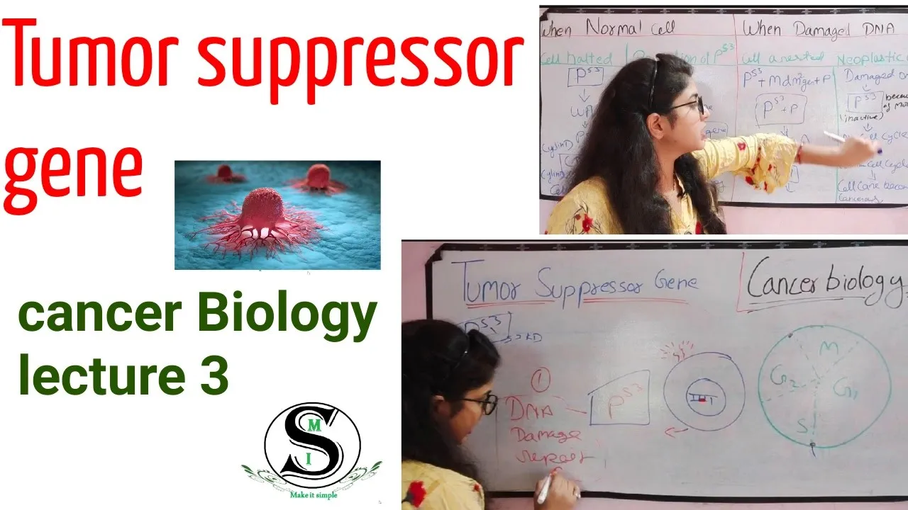
Tumor suppressor genes, often called 'guardians of the genome,' regulate cell division and promote cell death to prevent cancer development. The p53 gene is the most extensively studied tumor suppressor, functioning as a gatekeeper that responds to DNA damage by either halting the cell cycle to allow repair or triggering programmed cell death (apoptosis) when repair is impossible. Under normal conditions, p53 is continuously degraded by the MDM2 protein complex, but upon DNA damage, p53 activates the p21 gene to block cell cycle progression until repairs are completed. When p53 becomes mutated or dysfunctional, cells lose this protective mechanism, leading to uncontrolled cell proliferation and cancer development, particularly in breast and liver cancers.
Click to watch →
Oncogenetics - Mechanism of Cancer (tumor suppressor genes and oncogenes)
1.1M
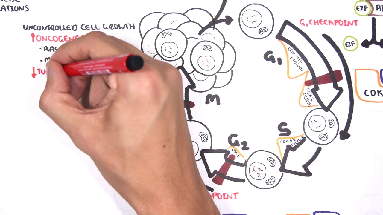
Cancer develops through genetic mutations that disrupt normal cell cycle regulation, specifically through the activation of oncogenes (such as RAS and MYC) which promote uncontrolled cell growth, and the inactivation of tumor suppressor genes (such as p53, APC, and BRCA1/2) which normally enforce cell cycle checkpoints, repair DNA damage, and trigger apoptosis; these mutations cause cells to bypass normal growth controls and proliferate continuously.
Click to watch →
6. Tumour Suppressor Genes (Retinoblastoma and the two hit hypothesis, p53)
197.8K
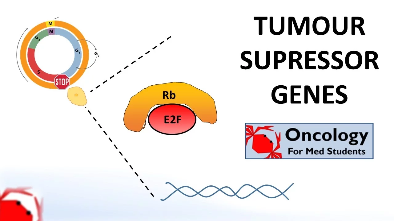
Tumor suppressor genes are genes that produce proteins acting as brakes on the cell cycle to prevent uncontrolled cell division; for these genes to be rendered inactive and contribute to cancer development, both copies (alleles) must be mutated through loss-of-function mutations, a principle known as the two-hit hypothesis. The retinoblastoma (RB) gene exemplifies this mechanism by binding to and inactivating the E2F transcription factor at the G1 checkpoint to prevent cells from entering S phase for DNA replication. Similarly, p53 is another critical tumor suppressor gene that halts damaged cells at checkpoints and triggers apoptosis, with mutations occurring in approximately 50% of all cancers.
Click to watch →
Prerequisite Knowledge
- Concept 01Understanding basic chromatin structure, including nucleosomes, histones, and the packaging of DNA within the nucleus.
- Concept 02The fundamental principles of gene expression, specifically how transcription factors access DNA and the difference between active (euchromatin) and inactive (heterochromatin) states.
- Concept 03The concept of histone acetylation mediated by Histone Acetyltransferases (HATs), which neutralizes positive charges on lysine residues to relax chromatin.
- Concept 04Basic cancer biology concepts, particularly the physiological role of tumor suppressor genes and how their inactivation leads to uncontrolled cell proliferation.
Subsequent Learning
- Step 01The classification of HDAC enzymes (Class I, II, III/Sirtuins, and IV) and their distinct sub-cellular localizations and physiological roles.
- Step 02The pharmacology, clinical applications, and limitations of FDA-approved HDAC inhibitors (such as Vorinostat/SAHA, Romidepsin, and Panobinostat).
- Step 03Synergistic epigenetic therapies, such as combining HDAC inhibitors with DNA Methyltransferase (DNMT) inhibitors or immunotherapies to treat resistant cancers.
- Step 04Advanced molecular techniques used to profile epigenetic landscapes, such as Chromatin Immunoprecipitation Sequencing (ChIP-seq) and Assay for Transposase-Accessible Chromatin (ATAC-seq).
Histone Deacetylation
HDAC Inhibition
Histone Deacetylation
0:03Playing Section
- 1
Histones regulate gene expression by controlling DNA tightness.
- 2
Excess HDAC enzyme over-deacetylates histones, silencing key DNA.
- 3
Silenced tumor suppressor genes, like p53, lead to uncontrolled cell growth.
Non-Histone Targets and Context-Dependent Dual Roles of HDACs
While the traditional model portrays HDACs strictly as epigenetic silencers of tumor suppressors, scientific consensus has shifted toward a more complex reality. First, HDACs deacetylate thousands of non-histone proteins—including p53, tubulin, and HSP90. Therefore, the therapeutic efficacy (and high toxicity) of HDAC inhibitors often stems from disrupting these non-epigenetic pathways rather than simple chromatin remodeling. Second, HDACs do not function solely as oncogenes; in several contexts, such as specific stages of breast, colorectal, and liver cancers, certain HDAC isoforms act as tumor suppressors. Inhibiting them can paradoxically promote epithelial-mesenchymal transition (EMT), metastasis, and chemotherapy resistance. This context-dependent behavior explains why HDAC inhibitors have historically shown disappointing results in solid tumors, challenging the simplistic narrative that HDACs are universal oncogenic targets.
Deep Dive
The classification of HDAC enzymes (Class I, II, III/Sirtuins, and IV) and their distinct sub-cellular localizations and physiological roles.

Mod-03 Lec-07 Role of chromatin in eukaryotic gene regulation
7.2K
Human HDACs are classified into four classes based on homology to yeast RPD3: Class I (HDAC1, 2, 3, 8), Class II (HDAC4, 5, 6, 7, 8, 9), Class III (sirtuins, NAD+-dependent), and Class IV (HDAC11). Aberrant HDAC activity is implicated in cancer: HDAC1 overexpression occurs in prostate, gastric, and colorectal cancers; HDAC2 in colorectal and gastric cancers; HDAC3 in lung cancer. Knockdown of HDAC8 inhibits tumor cell growth, suggesting HDACs silence tumor suppressor genes. The cAMP signaling pathway demonstrates how extracellular signals activate transcription: growth factors activate adenylyl cyclase, producing cAMP that binds protein kinase A. Phosphorylated CREB then recruits CBP/p300 HATs to cAMP response element promoters, causing histone acetylation and transcriptional activation. Nuclear receptor signaling demonstrates repression through HDAC dissociation: in the absence of thyroid hormone, the receptor binds to thyroid hormone response elements and recruits corepressor complexes containing HDACs, causing histone deacetylation and repression. Upon thyroid hormone binding, conformational changes prevent corepressor recruitment, releasing HDACs and enabling transcriptional activation.
Click to watch →

HDSA Research Webinar: Histone deacetylase HDAC inhibitors
1.2K
The HDAC family consists of 18 different enzymes divided into four classes: Class I includes HDACs 1, 2, 3, and 8; Class II includes HDACs 4, 5, 6, 7, 9, 10; Class III are sirtuins (NAD-dependent); Class IV contains only HDAC11. Research has revealed that different HDAC isoforms play distinct roles in the brain—some are neurotoxic while others are neuroprotective. This diversity explains why broad HDAC inhibition can cause toxicity and highlights the importance of isoform-selective inhibitors for therapeutic applications.
Click to watch →

Toolbox to evaluate biological function of histone deacetylases
164
There are 11 isozymes of metal-dependent HDACs divided into three classes (Class 1, 2, and 4), plus seven NAD-dependent HDACs (sirtuins, Class 3). Global loss of any Class 1 HDAC is lethal in mice, demonstrating their essential biological roles. However, the individual roles, regulation, and substrate specificity of individual HDAC isozymes remain poorly understood. This highlights the complexity of studying specific HDAC functions within the larger family of deacetylating enzymes.
Click to watch →
NAD+: Protokół zdrowia, energii, urody i długowieczności - Professor odc. 136
21K
Sirtuins are seven enzymes (SIRT1-SIRT7) present in cytosol, mitochondria, and nucleus. They function as histone deacetylases that control gene expression by removing acetyl groups from histones, tightening DNA packaging and inhibiting harmful gene transcription. Sirtuins also repair DNA, control metabolism, increase insulin sensitivity, stimulate fat burning, prevent muscle breakdown, stimulate mitochondrial production, and trigger autophagy. They act as sensors for biological, physical, chemical, and energetic stress, making them central to longevity mechanisms.
Click to watch →
Histone Acetylation | Part 2 | HATs and HDACs | Epigenetics
249
Histone acetylation is a reversible epigenetic modification where acetyl groups are added to lysine residues on histone tails, neutralizing their positive charge and weakening DNA-histone interactions to make chromatin more accessible; this process is catalyzed by Histone Acetyltransferases (HATs) which include nuclear Type A enzymes (GNAT, MiST, p300/CBP families) that modify nucleosomal histones, and cytoplasmic Type B enzymes that acetylate newly synthesized free histones to facilitate chromatin assembly; conversely, Histone Deacetylases (HDACs) remove acetyl groups and are classified into four classes (Class 1, 2, 3, and 4) with distinct sequence homology and catalytic requirements, where Classes 1, 2, and 4 require zinc ions while Class 3 (sirtuins) requires NAD+ as a cofactor.
Click to watch →
Deep Dive
The pharmacology, clinical applications, and limitations of FDA-approved HDAC inhibitors (such as Vorinostat/SAHA, Romidepsin, and Panobinostat).

Episode 352: Pharmacology 101: Epigenetics
325
HDAC inhibitors (histone deacetylase inhibitors) are drugs that block histone deacetylase enzymes, which remove acetyl groups from histones. By blocking these enzymes, HDAC inhibitors increase histone acetylation, which loosens the DNA around chromatin and allows tumor suppressor genes to be expressed. There are four FDA-approved HDAC inhibitors: vorinostat (2006), romidepsin (2009), belinostat (2014), and panobinostat (2015). Vorinostat, romidepsin, and belinostat are approved for cutaneous T-cell lymphoma, while panobinostat is approved for refractory multiple myeloma.
Click to watch →

Romas Geleziunas (Gilead)
985
Histone deacetylase (HDAC) inhibitors represent a promising class of latency reversal agents. HDAC enzymes suppress gene transcription by removing acetyl groups from histones; blocking them with inhibitors creates relaxed chromatin that favors transcription. Multiple laboratories have shown HDAC inhibitors activate latent HIV. Three HDAC inhibitors have reached clinical studies: vorinostat (FDA-approved for lymphomas), panobinostat (phase three), and romidepsin (FDA-approved). Clinical studies in the US, Australia, and Denmark have shown increased HIV expression in CD4+ cells, with some evidence of plasma viral blips, suggesting potential in vivo activity.
Click to watch →

2011 Bay Area Symposium on Viruses: Mini Balakrishnan & Closing Remarks
968
HDAC inhibitors reverse HIV latency by modifying chromatin structure—shifting from condensed, transcriptionally silent states to open, permissive configurations. These zinc metalloenzymes remove acetyl groups from histones and transcription factors. SAHA (FDA-approved for lymphoma) activates latent HIV in patient-derived resting CD4 T cells without inducing global T-cell activation, offering therapeutic advantage. Four HDAC classes exist with varying substrate specificities: pan-inhibitors (broad spectrum) versus class I-selective agents (romidepsin, mocetinostat). Clinical trials are underway in the US and Australia. Combination approaches targeting multiple signaling pathways may achieve more robust reactivation than single agents.
Click to watch →

Pharmacology | High Yield MCQ Revision Series for NEET PG || Dr. J. Thirunavukkarasu, M.D.,
3.9K
Histone deacetylase (HDAC) inhibitors are used in lymphoma treatment. Vorinostat and romidepsin are HDAC inhibitors approved for cutaneous T-cell lymphoma (mycosis fungoides). Panobinostat is another HDAC inhibitor used for multiple myeloma. Belinostat is used for peripheral T-cell lymphoma. These drugs work by modifying chromatin structure to reactivate tumor suppressor genes and induce cancer cell death.
Click to watch →
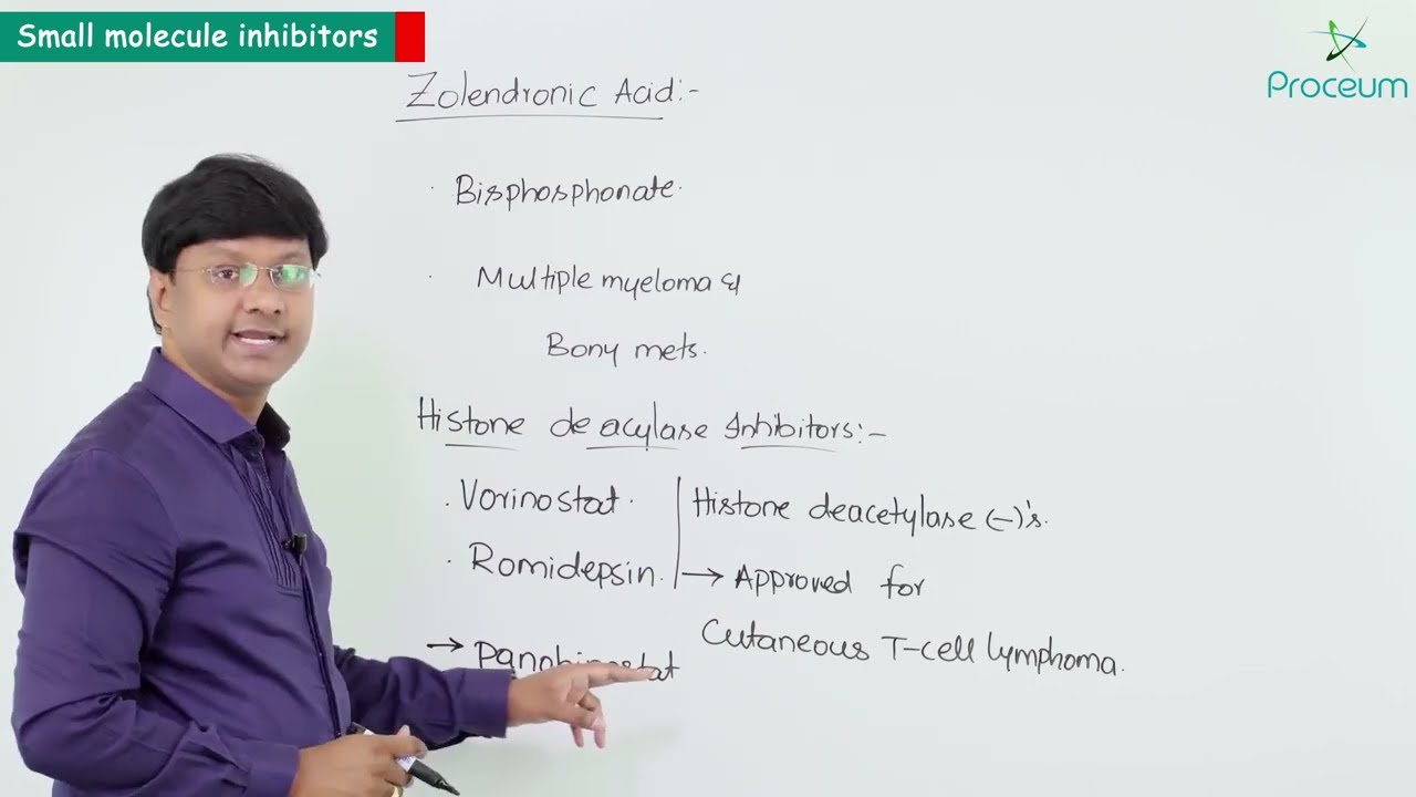
5. Anti- Cancer Drugs: Enginol Mebutate: Pharmacology Video Lectures
229
Histone deacetylase inhibitors (HDAC inhibitors) include vorinostat and romidepsin, which are approved for cutaneous T-cell lymphoma. Panobinostat is a newer HDAC inhibitor approved for multiple myeloma. Belinostat is approved for relapsed or refractory peripheral T-cell lymphoma.
Click to watch →
Deep Dive
Synergistic epigenetic therapies, such as combining HDAC inhibitors with DNA Methyltransferase (DNMT) inhibitors or immunotherapies to treat resistant cancers.

Blewitt ME (2011): Epigenetics and Cancer
2.4K
Four FDA-approved epigenetic drugs exist: two DNA methyltransferase inhibitors (5-azacytidine, decitabine) and two histone deacetylase inhibitors (vorinostat, romidepsin). DNA methyltransferase inhibitors are base analogs that deplete DNA methyltransferase 1, reducing global methylation. Challenges include acquired resistance, lack of specificity, and uncertainty about whether demethylation alone reactivates genes. Histone deacetylase inhibitors affect multiple classes and have unclear mechanisms. Synergism between HDAC and DNA methyltransferase inhibitors may be more effective than single agents. Future directions include targeting histone methyltransferases and using combination therapies to achieve greater therapeutic benefit.
Click to watch →
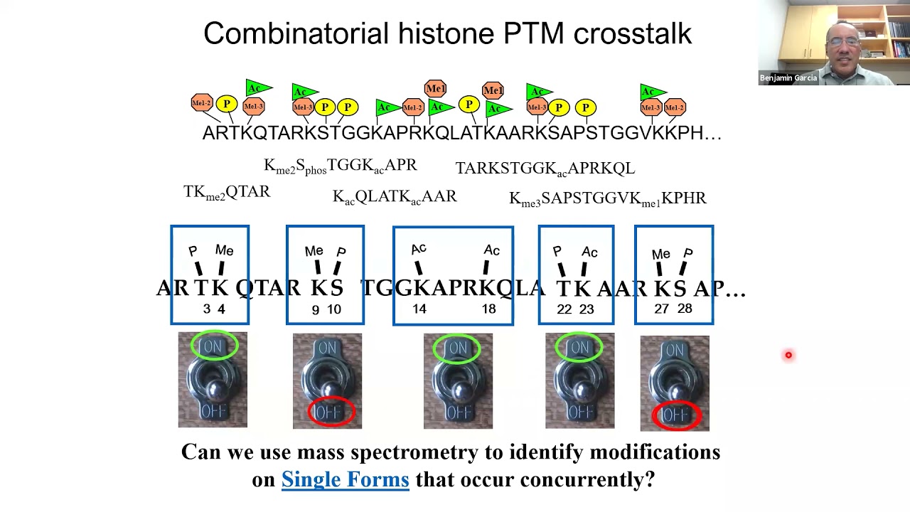
CCMB Seminar Series 09 22 2021 Benjamin A Garcia, PhD
207
PRC2-deficient MPNST cells show dramatically increased sensitivity to HDAC inhibitors compared to PRC2-retained cells, with panobinostat causing re-expression of interferon and immune response genes including HLA proteins. This sensitivity arises because PRC2-deficient cells already have elevated acetylation levels, making them more responsive to further HDAC inhibition that drives acetylation even higher. Combining HDAC inhibitors with DNA methyltransferase inhibitors produces synergistic anti-tumor effects, as the combination addresses both the elevated acetylation and compensatory hypermethylation characterizing PRC2-deficient tumors. Reintroducing functional PRC2 into PRC2-deficient cells reduces their sensitivity to HDAC inhibitors, confirming that heightened sensitivity relates directly to underlying epigenetic dysregulation. This research demonstrates that PRC2 loss creates a complex network of interdependent epigenetic changes—loss of silencing mark, gain of active marks, and compensatory DNA hypermethylation—that collectively drive tumor progression and immune evasion, providing multiple therapeutic targets for combinatorial epigenetic therapy.
Click to watch →
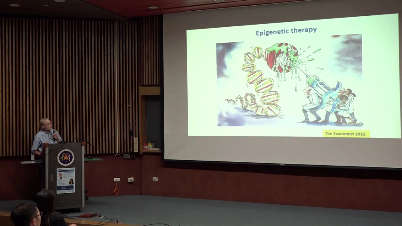
Cancer Epigenetics Special Lectures
764
Combining PARP inhibitors with DNA methyltransferase inhibitors enhances anti-cancer effects through synergistic mechanisms. Both drugs trap their respective targets (PARP and DNMT1) onto DNA, creating persistent protein-DNA complexes that cannot be efficiently repaired. This combination significantly increases cytotoxicity in triple-negative breast cancer and acute myeloid leukemia models. The drugs work together to maximize DNA repair pathway disruption while minimizing resistance mechanisms. Clinical trials are underway to translate these preclinical findings into effective treatments for patients who do not respond to single-agent therapy.
Click to watch →
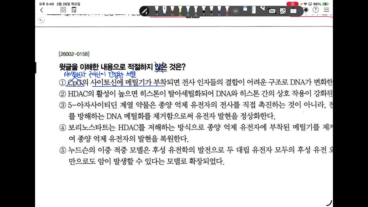
2027 수능특강 독서 과학기술1 후성유전학 항암치료
146
Conventional chemotherapy drugs target rapidly dividing cells but cause significant side effects by damaging healthy cells. Epigenetic drugs offer an alternative approach by correcting epigenetic errors rather than directly attacking cells. DNMT inhibitors, such as azacitidine and decitabine, work by inhibiting DNA methyltransferase enzymes. This reduces DNA methylation levels and restores the expression of tumor suppressor genes that were silenced by excessive methylation. HDAC inhibitors like vorinostat work by blocking histone deacetylase enzymes, preventing the removal of acetyl groups from histones. This maintains histone acetylation levels and promotes transcription of tumor suppressor genes. Both types of epigenetic drugs aim to restore normal cellular function by correcting epigenetic silencing of tumor suppressor genes, potentially allowing cancer cells to undergo programmed cell death.
Click to watch →

Mecanismos Epigenéticos
38.2K
Epigenetic modifications are reversible, unlike genetic mutations, making them promising targets for cancer therapy. Several epigenetic drugs have been developed, including DNA methyltransferase inhibitors and histone deacetylase inhibitors. For example, temozolomide is used to treat glioblastoma patients whose MGMT gene is methylated (and thus not expressed). Since MGMT normally repairs DNA damage caused by temozolomide, patients with methylated MGMT cannot repair the drug-induced damage and respond better to treatment. This demonstrates how understanding epigenetic patterns can guide personalized cancer treatment approaches.
Click to watch →
Deep Dive
Advanced molecular techniques used to profile epigenetic landscapes, such as Chromatin Immunoprecipitation Sequencing (ChIP-seq) and Assay for Transposase-Accessible Chromatin (ATAC-seq).
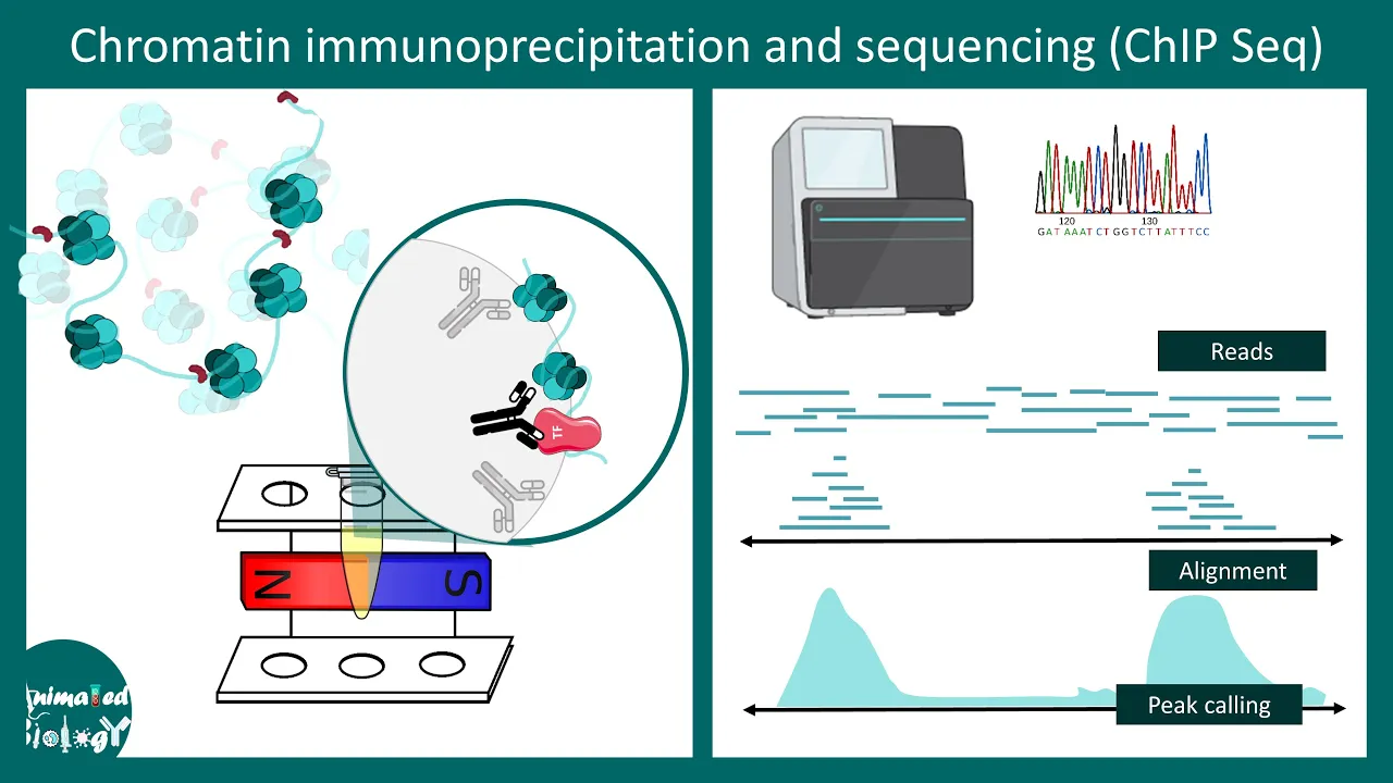
ChiP seq | Chromatin immunoprecipitation and sequencing | Methods in biology | CSIR NET
14.1K
ChIP-seq serves diverse applications including transcription factor binding mapping, epigenetic modification profiling, histone modification analysis, enhancer/promoter identification, and active transcription detection. Specific examples include H2A.Z variant mapping to promoters, H3.3 enrichment in actively transcribing gene bodies, and serine 5-phosphorylated RNA polymerase II distribution indicating transcriptional activity. The true power emerges when integrating ChIP-seq with complementary techniques: ATAC-seq measures chromatin accessibility revealing whether regions are open for transcription factor binding, while RNA-seq quantifies gene expression levels. This multi-omics approach enables understanding of how cell-type-specific transcription factor binding, chromatin accessibility changes, and resulting gene expression patterns coordinate during cellular differentiation and function.
Click to watch →
![[Wikipedia] ATAC-seq](https://i.ytimg.com/vi/sXa82E0Bxmk/maxresdefault.jpg)
[Wikipedia] ATAC-seq
2.3K
ATAC-seq (Assay for Transposase-Accessible Chromatin with high throughput sequencing) is a molecular biology technique that uses hyperactive transposase enzymes to identify accessible DNA regions in the genome; the transposase preferentially cuts and tags nucleosome-free DNA, and the resulting sequencing reads reveal regulatory elements like promoters and enhancers, with depleted signal regions indicating DNA-binding protein footprints.
Click to watch →
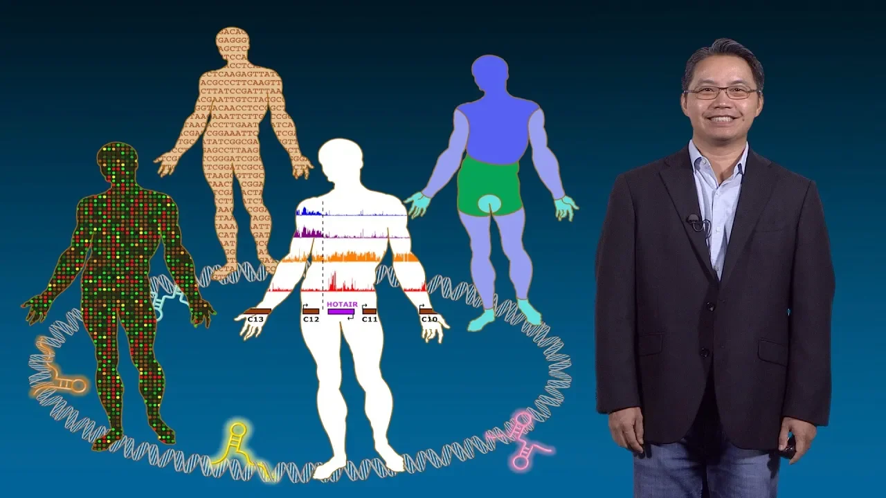
Howard Chang (Stanford, HHMI) 1: Epigenomic Technologies
32K
ATAC-seq uses a bacterial transposase enzyme to map accessible chromatin regions by inserting sequencing adapters only into open chromatin sites. This technology provides million-fold sensitivity improvements over previous methods. A key feature is transcription factor footprinting—approximately 8-10 base pair dips at peak summits where proteins block transposase insertion. By comparing these footprints against known transcription factor binding preferences, researchers can identify which factors occupy specific genomic locations, revealing the complete cast of regulatory proteins active in any cell type.
Click to watch →

Regulatory Genomics - Deep Learning in Life Sciences - Lecture 07 (Spring 2021)
10.2K
Three main technologies map regulatory elements: (1) ChIP-seq uses antibodies to precipitate protein-bound DNA, then sequences and maps it back to the genome; (2) DNase-seq uses DNase I to cut accessible chromatin, revealing nucleosome-free regions; (3) ATAC-seq uses Tn5 transposase to insert adapters only into accessible regions. Each technology provides complementary information about where transcription factors bind and where chromatin is accessible for regulation.
Click to watch →
Intro to ATAC-Seq: Method overview and comparison to ChIP-Seq
4.5K
ATAC-Seq (Assay for Transposase-Accessible Chromatin using sequencing) is a powerful, high-throughput method for detecting open chromatin regions across the genome, serving as a culmination of all ChIP-Seq histone modification peaks; unlike ChIP-Seq which requires specific antibodies and large cell numbers (millions of cells), ATAC-Seq needs minimal input (as few as 10,000 cells) and provides a rapid, one-step workflow (4-6 hours to library preparation) making it ideal for exploratory epigenetic studies and clinical applications where sample availability is limited.
Click to watch →
Histone Deacetylation
HDAC Inhibition
Histone Deacetylation
0:03- 1
Histones regulate gene expression by controlling DNA tightness.
- 2
Excess HDAC enzyme over-deacetylates histones, silencing key DNA.
- 3
Silenced tumor suppressor genes, like p53, lead to uncontrolled cell growth.
Non-Histone Targets and Context-Dependent Dual Roles of HDACs
While the traditional model portrays HDACs strictly as epigenetic silencers of tumor suppressors, scientific consensus has shifted toward a more complex reality. First, HDACs deacetylate thousands of non-histone proteins—including p53, tubulin, and HSP90. Therefore, the therapeutic efficacy (and high toxicity) of HDAC inhibitors often stems from disrupting these non-epigenetic pathways rather than simple chromatin remodeling. Second, HDACs do not function solely as oncogenes; in several contexts, such as specific stages of breast, colorectal, and liver cancers, certain HDAC isoforms act as tumor suppressors. Inhibiting them can paradoxically promote epithelial-mesenchymal transition (EMT), metastasis, and chemotherapy resistance. This context-dependent behavior explains why HDAC inhibitors have historically shown disappointing results in solid tumors, challenging the simplistic narrative that HDACs are universal oncogenic targets.
Understanding basic chromatin structure, including nucleosomes, histones, and the packaging of DNA within the nucleus.
The fundamental principles of gene expression, specifically how transcription factors access DNA and the difference between active (euchromatin) and inactive (heterochromatin) states.
The concept of histone acetylation mediated by Histone Acetyltransferases (HATs), which neutralizes positive charges on lysine residues to relax chromatin.
Basic cancer biology concepts, particularly the physiological role of tumor suppressor genes and how their inactivation leads to uncontrolled cell proliferation.
The classification of HDAC enzymes (Class I, II, III/Sirtuins, and IV) and their distinct sub-cellular localizations and physiological roles.
The pharmacology, clinical applications, and limitations of FDA-approved HDAC inhibitors (such as Vorinostat/SAHA, Romidepsin, and Panobinostat).
Synergistic epigenetic therapies, such as combining HDAC inhibitors with DNA Methyltransferase (DNMT) inhibitors or immunotherapies to treat resistant cancers.
Advanced molecular techniques used to profile epigenetic landscapes, such as Chromatin Immunoprecipitation Sequencing (ChIP-seq) and Assay for Transposase-Accessible Chromatin (ATAC-seq).
56.7K views172likes3:18@URIanimationOriginal Release: 2007-07-03
Histones are proteins that play a role in the regulation of transcription by helping to condense the genetic material DNA into its compact form.
Histones play a major role in helping transcription factors to bind to a specific area of the DNA.
[Music] As you can see, the DNA wraps around the histones.
Modification of histone proteins can control the tightness of DNA around the histone proteins and consequently control the expression of the genes.
[Music] An enzyme called HDAC performs deacilation.
[Music] HD deck interacts with the acetilated lysene on the surface of the histone causing the removal of acetil from the structure.
[Music] So long as normal levels of HDAC are present, there isn't a problem. However, when abnormally high levels of HDAC exist, as in cancer cells, deacilation becomes out of control. An overexpressed HDAC causes too much deacilation to a histone, which in turn grasps too tightly to the DNA.
The result is the segment of DNA associated to the over deacitilated histone is squeezed so tightly that it becomes silenced. In other words, the DNA is no longer readable. This creates a problem when the piece of DNA that is silenced is vital to maintain the cell cycle. One such example is p-53, a tumor suppressor gene. Excessive deacilation causes the associated histone to silence the tumor suppressing DNA segment resulting in uncontrollable cell growth i.e. cancer cells.
The solution to this problem was to design HDAC inhibitors which block out the HDAC enzyme. One such drug Zalinsza also known as SAHA docks into the active site of the enzyme.
Further deacitilation is prevented and the histone does not increase its grip on the DNA segment. Consequently, the DNA segment remains accessible for transcription and the cells can continue working with the DNA segment as it naturally would [Music] [Music]
Up Next

18:20
Chromosome Structure and Chromatin Organization Explained
@animatedbiologywitharpan
128.8K views•2023-01-28

50:12
Bacterial Communication: Quorum Sensing and Biofilm Formation Explained
@JHUAAP
12.6K views•2011-06-28

5:35
Enteric Nervous System Explained: The Gut's Brain | Neurobiology Lecture
@alumniu6029
438 views•2018-09-12

7:08
Bacteriophages: Earth's Deadliest Killers and Future Antibiotics
@kurzgesagt
34.6M views•2018-05-13
Related Study Plans & Knowledge Roadmaps
Structured learning paths in Biology
biology
RNAi: Biogenesis, RISC & Gene Silencing
01Molecular Biology Foundations: DNA to Protein
02Introduction to RNA Interference (RNAi)
03Molecular Biogenesis of miRNA and siRNA
+ 2 more learning modules
10h5 modules
Explore Roadmap
biology
Mycorrhizae: Signaling, Biophysics & Ecology
01Foundations of Mycorrhizal Symbiosis
02Molecular Signaling and Root Colonization
03Biophysics and Bioenergetics of Nutrient Exchange
+ 1 more learning modules
11h4 modules
Explore Roadmap
biology
Circadian Clocks: TTFL, SCN & Periphery
01Foundations of Molecular Biology & Gene Expression
02Introduction to Chronobiology and Actograms
03The Core TTFL: Molecular Architecture of the Clock
+ 3 more learning modules
14h6 modules
Explore Roadmap
biology
Population Genetics: Math, Drift & Selection
01Genetic & Probability Foundations
02The Hardy-Weinberg Principle and Baseline Models
03Mathematical Modeling of Natural Selection
+ 3 more learning modules
18h6 modules
Explore Roadmap