The olfactory pathway transmits smell information through a series of specialized structures: olfactory receptor cells in the nasal mucosa detect odorants and send signals via olfactory rootlets and nerves that pass through the cribriform plate into the olfactory bulb, where they synapse with mitral and tufted cells; these cells project as the olfactory tract to the primary olfactory cortex (medial and lateral olfactory striae) for basic smell perception, and then to the secondary olfactory cortex (entorhinal area and uncus) for memory and emotional associations related to smell.
Olfactory Nerve and Pathway | Nasal Cavity to Cortex
Added:Deep Dive
Basic anatomy of the nasal cavity, including the cribriform plate of the ethmoid bone and the nasal mucosa.

Lecture:2 P1 lateral view of skull + pterion + Clinical
3.5K
The ethmoid bone contains the cribriform plate (sieve-like structure forming the nasal cavity roof), lamina papyracea (paper-thin lateral wall), and three pairs of conchae (superior, middle, inferior). The cribriform plate contains openings for olfactory nerve passage to the brain. The perpendicular plate forms the superior nasal septum. The ethmoid bone contributes to both lateral and medial orbital walls. The lamina papyracea separates the nasal cavity from ethmoidal air cells. The inferior concha is a separate bone, not part of the ethmoid.
Click to watch →

MOSTRACIÓN ANATÓMICA DE CABEZA Y CUELLO
293
The ethmoid bone is a complex bone located in the medial skull base, approximately thumb-sized. It contains the cribriform plate (roof of nasal cavity with olfactory foramina), perpendicular plate (forms superior nasal septum), and conchae (superior and middle turbinates). The perpendicular plate articulates with the vomer to complete the septum. The conchae create meatuses for sinus drainage. The ethmoidal cells (anterior, middle, posterior) are air-filled spaces lined with mucosa that drains into the middle and superior meatuses.
Click to watch →
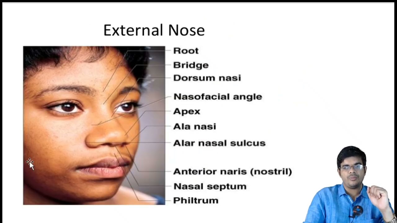
#anatomy #nosedoctor #nose #pns #doctor #eardoctor #education #neet #neetpg #otorhinolaryngology
102
The nasal cavity divides into the vestibule (anterior-inferior, skin-lined with hair follicles prone to fungal infections) and proper cavity. The ethmoid bone contributes the cribriform plate (perforated for olfactory nerve passage), perpendicular plate (nasal septum component), lamina papyracea (separating nasal cavity from orbit), and superior/middle turbinates. Olfactory sensation requires sniffing to direct air currents to the upper one-third where olfactory epithelium resides. The ethmoid sinuses include cribriform plate, perpendicular plate, lamina papyracea, superior/middle turbinates, and air cells. Ethmoid air cells divide into anterior and posterior groups separated by the ground lamella (horizontal middle turbinate portion). Anterior cells drain into middle meatus; posterior cells drain into superior meatus and ethmoidal recess.
Click to watch →

الاشعة | المرحلة الثانية | التشريح |Normal anatomy of the skull (cranial bones) | المحاضرة 1
3K
The ethmoid bone is a single spongy bone between the orbits containing multiple thin layers separated by air spaces. Key structures include: the cribriform plate (thin depressed portion separating nasal cavity from cranial cavity with openings for olfactory nerves), the crista galli (triangular upward projection serving as attachment for falx cerebri), ethmoidal air cells (3-18 in number), and the nasal conchae (medial and superior). The nasal conchae create turbulence to filter, warm, and humidify incoming air before it reaches the respiratory tract.
Click to watch →
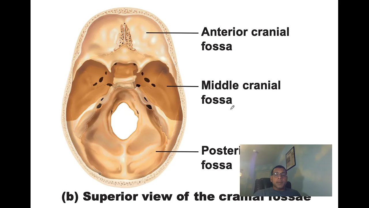
AnatomyAxialSkeletonCh7
6.3K
The ethmoid bone is located between the sphenoid, frontal, and nasal bones, forming most of the nasal cavity and helping form the orbit. The cribriform plate forms the roof of the nasal cavity and floor of the anterior cranial fossa, containing olfactory foramina for CN I. The crista galli serves as a meningeal anchoring site. The perpendicular plate forms the superior nasal septum. The lateral masses contain ethmoidal air cells. The superior and middle nasal conchae create turbulence in inspired air.
Click to watch →
Deep Dive
The general concept and organization of the 12 pairs of cranial nerves, particularly their roles in sensory versus motor function.

Madhyamik Test Exam COMPLETE REVISION MARATHON 🏃♂️🏆 মাধ্যমিক 2026 | Life Science - জীবন বিজ্ঞান
22.7K
There are 12 pairs of cranial nerves. Sensory cranial nerves include: Olfactory (I), Optic (II), Oculomotor (III), Trochlear (IV), Trigeminal (V), Abducens (VI), Facial (VII), Vestibulocochlear (VIII), Glossopharyngeal (IX), Vagus (X), Spinal Accessory (XI), and Hypoglossal (XII). Motor cranial nerves include: Oculomotor (III), Trochlear (IV), Trigeminal (V), Abducens (VI), Facial (VII), Glossopharyngeal (IX), Vagus (X), Spinal Accessory (XI), and Hypoglossal (XII). Mixed cranial nerves include: Trigeminal (V), Facial (VII), Vagus (X), and Hypoglossal (XII).
Click to watch →

12 Cranial Nerves
258.9K
The 12 cranial nerves are pairs of nerves that bypass the spinal cord and originate directly from the brain and brainstem, classified as sensory (carrying information into the brain), motor (sending signals out to control muscles), or mixed (containing both types). They include: Olfactory (smell, sensory), Optic (vision, sensory), Oculomotor (eye movement, motor), Trochlear (eye rotation, motor), Trigeminal (facial sensation and chewing, mixed), Abducens (outward eye movement, motor), Facial (facial expressions and tongue sensation, mixed), Vestibulocochlear (hearing and balance, sensory), Glossopharyngeal (swallowing and tongue sensation, mixed), Vagus (wandering nerve controlling heart, lungs, and digestion, mixed), Accessory (shoulder and head movements, motor), and Hypoglossal (tongue movements, motor). An easy acronym to remember them is 'O to touch and feel a very good Velvet' (Olfactory, Optic, Oculomotor, Trochlear, Trigeminal, Abducens, Facial, Vestibulocochlear, Glossopharyngeal, Vagus, Accessory, Hypoglossal).
Click to watch →
CRANIAL NERVE | Important Short Topic | NORCET & Other Nursing Exams | By Raju Sir
103.9K
Cranial nerves are 12 pairs (24 total) of nerves that connect the brain to the head, neck, and trunk, classified into three types: sensory (carrying messages to the brain), motor (carrying messages from the brain), and mixed (performing both functions). The 12 cranial nerves are: Olfactory (I, sensory), Optic (II, sensory), Oculomotor (III, motor), Trochlear (IV, motor), Trigeminal (V, mixed), Abducens (VI, motor), Facial (VII, mixed), Vestibulocochlear (VIII, sensory), Glossopharyngeal (IX, mixed), Vagus (X, mixed), Accessory (XI, motor), and Hypoglossal (XII, motor).
Click to watch →
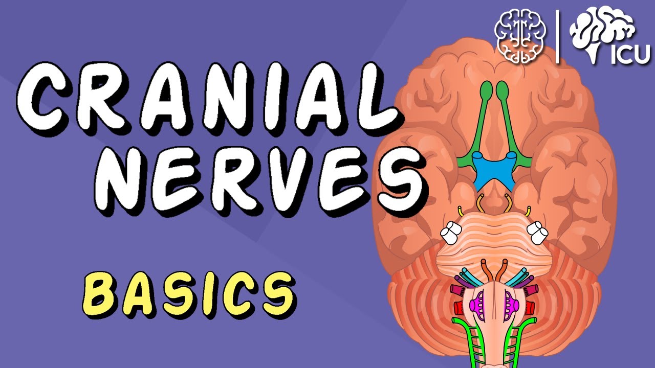
Cranial Nerve BASICS - The 12 cranial nerves and how to REMEMBER them!
1.9M
The 12 cranial nerves are paired nerves originating from the brain or brainstem, numbered I-XII based on their emergence point from front to back; they include sensory (afferent), motor (efferent), and combined nerves, with key examples being the olfactory nerve (smell, CN I), optic nerve (vision, CN II), trigeminal nerve (facial sensation and chewing, CN V), facial nerve (facial expressions and taste, CN VII), vestibulocochlear nerve (hearing and balance, CN VIII), glossopharyngeal nerve (tongue and throat sensation, CN IX), vagus nerve (swallowing, speech, and parasympathetic functions, CN X), accessory nerve (shoulder shrugging, CN XI), and hypoglossal nerve (tongue movement, CN XII); effective memorization strategies include drawing a face to associate nerves with body regions, using mnemonics like 'On Old Olympus's Towering Top A Finn and German Viewed Some Hops' for order and 'Some Say My Mother Bought My Brother Some Bad Beer' for sensory/motor classification.
Click to watch →
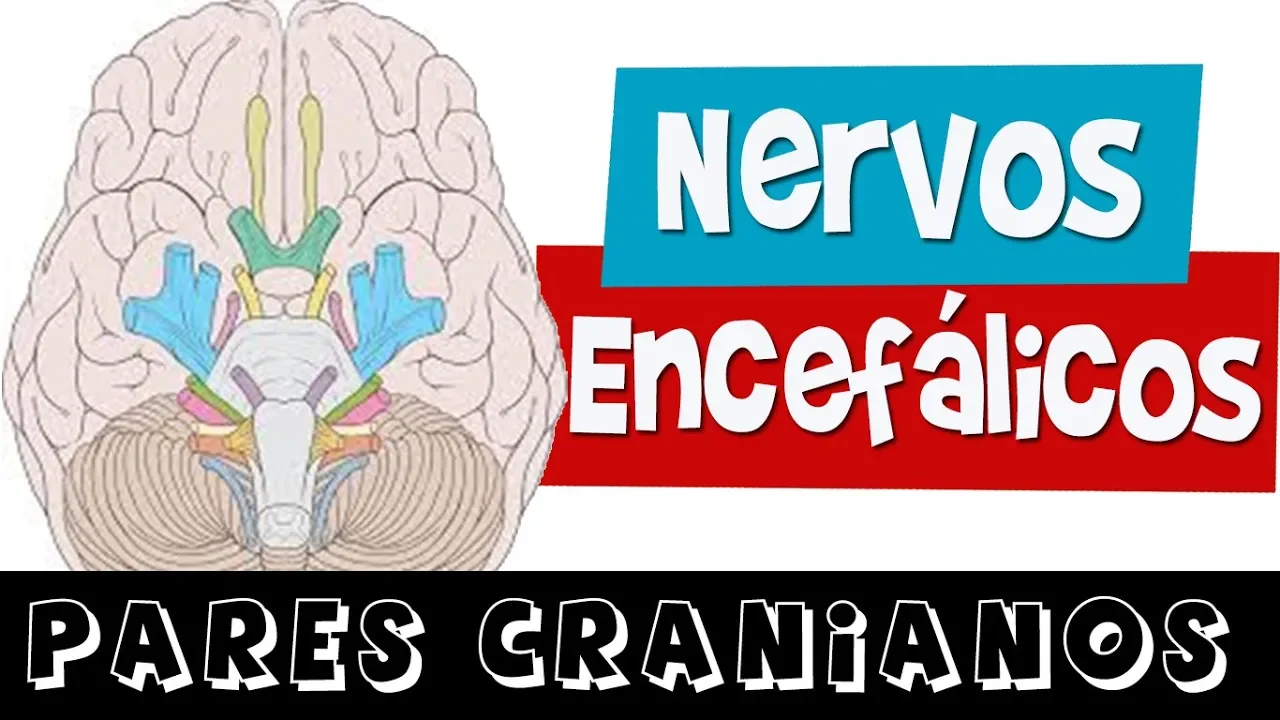
NERVOS ENCEFÁLICOS / PARES CRANIANOS (Aula completa) - Rogério Souza
97.6K
The 12 cranial nerves are: 1) Olfactory (I), 2) Optic (II), 3) Oculomotor (III), 4) Trochlear (IV), 5) Trigeminal (V), 6) Abducens (VI), 7) Facial (VII), 8) Vestibulocochlear (VIII), 9) Glossopharyngeal (IX), 10) Vagus (X), 11) Accessory (XI), and 12) Hypoglossal (XII). Of these 12 pairs, 10 emerge from the brainstem, while the first two emerge from the forebrain.
Click to watch →
Deep Dive
Fundamentals of sensory transduction, specifically how chemoreceptors detect chemical stimuli and generate action potentials.
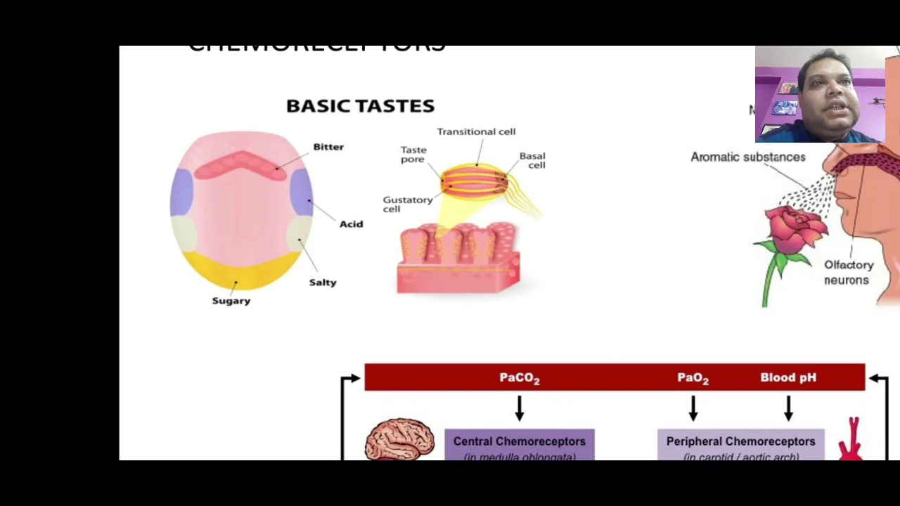
SENSORY TRANSDUCTION
267
Chemoreceptors detect chemical concentration changes in the body. Taste receptors (gustatory cells) on the tongue detect dissolved molecules in saliva, generating receptor potentials that trigger action potentials for taste perception. Olfactory receptors in the olfactory epithelium detect airborne chemical molecules, converting them into electrical signals for smell perception. Central chemoreceptors in the brainstem detect carbon dioxide levels and regulate breathing rate and depth. Peripheral chemoreceptors in carotid and aortic bodies detect oxygen levels and trigger ventilation responses. These systems maintain chemical homeostasis and provide sensory information about the body's internal and external chemical environment.
Click to watch →

Chemoreceptor
2.3K
Chemoreceptors (chemosensors) transduce chemical signals into action potentials to detect environmental stimuli. Two main classes exist: distance chemoreceptors (like olfactory receptors detecting gaseous chemicals) and direct chemoreceptors requiring physical contact (like taste buds). In vertebrates, the olfactory system includes the main olfactory epithelium and vomeronasal organ, both capable of detecting odorants and pheromones. Gustation detects aqueous tasteants through receptors on the tongue, triggering appetitive or defensive responses. Contact chemoreception enables fish and crustaceans to identify food chemicals in water, while insects use it to recognize cuticular hydrocarbons and host plant chemicals. Insects possess feathery antennae with sensilla for enhanced olfaction, particularly advantageous for nocturnal species like moths.
Click to watch →
Overview Sensory System Part 1
330
Different receptor types use specific mechanisms: chemoreceptors bind chemicals to ion channels, thermoreceptors have temperature-sensitive channels activated by warming or cooling, photoreceptors (rods and cones) respond to light photons, and mechanoreceptors respond to membrane stretch. Pain receptors combine multiple types. Graded potentials (receptor potentials) summate from resting potential (-70mV) toward threshold (-50mV). Once threshold is reached, action potentials are generated, depolarizing to +30mV for transmission to the central nervous system.
Click to watch →
Los sistemas Sensoriales
130
Chemoreceptors detect chemical stimuli. Taste buds contain specialized cells that release neurotransmitters when chemicals bind to receptors, generating receptor potentials. Olfactory receptors are neurons with cilia that detect airborne chemicals, generating graded potentials that summate in the axon hillock. Pain receptors (nociceptors) detect harmful chemicals released during tissue damage, transmitting signals to the brain to indicate injury location and intensity.
Click to watch →

Chemoreceptor
457
Chemoreceptors are sensory receptors that transduce chemical signals into action potentials, essential for survival in detecting food, habitat, mates, and predators. There are two main classes: distance chemoreceptors (detecting chemicals from a distance, like olfactory neurons and insect antennae) and direct chemoreceptors (detecting through physical contact, like taste buds). In vertebrates, olfaction occurs in the nose where volatile stimuli reach the olfactory cleft, with olfactory sensory neurons transmitting signals through the cribriform plate to the olfactory bulb. The tongue serves as the primary gustatory organ, with taste receptors falling into two G-protein coupled receptor superfamilies: ion channels for salty/sour tastes and GPCRs for sweet/bitter tastes. Contact chemoreceptors are uniporous receptors with dendrites near pores. Primary cilia serve as cellular antenna coordinating signaling pathways. Carotid and aortic bodies detect blood gas levels, with central chemoreceptors on the medulla oblongata detecting pH changes in cerebrospinal fluid. Stimulation of peripheral chemoreceptors activates the medullary vagal center, slowing heart rate, though this response is often modulated by other factors.
Click to watch →
Deep Dive
General structural neuroanatomy, including the basic organization of the cerebral cortex, thalamus, and synaptic transmission.
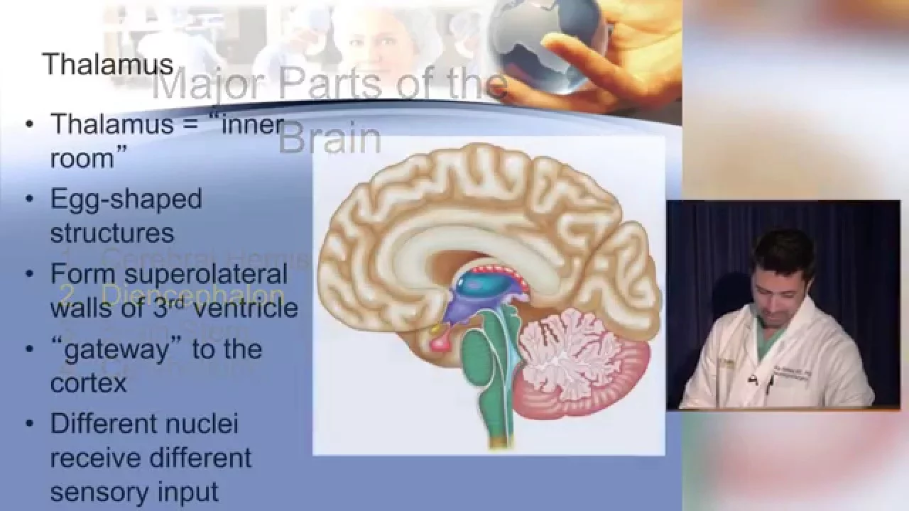
Neuroanatomy Basics: A Simplified Guide for Medical Students
1.2M
The brain is organized into major regions including the cerebral hemispheres (83% of brain mass) divided into frontal, parietal, temporal, and occipital lobes by anatomical sulci; the diencephalon containing the thalamus (sensory relay center) and hypothalamus (autonomic control); the brainstem (midbrain, pons, medulla) controlling reflexive functions and cranial nerves; and the cerebellum coordinating movement. The cerebral cortex contains primary sensory and motor areas with somatotopic organization (homunculus), association areas for higher-order processing, and Broca's area for speech production. Deep gray matter structures like the basal ganglia regulate movement, while white matter tracts connect different brain regions.
Click to watch →
Biology | senior 2 second term | final revision | دكتور مصطفي خضر
35.5K
A neuron (النيرف سيل) consists of the cell body (سيل بدي), dendrites (الشعيرات), and axon (الاجزون). The cell body contains organelles like the Golgi apparatus. Neurons are covered by myelin sheaths (مايلين شيس) produced by Schwann cells (الشوان). Nerve impulses always travel from the cell body to the axon terminal. Neurons cannot regenerate themselves. Synaptic transmission involves calcium (كالسيوم) and potassium (كولين) ions. The action potential has a value of 110 mV. Calcium is released from storage vesicles when a nerve impulse arrives, triggering neurotransmitter release. The cerebral cortex (السيريبرال كورتكس) has four lobes: frontal lobe (فرونتال) at the front controlling voluntary movements, parietal lobe (برايت) at the top processing sensory information, temporal lobe (تمبورال) at the side involved in hearing and smell, and occipital lobe (اوكسيبيتال) at the back controlling vision.
Click to watch →
◄╣ Neuroanatomy ♦ 1 Topography of the Nervous System ╠► YouTubevia torchbrowser com
106K
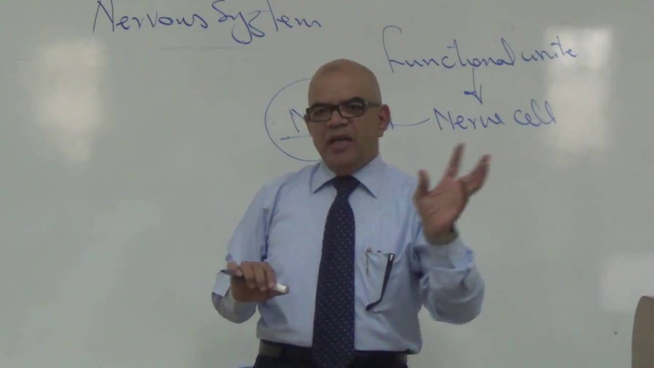
Neuroanatomy is a branch of medical education focusing on nervous system structure and organization. The nervous system is defined as an information system that processes stimuli from both external and internal sources, generating appropriate responses through neurons. Neurons are the functional units, consisting of a cell body (soma) containing the nucleus with nucleolus, dendrites for receiving signals, and an axon for transmitting signals. Nissl granules are characteristic structures in the cell body consisting of rough endoplasmic reticulum with ribosomes for protein synthesis. Nerve fibers form when axons of multiple neurons bundle together. Synaptic transmission involves chemical neurotransmitters released from presynaptic neurons binding to receptors on postsynaptic neurons. Gray matter consists of neuronal cell bodies and appears gray due to Nissl granules and lack of myelin. White matter consists of myelinated axons and appears white due to lipid-rich myelin sheaths formed by glial cells (Schwann cells in PNS, oligodendrocytes in CNS). The spinal cord gray matter is organized into dorsal horns (sensory) and ventral horns (motor). Neurons are classified by process number: pseudounipolar (sensory), bipolar (retina), and multipolar (cerebral cortex). Each cerebral hemisphere is divided into four lobes: frontal (movement, speech, personality), parietal (sensory processing, spatial orientation), temporal (auditory processing, memory), and occipital (visual processing). The two hemispheres communicate through the corpus callosum.
Click to watch →
Neuroanatomia: Clase 2 "Generalidades" (parte 1)
609
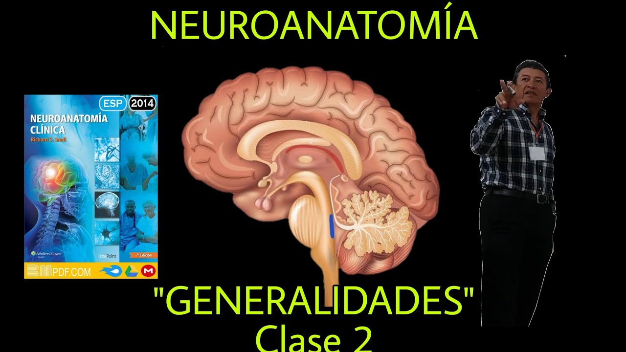
The brain consists of the cerebral cortex (with 23 gyri and sulci), brainstem (midbrain, pons, and medulla), and cerebellum, containing white matter organized into three cords (anterior motor, lateral mixed, posterior sensory) and gray matter forming the outer cortex; the brainstem serves as a critical pathway for sensory and motor tracts connecting the spinal cord to the cortex, while the autonomic nervous system (sympathetic and parasympathetic divisions) regulates involuntary body functions through cranial and spinal nerves.
Click to watch →
thalamus anatomy 3d | anatomy of thalamus nuclei anatomy
176.5K
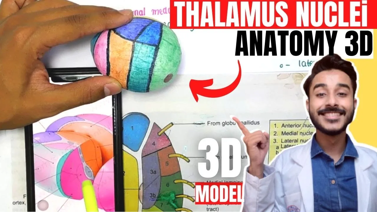
This section introduces fundamental neuroanatomical concepts including spinal cord structure with grey matter (composed of neuron cell bodies) and white matter organization. It explains that nuclei are clusters of nerve cells performing specific functions. The thalamus is presented as a critical brain structure serving dual roles: acting as a relay station for transmitting sensory and motor signals between brain regions, and functioning as a regulator that controls which signals are transmitted and which are suppressed. All sensory nerves from the body must pass through the thalamus before reaching their cortical destinations, establishing its central role in sensory processing.
Click to watch →
Prerequisite Knowledge
- Concept 01Basic anatomy of the nasal cavity, including the cribriform plate of the ethmoid bone and the nasal mucosa.
- Concept 02The general concept and organization of the 12 pairs of cranial nerves, particularly their roles in sensory versus motor function.
- Concept 03Fundamentals of sensory transduction, specifically how chemoreceptors detect chemical stimuli and generate action potentials.
- Concept 04General structural neuroanatomy, including the basic organization of the cerebral cortex, thalamus, and synaptic transmission.
Subsequent Learning
- Step 01Clinical disorders of olfaction, such as anosmia, hyposmia, and phantosmia, and their diagnostic relevance to neurodegenerative diseases.
- Step 02The functional connection between the primary olfactory cortex and the limbic system (specifically the amygdala and hippocampus) in processing emotions and memories.
- Step 03The physiological integration of olfaction and gustation (taste) in the orbitofrontal cortex to produce the cognitive perception of flavor.
- Step 04The mechanisms of olfactory adaptation and receptor desensitization at the cellular level during prolonged exposure to an odorant.
Pathway Overview
Cortex Functions
Pathway Overview
0:00Playing Section
- 1
Traces olfactory nerve from nasal rootlets to cortex.
- 2
Details passage through cribriform plate into bulb.
- 3
Bulb synapses with mitral and tufted cells.
The Active Thalamic Pathway in Olfactory Processing
Traditional neuroanatomy curricula emphasize that the olfactory pathway is unique among sensory systems because it bypasses the thalamus, projecting directly from the olfactory bulb to the primary olfactory cortex. However, modern neuroscience presents an important counterpoint: the thalamus (specifically the mediodorsal nucleus) is not functionally bypassed, but rather plays an essential role in downstream olfactory processing. Research indicates that the pathway from the primary olfactory cortex to the mediodorsal thalamus, and subsequently to the orbitofrontal cortex, is crucial for olfactory attention, odor discrimination, and the cognitive integration of smell with other sensory modalities. Treating olfaction as a purely 'thalamus-free' pathway oversimplifies sensory processing and ignores how the brain coordinates complex, feedback-driven olfactory perception.
Deep Dive
Clinical disorders of olfaction, such as anosmia, hyposmia, and phantosmia, and their diagnostic relevance to neurodegenerative diseases.
CN 1: Olfactory Nerve (Scheme, Pathway, Clinical Relevance) | Neuroanatomy
104.3K
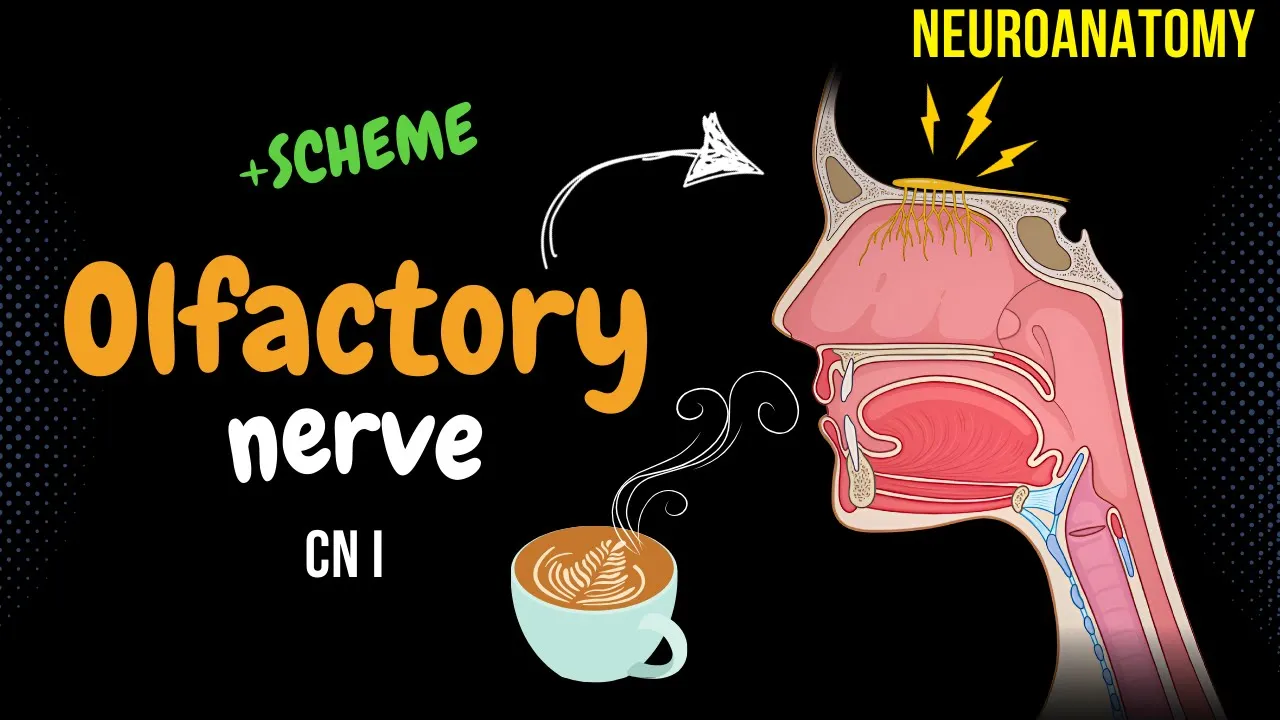
Olfactory dysfunction manifests through several clinical conditions. Anosmia represents temporary or permanent loss of smell, commonly caused by inflammation/infections leading to mucus buildup that impedes odor molecule transport to receptors, trauma damaging the cribriform plate or ethmoidal bone, or degenerative CNS diseases. Since taste is 80% dependent on olfaction, anosmia significantly impairs flavor perception. Dysosmia involves distorted smell perception where familiar objects smell different or unpleasant, including parosmia (normal smells perceived as unpleasant) and phantosmia (detecting non-existent odors). Parosmia results from chronic conditions like sinusitis or head trauma reducing receptor diversity. Phantosmia may arise from inflamed sinuses, certain medications (antipsychotics, antimigraine drugs, antiseizure medications), or brain tumors, potentially serving as a clinical indicator of CNS pathology.
Click to watch →
Visiting Lecture: Olfactory Changes & Early Detection of Neurodegenerative Disorders
137
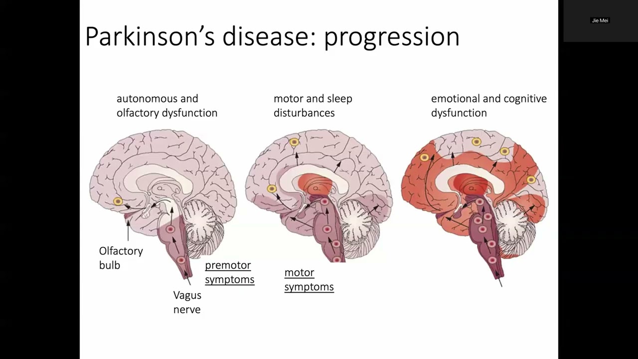
Neurodegenerative diseases including Alzheimer's and Parkinson's involve progressive neuronal degeneration leading to disability and death. Both diseases share a common feature: olfactory dysfunction appears 10-15 years before typical clinical symptoms. Parkinson's involves dopamine neuron degeneration in the substantia nigra due to alpha-synuclein aggregation, causing motor symptoms. Alzheimer's involves amyloid-beta plaques and tau tangles causing temporal cortex degeneration, leading to memory loss. The olfactory system detects environmental volatiles through receptors in the olfactory epithium, sending signals to the olfactory bulb. Two main dysfunction types exist: quantitative (hyposmia/anosmia) and qualitative (parosmia/phantosmia). Factors influencing olfactory function include age, sex, education, smoking, and alcohol consumption. Meta-analysis reveals that both Alzheimer's and Parkinson's cause significant olfactory impairment across multiple tasks, but with distinct patterns: Parkinson's affects all tasks uniformly while Alzheimer's shows particular impairment in memory-dependent tasks like identification and recognition. This distinction may help differentiate between conditions.
Click to watch →

Olfactory Nerve (I CN) in detail by Dr. Azmi Mohsin
464
Olfactory dysfunction presents with several clinical manifestations: anosmia (complete loss of smell, unilateral or bilateral), hyposmia/microsmia (partial loss), dysosmia (distorted smell perception), phantosmia (spontaneous smell sensations without odorant), and olfactory agnosia (inability to recognize odors despite intact function, often due to nasal congestion). Hyperosmia (heightened sense of smell) is rare. Destruction of the olfactory bulb or primary cortex (Brodmann area 34) causes ipsilateral anosmia. Causes include aging (strongest factor, with progressive neuron loss and cribriform plate osteophyte formation), viral infections (most common cause of permanent hyposmia), toxic chemical exposure (herbicides, pesticides, solvents), head trauma (occipital/side impacts cause more damage than frontal impacts; CSF rhinorrhea indicates anterior cranial fossa fracture), and neurodegenerative diseases (Alzheimer's, Parkinson's).
Click to watch →
Olfaction in neurodegenerative disease
367
Comprehensive diagnostic evaluation includes detailed patient history, ENT examination with nasal endoscopy to exclude structural pathology, psychophysical testing using validated tools like Sniffin' Sticks (TDI test) for threshold, discrimination, and identification, and MRI assessment evaluating olfactory bulb volume (normal 40-60 mm³) and olfactory sulcus depth. Classification uses Sniffin' Sticks: identification scores of 6 or less indicate functional anosmia; 7-10 indicate hyposmia; 11-12 indicate normosmia. Post-infectious olfactory loss typically presents with parosmia and has the best prognosis, with 40-60% recovering normal function through olfactory training (daily practice for 12+ weeks using four essential oils). Currently, no treatments improve olfactory function in neurodegenerative diseases, though olfactory training shows promise. Patients with olfactory dysfunction face real dangers including inability to detect spoiled food, gas leaks, or smoke, requiring patient and family education about safety concerns.
Click to watch →

Quick Macleod CNS Examination ; Cranial nerves .
445
The olfactory nerve (CN I) is purely sensory and responsible for smell. Disorders include hyposmia (reduced smell) and anosmia (complete loss of smell), most commonly caused by upper respiratory tract infections. Parosmia (distorted smell perception) commonly occurs in Parkinson's disease, while phantosmia (perception of non-existent smells) can occur in Alzheimer's disease and focal seizures. The examination involves testing smell function bilaterally using familiar substances like coffee.
Click to watch →
Deep Dive
The functional connection between the primary olfactory cortex and the limbic system (specifically the amygdala and hippocampus) in processing emotions and memories.

Neurology | Cerebrum: Temporal Lobe Anatomy & Function
211.2K
The olfactory pathway begins with odorant activation of nasal receptors, stimulating olfactory nerves that travel through the cribiform plate to the olfactory bulb. Signals follow the olfactory tract and striae to the primary olfactory cortex in the temporal lobe. The olfactory association cortex performs three critical functions: storing smell memories for future recognition, analyzing and identifying specific odors by comparing with stored patterns, and connecting with the limbic system (particularly the amygdala) to associate smells with emotional experiences. This explains why certain odors can trigger vivid emotional memories and how the brain creates lasting associations between sensory experiences and emotional states.
Click to watch →

The Anatomy of Smell - Imaging of the the olfactory system
3.1K
The primary olfactory cortex is a collection of structures receiving direct input from ipsilateral olfactory bulbs, including the piriform cortex (C-shaped, anterior and medial to the amygdala), amygdala, and hippocampus. The piriform cortex can be located on MRI by identifying the amygdala as a reference point. Secondary olfactory cortical systems include the orbital frontal cortex (multisensory integration area crucial for taste, since 80-90% of flavor perception relies on smell), amygdala, hippocampus, basal ganglia, and thalamus. The amygdala links smells with emotions, while the hippocampus links smells with memories, explaining why odors can immediately evoke strong emotional and mnemonic responses. This neural architecture provides the foundation for understanding how olfactory experiences become deeply embedded in emotional and memory networks.
Click to watch →

Sensation vs. Perception: What's the Difference?
143.5K
Unlike other senses, smell directly connects to the limbic system—the brain's center for emotions, memories, and survival instincts. The olfactory cortex communicates with the hippocampus (for recognizing familiar smells and explicit memories) and the amygdala (for triggering fear and strong emotional responses to potentially dangerous odors like smoke or rotting food).
Click to watch →

100% Realtalk 181 | Michael Nehls | Impfungen | Orwell & Huxley | Maßnahmen & Weltregierung | A.I.
124.9K
The olfactory system (smell) has a direct connection to the hippocampus, which is why smells can trigger vivid memories. This connection is evolutionarily ancient, as even fish use smell to navigate back to their birthplace. The hippocampus and olfactory system are both part of the limbic system, which processes emotions and memories.
Click to watch →
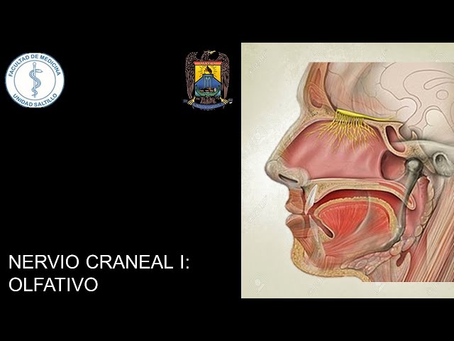
NERVIO OLFATIVO (NERVIO CRANEAL I)
1.2K
The olfactory tract projects to two distinct cortical areas in the temporal lobe. The primary olfactory cortex (Brodmann area 34) in the periamygdaloid and prepiriform areas connects to the limbic system, explaining why olfactory stimuli evoke strong emotional memories. The secondary olfactory cortex (Brodmann area 28) in the entorhinal area handles basic survival behaviors like detecting spoiled food and generating responses to odors. Both pathways integrate olfactory information in the temporal lobe, with the primary pathway linking smell to emotion and memory, and the secondary pathway managing instinctive behavioral responses.
Click to watch →
Deep Dive
The physiological integration of olfaction and gustation (taste) in the orbitofrontal cortex to produce the cognitive perception of flavor.
Sensation and Perception - Chemical Senses Pt4 - Flavor Perception
295
Flavor is a multisensory perceptual experience that combines taste (sweet, salty, sour, bitter, umami) with smell through retronasal olfaction, along with contributions from vision, audition, somatosensation, and top-down cognitive factors; the brain integrates these diverse inputs primarily in the orbital frontal cortex, where taste signals from the primary gustatory cortex and smell signals from the olfactory bulb converge to create our unified flavor experience, which is why blocking nostrils during eating dramatically reduces perceived flavor despite unchanged taste receptor activation.
Click to watch →
30-Respiratory disordersإضطرابات الجهاز التنفسى
583
Flavor perception combines taste (sweet, salty, sour, bitter, umami) and smell (aroma). Volatile compounds from food reach olfactory receptors through nasal passages or retro-nasal pathway. Both signals integrate in the orbitofrontal cortex. Olfactory dysfunction reduces flavor perception because volatile compounds cannot reach receptors, making food taste bland despite intact basic taste.
Click to watch →
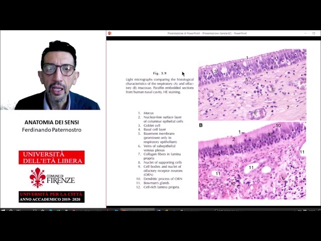
ANATOMIA DEI SENSI (5) - OLFATTO E GUSTO
8.1K
The olfactory cortex contains three functional areas: the anterior olfactory nucleus for feedback control, the amygdala and tuberculum olfactorium for behavioral regulation related to food, and the entorhinal cortex and piriform cortex for odor discrimination and integration with gustatory information. Taste and olfaction work together to create the perception of flavor, with taste detecting basic chemical properties and olfaction providing complex aromatic information. Taste receptors are specialized cells located in taste buds, which are found on taste papillae on the tongue, palate, pharynx, and inner cheeks. Each taste papilla contains up to 100 taste buds, which are the functional units for taste detection.
Click to watch →

«Мозг гурман: нейрофизиология гастрономических удовольствий» — Анна Хоружая
175
The orbitofrontal cortex integrates all sensory information about food, including taste, smell, texture, temperature, and visual appearance. This integration creates the complete perception of flavor and influences food preferences. The brain combines olfactory, gustatory, and visual information to create the comprehensive experience of eating, demonstrating the multisensory nature of flavor perception.
Click to watch →

The Good, the Bad, and the Incredibly Nuanced!
259
The host explains that taste and smell are distinct chemical senses but together create the experience of flavor. She discusses the primary gustatory cortex in the insula and the primary olfactory cortex in the temporal lobe. The host explains that 80% of what we call flavor actually comes from aroma, as odors released while chewing travel up to the nasal cavity. Retro-nasal smell is how we experience much of what we call flavor - when we breathe out, the breath heats up what's on the tongue, allowing us to taste what we're eating. The host also discusses the orbital frontal cortex (which assigns reward value) and the amygdala (which ties food experience to emotion and memory).
Click to watch →
Deep Dive
The mechanisms of olfactory adaptation and receptor desensitization at the cellular level during prolonged exposure to an odorant.

Why Can't People Smell Themselves
138.4K
Olfactory fatigue occurs through a negative feedback mechanism involving calcium-calmodulin binding to open CNG channels. When odor molecules cause the rapid influx of calcium, this initiates a chemical cascade that causes the CNG channels to close. Once closed, similar odor molecules cannot activate the receptor and no longer send electrical signals to the brain. The specific CNG channels involved in olfactory adaptation are CNGA4 and CNGB1b. This closure prevents continuous stimulation by the same odorant, allowing the system to reset and respond to new odors more effectively.
Click to watch →

Receptor Desensitization Part 1
36.3K
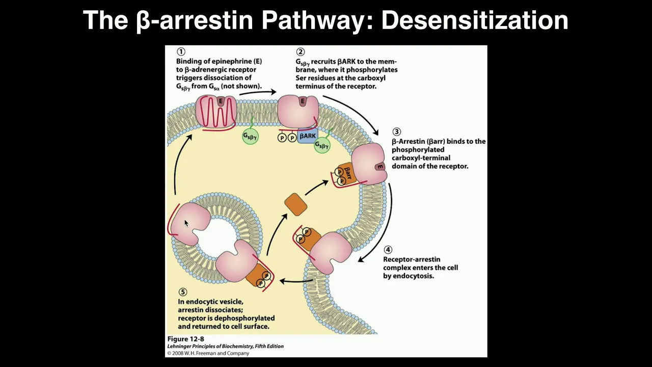
Receptor desensitization is a cellular phenomenon where prolonged exposure to an agonist causes the cell's response to that agonist to decrease over time, achieved through three primary mechanisms: uncoupling (reducing receptor interaction with downstream pathways), sequestration (internalizing receptors into early endosomes), and downregulation (permanently destroying receptors); this concept is best understood through the well-studied beta-2 adrenergic receptor, which responds to adrenaline and noradrenaline with adrenaline having greater affinity for this receptor.
Click to watch →
Biosignaling | Receptor Desensitization by Beta-arrestin
24.4K
Receptor desensitization is a cellular mechanism where prolonged activation of G-protein coupled receptors (GPCRs) triggers their internalization into the cell, thereby reducing their responsiveness to signals; this occurs when beta-arrestin and receptor kinase phosphorylate serine residues on the receptor's cytoplasmic domain, attracting beta-arrestin which facilitates endocytosis, and this process has important implications for drug tolerance (e.g., opioid addiction) and sensory adaptation (e.g., olfactory desensitization).
Click to watch →

Nobel Laureate in chemistry Professor Robert J. Lefkowitz – Nobel Lectures in Uppsala 2012
8.4K
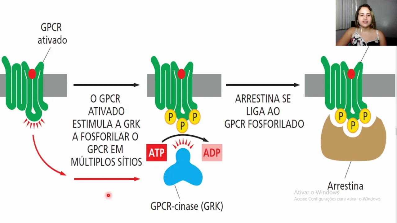
Receptor desensitization represents a universal homeostatic mechanism where prolonged stimulation reduces cellular response. Photoaffinity labeling revealed receptor phosphorylation as the molecular basis. Discovery of GRK kinases and beta-arrestins established a universal paradigm: GRKs phosphorylate activated receptors, facilitating beta-arrestin binding which sterically blocks further G protein signaling. This system also enables independent signaling through scaffolding various pathways and mediates clathrin-dependent endocytosis.
Click to watch →
Aula 10 - Dessensibilização de receptores
4.4K
Receptor desensitization is a cellular mechanism that reduces receptor responsiveness to prevent overstimulation, involving five main processes: receptor sequestration (removal from cell surface), receptor degradation (complete destruction), receptor recycling (reutilization), inactivation of signaling proteins, and production of inhibitory proteins; this is particularly important in pharmacology where repeated drug exposure can diminish therapeutic effects, and in GPCR signaling where phosphorylation by GRK and arrestin recruitment leads to receptor internalization and downregulation.
Click to watch →
Pathway Overview
Cortex Functions
Pathway Overview
0:00- 1
Traces olfactory nerve from nasal rootlets to cortex.
- 2
Details passage through cribriform plate into bulb.
- 3
Bulb synapses with mitral and tufted cells.
The Active Thalamic Pathway in Olfactory Processing
Traditional neuroanatomy curricula emphasize that the olfactory pathway is unique among sensory systems because it bypasses the thalamus, projecting directly from the olfactory bulb to the primary olfactory cortex. However, modern neuroscience presents an important counterpoint: the thalamus (specifically the mediodorsal nucleus) is not functionally bypassed, but rather plays an essential role in downstream olfactory processing. Research indicates that the pathway from the primary olfactory cortex to the mediodorsal thalamus, and subsequently to the orbitofrontal cortex, is crucial for olfactory attention, odor discrimination, and the cognitive integration of smell with other sensory modalities. Treating olfaction as a purely 'thalamus-free' pathway oversimplifies sensory processing and ignores how the brain coordinates complex, feedback-driven olfactory perception.
Basic anatomy of the nasal cavity, including the cribriform plate of the ethmoid bone and the nasal mucosa.
The general concept and organization of the 12 pairs of cranial nerves, particularly their roles in sensory versus motor function.
Fundamentals of sensory transduction, specifically how chemoreceptors detect chemical stimuli and generate action potentials.
General structural neuroanatomy, including the basic organization of the cerebral cortex, thalamus, and synaptic transmission.
Clinical disorders of olfaction, such as anosmia, hyposmia, and phantosmia, and their diagnostic relevance to neurodegenerative diseases.
The functional connection between the primary olfactory cortex and the limbic system (specifically the amygdala and hippocampus) in processing emotions and memories.
The physiological integration of olfaction and gustation (taste) in the orbitofrontal cortex to produce the cognitive perception of flavor.
The mechanisms of olfactory adaptation and receptor desensitization at the cellular level during prolonged exposure to an odorant.
191.9K views3Klikes3:11@MEDSimplifiedOriginal Release: 2015-10-28
[Music] In this video we will discuss the alfactory nerve. Alfactory nerve or the cranial nerve one is associated with the sense of smell and starts from the nose and ends into the primary and the secondary alfactory cortex.
So this is a section of the nose which also shows the sinuses.
This is the frontal sinus. This is the sppheninoidal sinus.
This is the pallet and above lies the cribform plate.
So the alfactory pathway begins with the alfactory rootlets. Now these alfactory rootlets at their nasal end have the alfactory cells which pick up the smell and they continue posteriorly as the alfactory nerves. The alfactory nerves pass through the perforations in the cribform plate and they enter into the alfactory bulb. The alfactory bulb lies on the superior part of the cribform plate. The alfactory nerves enter the alfactory bulb and here the signapse with the mitral and the tufted cells.
And the processes of the mital and the tufted cells continue posteriorly as the alfactory tract.
And this alfactory tract continues posteriorly and enters into the primary and the secondary alfactory cortex. So let's look at the section of a brain to understand the central portion of the alfactory pathway. On the top right corner of the screen, you will see a diagram of the brain. And if we cut the brain, this point and look at it from below upwards. This is the current section that I'm drawing here.
So this is the alfactory bulb.
And the rest of the portion that I'm drawing here is the rest of the alfactory pathway.
So here are the alfactory rootlets which enter into the alfactory bulb and the alfactory bulb sends the alfactory tract which continues posteriorly. So this is the alfactory tract.
Now the areas you are seeing here are the medial alfactory stria, the lateral alfactory stria and the area just posterior to them is the anterior perforated substance and this area here forms the primary alfactory cortex and the primary alfactory cortex is associated with the active perception of sense of smell. These neurons continue posteriorly and they enter into a area known as the entrinal area and the area just medial to it is the uncus. And these two areas form the secondary alfactory cortex. The secondary alfactory cortex is associated with functions like memory and emotions associated with the sense of smell. So this was a short description of the alfactory nerve and the alfactory pathway. Also, if you want to see more videos on cranial nerves and other topics, don't forget to hit the subscribe button below.
Up Next
10:55
Cardiac Excitation-Contraction Coupling Explained
@StrongMed
167.1K views•2021-11-03

50:12
Bacterial Communication: Quorum Sensing and Biofilm Formation Explained
@JHUAAP
12.6K views•2011-06-28

5:35
Enteric Nervous System Explained: The Gut's Brain | Neurobiology Lecture
@alumniu6029
438 views•2018-09-12

7:08
Bacteriophages: Earth's Deadliest Killers and Future Antibiotics
@kurzgesagt
34.6M views•2018-05-13
Related Study Plans & Knowledge Roadmaps
Structured learning paths in Biology
biology
RNAi: Biogenesis, RISC & Gene Silencing
01Molecular Biology Foundations: DNA to Protein
02Introduction to RNA Interference (RNAi)
03Molecular Biogenesis of miRNA and siRNA
+ 2 more learning modules
10h5 modules
Explore Roadmap
biology
Mycorrhizae: Signaling, Biophysics & Ecology
01Foundations of Mycorrhizal Symbiosis
02Molecular Signaling and Root Colonization
03Biophysics and Bioenergetics of Nutrient Exchange
+ 1 more learning modules
11h4 modules
Explore Roadmap
biology
Circadian Clocks: TTFL, SCN & Periphery
01Foundations of Molecular Biology & Gene Expression
02Introduction to Chronobiology and Actograms
03The Core TTFL: Molecular Architecture of the Clock
+ 3 more learning modules
14h6 modules
Explore Roadmap
biology
Population Genetics: Math, Drift & Selection
01Genetic & Probability Foundations
02The Hardy-Weinberg Principle and Baseline Models
03Mathematical Modeling of Natural Selection
+ 3 more learning modules
18h6 modules
Explore Roadmap