CRISPR-Cas9 is a revolutionary genome editing technology derived from a bacterial immune system that enables precise modification of DNA sequences in living cells; it works by using a guide RNA to direct the Cas9 enzyme to a specific location in the genome, where it creates a double-strand break that the cell attempts to repair, allowing researchers to introduce targeted mutations, replace defective genes with healthy copies, or study gene function with unprecedented precision and efficiency across diverse applications including biomedical research, agriculture, and potential human genetic disease treatment.
CRISPR-Cas9 Genome Editing: Mechanism & Applications
Added:Deep Dive
The structure of DNA and the rules of complementary base pairing (A-T, C-G).

Molecular Basis of Inheritance | NEET | Structure & Types of DNA - Part 2 | Neela Bakore Tutorials
284.6K
This comprehensive section covers DNA fundamentals including the Watson-Crick double helix structure, complementary base pairing (A-T with 2 H-bonds, C-G with 3 H-bonds), Chargaff's rules (A=T and C=G), and DNA classification systems. Functionally, DNA divides into trophic DNA (metabolic activities) and genetic DNA (heredity/reproduction), exemplified by Paramecium's macronucleus and micronucleus. Structurally, DNA exists in five forms: A-DNA (11 bp, right-handed), B-DNA (10 bp, right-handed - standard form), C-DNA (9.33 bp, right-handed), D-DNA (8 bp, right-handed), and Z-DNA (12 bp, left-handed). These principles enable determination of unknown DNA sequences and form the foundation for molecular biology.
Click to watch →
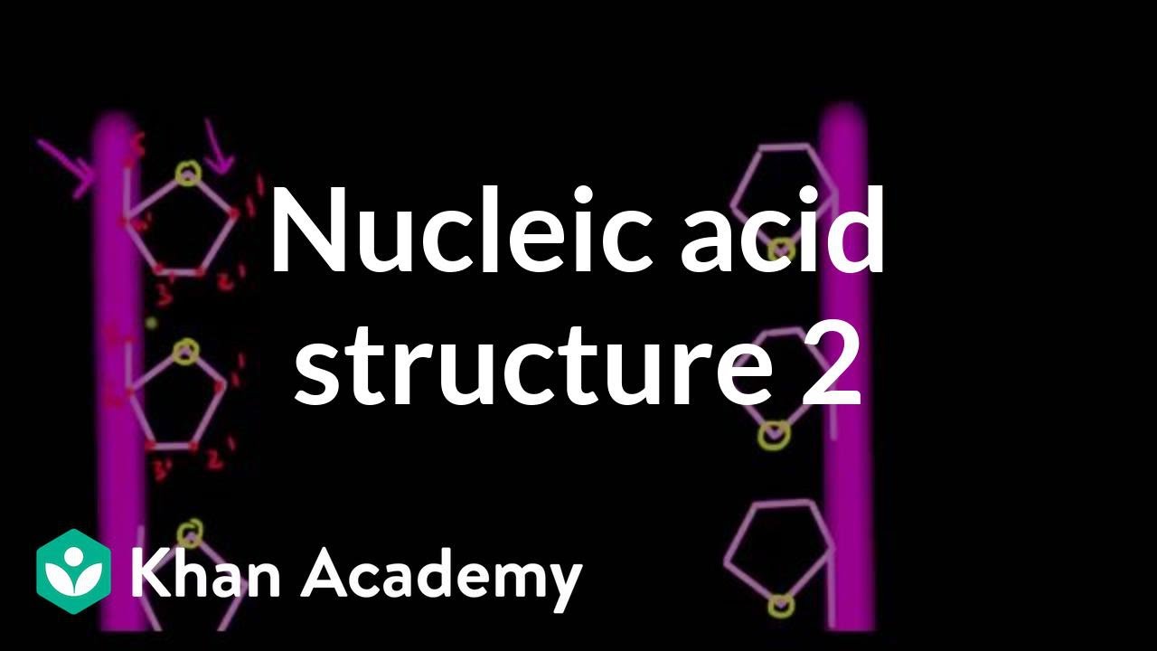
Nucleic acid structure 2
78.3K
DNA forms a double helix with two antiparallel strands running in opposite directions (5' to 3' and 3' to 5'). The sugar-phosphate backbones form the outer framework, while nitrogenous bases face inward. Purines (adenine, guanine) pair with pyrimidines (thymine, cytosine) through hydrogen bonds: A-T with two bonds, C-G with three bonds. This complementary base pairing allows DNA to serve as a template for replication. The Watson-Crick model describes this elegant structure where genetic information is stored in the sequence of bases along the helical strands.
Click to watch →

⚡NEET 2026: NCERT 360: Molecular Basis Of Inheritance - One Shot Part - 1| Seep Pahuja
216.3K
James Watson and Francis Crick proposed the double helix structure of DNA in 1953, for which they received the Nobel Prize. Their model was based on X-ray diffraction data from Rosalind Franklin and Maurice Wilkins, as well as chemical analysis by Erwin Chargaff. The double helix consists of two antiparallel strands wound around a common axis. Chargaff's rules state that in DNA: A = T, G = C, and (A + G) = (T + C). The ratio of (A + T)/(C + G) is constant for a given species but varies between species. In the double helix, Adenine forms 2 hydrogen bonds with Thymine, while Guanine forms 3 hydrogen bonds with Cytosine. Purines always pair with pyrimidines, maintaining constant helix width. The DNA double helix has two antiparallel strands with sugar-phosphate backbones and nitrogenous bases projecting inward. The strands are coiled around a common axis, not wound around each other. The bases are stacked perpendicular to the backbone, providing stability. The 3.4 nm distance between base pairs and 20 Å diameter are key structural features.
Click to watch →
DNA Structure | A-level Biology | OCR, AQA, Edexcel
128.7K
In DNA, the four nitrogenous bases pair up specifically: adenine (A) always pairs with thymine (T), and guanine (G) always pairs with cytosine (C). This complementary base pairing means that in any rung of the DNA ladder, you will either find an A paired with a T or a G paired with a C. This specificity ensures accurate genetic information transfer during DNA replication and protein synthesis.
Click to watch →
CHAPTER 5 | Lesson 2 | ELMARGE3 | Structure and replication of DNA | Exam 26
163
In DNA, adenine (A) always pairs with thymine (T) through 2 hydrogen bonds, while guanine (G) always pairs with cytosine (C) through 3 hydrogen bonds. This complementary base pairing ensures accurate DNA replication. Valid DNA base pair combinations are only A-T and G-C; combinations like A-A, T-T, G-G, or C-C are not possible. The two DNA strands run in opposite directions (antiparallel), with one strand running 5' to 3' and the other 3' to 5'.
Click to watch →
Deep Dive
The Central Dogma of Molecular Biology, specifically how DNA is transcribed into RNA and translated into proteins.

SESIÓN 13: GENÉTICA MENDELIANA
440
The central dogma of molecular biology describes the flow of genetic information: DNA is transcribed into RNA, and RNA is translated into proteins. Transcription is the process by which genetic information in DNA is copied into RNA. Translation is the process by which ribosomes synthesize proteins using the information in messenger RNA (mRNA).
Click to watch →

Transcrição e Dogma Central da Biologia - Gabmed
161
The Central Dogma describes the flow of genetic information: DNA → RNA → Protein. Transcription synthesizes RNA from DNA template. Translation produces proteins from mRNA. Only mRNA undergoes translation. The nucleotide sequence of DNA determines mRNA sequence, which determines amino acid sequence of proteins. Each three nucleotides (codon) specifies one amino acid. Transcription produces different RNA types: mRNA, rRNA, tRNA.
Click to watch →

الدرس الرابع بناء البروتين أحياء كامبردج 12 الفصل الدراسي الأول
58.4K
The central dogma of molecular biology describes the flow of genetic information: DNA is transcribed into RNA, and RNA is translated into protein. This two-stage process explains how genetic information stored in DNA is used to build proteins. The first stage (transcription) copies genetic information from DNA to RNA, and the second stage (translation) converts the RNA code into a polypeptide chain.
Click to watch →

Transcription 1
431.4K
The central dogma of molecular biology states that DNA makes RNA, which then makes protein. The information in DNA is transmitted or copied onto RNA through a process called transcription. The information in RNA is then used to make the corresponding polypeptide through a process called translation. Three nucleotides on DNA code for one amino acid, and similarly, three nucleotides on RNA code for one amino acid. These amino acids are then put together to form polypeptides, which make up proteins.
Click to watch →

Chapter 10 Molecular Biology
14.7K
The central dogma of biology describes the flow of genetic information: DNA is transcribed into RNA (specifically mRNA), and then mRNA is translated into protein. Transcription is the process of copying DNA into RNA (both are nucleic acids), while translation is the process of converting RNA language into protein language (amino acids).
Click to watch →
Deep Dive
The nature of genetic mutations and how alterations in the genetic code can lead to inherited diseases.

NDD#6 Genetic Diseases
635
Genetic or inherited diseases result from errors in genetic information. These mutations can occur in the germ line (reproductive cells) and will be passed on to future generations, or in somatic cells (body cells) which cause problems for the individual but are not inherited. Mutations can be chromosomal type (affecting entire chromosomes) or genic mutations (affecting single genes or even single amino acids in polypeptide chains). These changes can have either small but significant or very large consequences on individuals.
Click to watch →
أحياء التوجيهي ٢٠٠٥ / الطفرات و الاختلالات / الطفرات الجينية
11K
A mutation is a change in genetic material occurring during DNA replication or cell division. Genetic material exists in the nucleus as chromosomes (DNA wrapped around histones). Gene mutations involve nucleotide sequence changes at the gene level, while chromosomal mutations alter chromosome structure or number. Mutations are inherited only if occurring in gametes; somatic mutations are not heritable. Factors increasing mutation probability include chemical agents (fungal toxins, tobacco) and physical factors (X-rays, UV radiation). Gene mutations are classified into substitution (replacing one nucleotide pair with another, affecting one codon) and frameshift (adding/deleting nucleotide pairs in numbers not divisible by three, shifting reading frame and altering all subsequent codons). The genetic code's redundancy means some substitutions produce the same amino acid (silent mutation), while others change the amino acid sequence (missense mutation) or create premature stop codons (nonsense mutation).
Click to watch →

Vidéo N°7 Term ST2S Transcription-Traduction-Mutations - 2 décembre 2020
3.7K
This section explores the genetic code's properties and how mutations cause disease. The genetic code has redundancy (64 codons for 20 amino acids, meaning multiple codons code for the same amino acid) and universality (nearly identical across all organisms). Mutations are DNA sequence changes: substitutions (one nucleotide replaced), deletions (nucleotides removed), and insertions (nucleotides added). Silent mutations change DNA but not amino acid sequence due to code redundancy. Missense mutations change one amino acid to another. Nonsense mutations create premature stop codons, producing truncated proteins. Frameshift mutations (insertions/deletions not divisible by 3) shift the reading frame, altering all subsequent codons. Genetic diseases result from mutations causing protein dysfunction: cystic fibrosis (CFTR gene mutation affecting chloride channel function) and Duchenne muscular dystrophy (dystrophin gene mutation affecting muscle fiber integrity). The relationship between protein structure and function is fundamental: if structure changes, function changes, leading to pathology.
Click to watch →

ΓΟΝΙΔΙΑ, DNA, ΚΑΙ ΚΛΗΡΟΝΟΜΙΚΕΣ ΑΣΘΕΝΕΙΕΣ
225
Genetic mutations can cause inherited diseases. For example, a child born with mutations in both copies of the beta-globin gene develops sickle cell anemia or thalassemia. Other mutations in different genes can cause congenital heart diseases, hypertrophic cardiomyopathy, or arrhythmias. These diseases can be considered geographically influenced, as certain mutations may have been selected and preserved over generations due to environmental pressures.
Click to watch →

Una lezione molecolare | ASMR ITA | Biology Lesson • DNA 🧬
32.3K
Genetic mutations occur during DNA duplication when copying errors alter information. Somatic mutations affect only the individual, while mutations in sexual cells can be hereditary. Mutations can be harmful (causing genetic diseases), neutral, or advantageous (improving adaptation). Mutations may be spontaneous or caused by environmental factors like radiation or chemicals. Genetic diseases include: predisposition diseases (genes combined with environmental factors like hypertension/diabetes), chromosomal abnormalities (alterations in chromosome number/structure), and monogenic diseases (single gene mutations). Many chromosomal abnormalities are incompatible with life.
Click to watch →
Deep Dive
The cell's endogenous DNA repair pathways, specifically Non-Homologous End Joining (NHEJ) and Homology-Directed Repair (HDR).
GENE EDITING EXPLAINED! - A comprehensive guide to the principles, methods and technologies!
25.6K
Two major DNA repair pathways govern genome editing outcomes: Non-Homologous End Joining (NHEJ) is fast and error-prone, joining breaks without templates and producing random insertions/deletions (indels); Homology-Directed Repair (HDR) is slower but accurate, using sister chromatids or donor templates to make precise copies. NHEJ dominates throughout the cell cycle (especially G1/S), while HDR is restricted to S/G2 phases when replication occurs. This fundamental difference explains why knockout strategies favor NHEJ while precise editing requires HDR approaches, creating a critical consideration for experimental design.
Click to watch →

Daniel Durocher- Manipulating DNA Repair To Improve Gene Editing
1.1K
Cells repair DNA double-strand breaks through two main pathways. NHEJ uses DNA ligase IV and Ku70/Ku80 proteins, is error-free in most cases, and is the predominant repair mechanism. Alternative NHEJ pathways use different ligases and are always error-prone. HDR uses homologous templates (sister chromatids) and is error-free but requires DNA end resection, Rad51, and BRCA1/2. HDR is restricted to S and G2 phases of the cell cycle. 53BP1 sits at the center of pathway choice, suppressing HDR and promoting NHEJ when recruited to damage sites.
Click to watch →
Patric Hsu - Research Connections For Teachers Symposium at the Salk Institute
836
Cells repair broken DNA through two major pathways. Non-homologous end joining (NHEJ) stitches DNA back together but can introduce insertions or deletions (indels), causing frameshifts and gene knockouts. Homology-directed repair (HDR) uses the sister gene copy as a template for precise repair. Scientists can introduce repair templates containing desired changes, allowing precise mutations, corrections, or insertions. Zinc finger proteins and TALEN proteins serve as DNA binding domains that can be fused to molecular scissors, but creating these Lego-like blocks remains challenging. The key insight is that precise genome editing relies on hijacking the cell's natural repair machinery to make targeted modifications.
Click to watch →
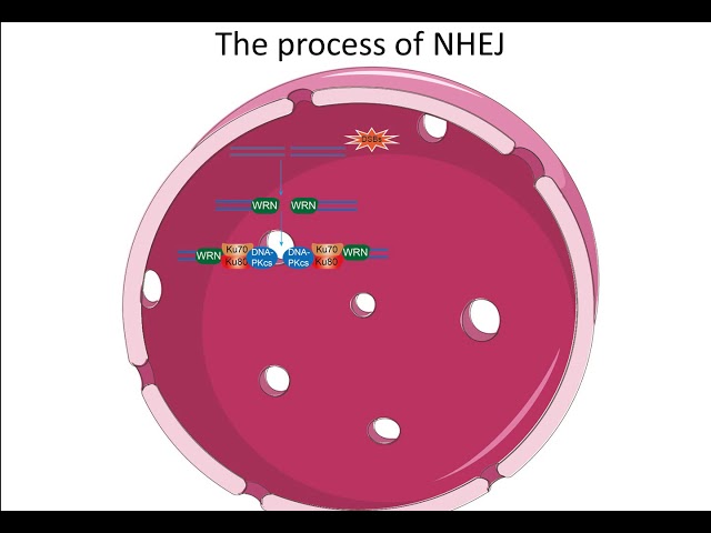
Non homologous end joining pathway Creative Diagnostic
1.9K
Non-homologous end joining (NHEJ) is the primary DNA repair pathway in eukaryotic cells for fixing double-strand breaks, functioning throughout the entire cell cycle without requiring a homologous template; the pathway involves sequential steps including recruitment of WRN helicase, Ku70/80 heterodimer binding to form a ring structure that slides along DNA, phosphorylation of DNA-PKcs to induce conformational changes, processing of DNA ends by enzymes like FEN1, PNKP, and Artemis, and finally ligation by DNA ligase 4 stabilized by XRCC4, with dissociation regulated by E3 ubiquitin ligase RNF8 and phosphorylation-induced conformational changes in DNA-PKcs.
Click to watch →
DNA Repair | Double Strand Break Repair | Non Homologous End Joining Repair Pathway
107
Non-homologous end joining (NHEJ) is a DNA repair pathway that fixes double strand breaks without using a template, making it mutagenic but essential for cell survival; the pathway involves key proteins including Ku70/Ku80 heterodimer that recognizes breaks, DNA-PKcs kinase, Artemis endonuclease for strand processing, and DNA ligases (XRCC4, XLF) that join the strands, with the original DNA sequence not being restored.
Click to watch →
Prerequisite Knowledge
- Concept 01The structure of DNA and the rules of complementary base pairing (A-T, C-G).
- Concept 02The Central Dogma of Molecular Biology, specifically how DNA is transcribed into RNA and translated into proteins.
- Concept 03The nature of genetic mutations and how alterations in the genetic code can lead to inherited diseases.
- Concept 04The cell's endogenous DNA repair pathways, specifically Non-Homologous End Joining (NHEJ) and Homology-Directed Repair (HDR).
Subsequent Learning
- Step 01Advanced gene-editing technologies that build upon CRISPR-Cas9, such as base editors and prime editors.
- Step 02In vivo and ex vivo delivery systems for CRISPR components, including viral vectors and lipid nanoparticles (LNPs).
- Step 03The ethical, legal, and societal implications (ELSI) of germline versus somatic gene editing in humans.
- Step 04Current clinical breakthroughs, active clinical trials, and real-world therapeutic applications of CRISPR (e.g., sickle cell disease treatments).
- Step 05The mechanism and ecological consequences of 'gene drives' to control vector-borne diseases or invasive species.
DNA & Genes
CRISPR Editing
Future Scope
DNA & Genes
0:01Playing Section
- 1
All cells contain a genome with 20,000 genes.
- 2
DNA pairing rules and genes influence health risks.
- 3
Sequencing advances identify disease-related genes.
Limitations of Double-Stranded Breaks and the Shift to Precision Editing Alternatives
While CRISPR-Cas9 is celebrated for its gene-cutting abilities, a major critical perspective in genomic science focuses on the biological risks of introducing double-stranded DNA breaks (DSBs). When CRISPR-Cas9 cleaves DNA, the cell's error-prone natural repair mechanisms can cause unintended large deletions, insertions, or chromosomal rearrangements. Furthermore, inducing DSBs can trigger a p53-mediated DNA damage response, which may kill healthy cells or inadvertently select for cancer-prone cells. Additionally, 'off-target' effects remain a persistent concern, where the system cuts unintended genomic sites. Because of these safety risks and unpredictable outcomes, many scientists advocate for newer alternatives like Base Editing and Prime Editing. These next-generation technologies allow for precise, single-nucleotide alterations or search-and-replace editing without severing the DNA backbone, offering a safer and more controlled pathway for therapeutic applications.
Deep Dive
Advanced gene-editing technologies that build upon CRISPR-Cas9, such as base editors and prime editors.

Raymond Schiffelers and colleagues | NANOSPRESSO: personalized local nucleic acid nanomedicines
130.9K
Base editing uses a modified Cas9 enzyme (nicking Cas9) fused to a DNA-modifying enzyme to convert specific nucleotides (e.g., A to G or C to T) without inducing double-strand breaks. Prime editing combines nicking Cas9 with reverse transcriptase to rewrite DNA sequences up to 20 nucleotides long, enabling correction of virtually any point mutation with high precision.
Click to watch →
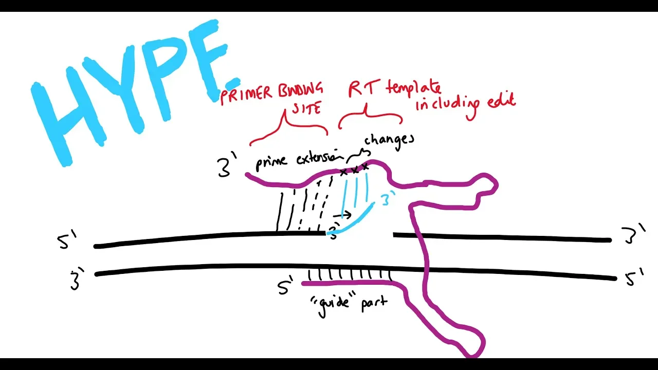
Prime editing (another way to edit genes)
36.9K
Genome editing has evolved from CRISPR-Cas9, which uses Cas9 nuclease to create double-stranded breaks requiring donor DNA templates, through base editing (dead Cas9 with deaminases avoiding breaks but limited to specific base conversions), to prime editing—a revolutionary 'search and replace' technique. Prime editing eliminates donor DNA needs and avoids double-stranded breaks by combining Cas9 nickase with reverse transcriptase and extended guide RNAs. This evolution represents a progression toward safer, more efficient genome modification, though challenges remain in optimizing efficiency, minimizing off-target effects, and translating these technologies to clinical applications for treating genetic diseases.
Click to watch →

Ils ont "hacké" l’ADN d’un bébé pour le sauver !
37.7K
CRISPR-Cas9 is a revolutionary gene editing technology functioning like molecular scissors, consisting of CRISPR (which identifies target DNA locations) and Cas9 (which cuts DNA). This technology earned the 2020 Nobel Prize in Chemistry. Base editing represents an advanced variation that allows single-letter DNA corrections without cutting the DNA strand, functioning more like a genetic eraser. The primary challenge is achieving precise targeting to avoid unintended modifications. This technology enables scientists to correct single-letter mutations that cause rare genetic diseases, representing a paradigm shift in medical treatment approaches.
Click to watch →
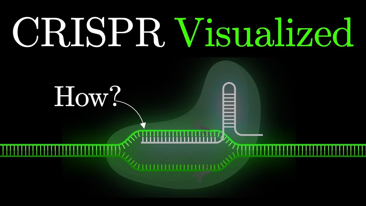
But what is CRISPR-Cas9? An animated introduction to Gene Editing. #some2
478.5K
CRISPR-Cas9 is a revolutionary gene editing technology derived from a bacterial immune system that enables precise modification of DNA sequences; originally discovered in bacteria as a defense mechanism against viruses, it was adapted by scientists Jennifer Doudna and Emmanuelle Charpentier to allow targeted DNA cutting through a guide RNA that directs the Cas9 enzyme to specific locations, with subsequent developments including base editing and prime editing techniques that further expand its capabilities for treating genetic diseases and advancing biotechnology.
Click to watch →

UPSC Prelims Test Series GS FLT 9 : Environment & Science and Technology Explanation | LTX IAS |
195
This section covers two major gene editing technologies. CRISPR-Cas9 uses a modified Cas9 protein guided by RNA to create double-strand breaks in DNA, enabling precise genetic modifications. Prime editing represents an advancement that does not require creating double-strand breaks, making it safer and reducing unintended mutations. Prime editing uses a modified Cas9 protein fused with reverse transcriptase enzyme, guided by prime editing guide RNA (pegRNA). It can perform insertions, deletions, and all 12 types of base-to-base conversions, offering greater versatility than traditional CRISPR-Cas9 which is primarily limited to insertions and deletions.
Click to watch →
Deep Dive
In vivo and ex vivo delivery systems for CRISPR components, including viral vectors and lipid nanoparticles (LNPs).
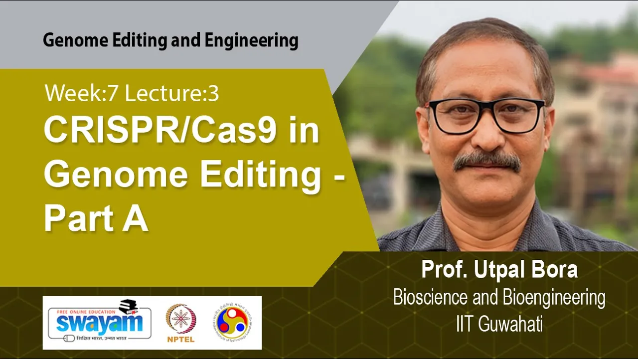
Lec 22: CRISPR/Cas9 in Genome Editing - Part A
1.9K
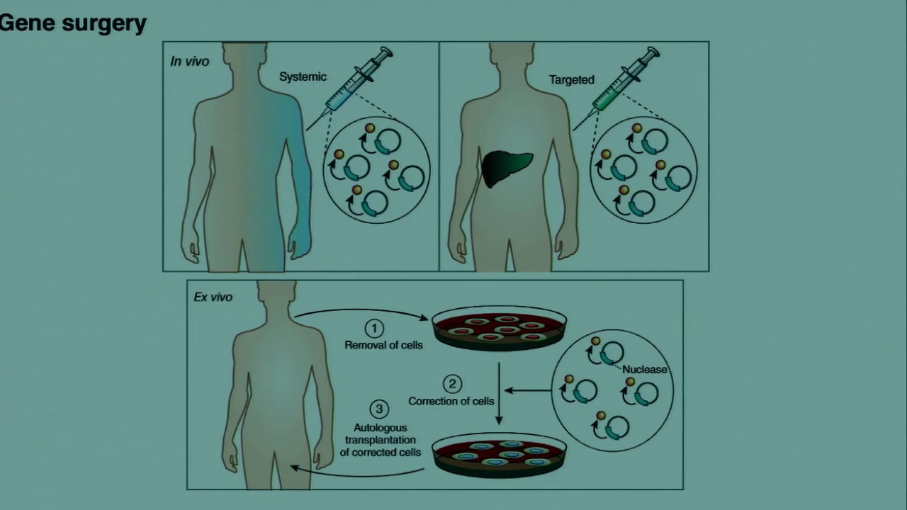
Ex vivo delivery involves extracting target cells from patients, culturing and expanding them in vitro, delivering CRISPR components, selecting and expanding edited cells, and reintroducing them into patients. In vivo delivery involves direct delivery to the body through systemic administration via intravenous infusion (cargo travels through bloodstream to target tissue) or local delivery via direct injection. The ex vivo approach allows for cell selection and expansion before reimplantation, while in vivo delivery targets tissues directly within the body. Both approaches have distinct advantages depending on the therapeutic context and target tissue accessibility.
Click to watch →
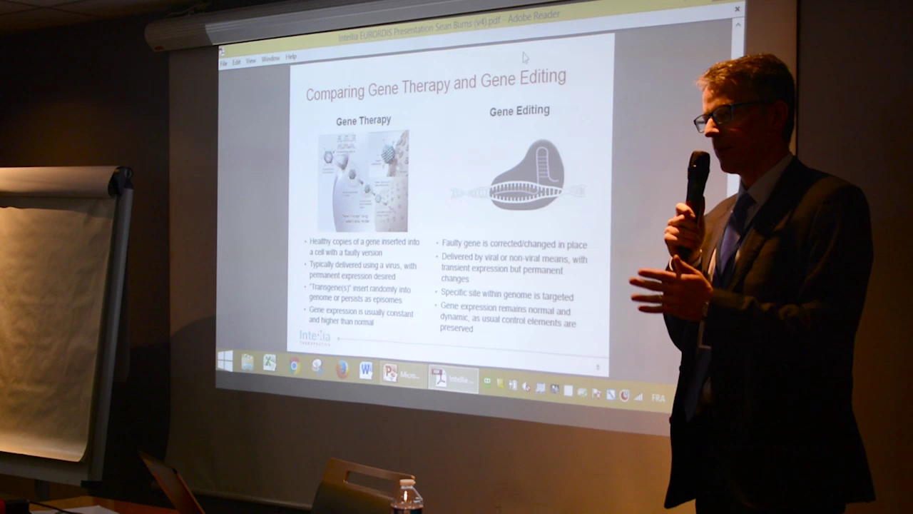
Genome Editing Workshop: 3. Perspectives of the biotech sector and regulatory aspects
303
CRISPR delivery strategies are categorized into ex vivo and in vivo approaches. Ex vivo editing involves extracting cells, editing them outside the body, and reintroducing them, which is more straightforward and well-understood. In vivo delivery requires injecting CRISPR components directly into the human body, facing challenges including immune system survival, tissue targeting, and achieving sufficient editing efficiency. Two primary in vivo platforms are lipid nanoparticles (LNPs) and adeno-associated viruses (AAVs). LNPs have been successfully used for siRNA delivery and can carry larger genetic payloads, with preclinical studies showing 50-75% editing efficiency in liver cells. AAVs offer tissue-specific targeting through different serotypes but face challenges with pre-existing immunity and limited cargo capacity. CRISPR has diverse therapeutic applications including transthyretin amyloidosis (single point mutation correction), hepatitis B virus (clearing latent viral DNA), and inborn errors of metabolism. Technical challenges include delivery efficiency, achieving sufficient editing percentages, and ensuring both gene copies are edited. Safety concerns include off-target effects and immunogenicity of bacterial Cas9 protein.
Click to watch →
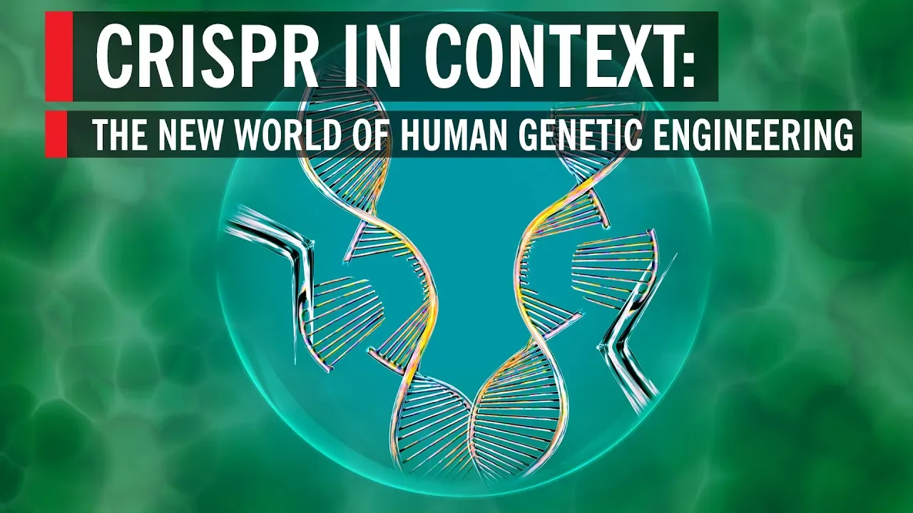
CRISPR in Context: The New World of Human Genetic Engineering
1.3M
CRISPR-Cas9 represents a revolutionary gene-editing technology functioning as a molecular scalpel. It consists of guide RNA (identifying specific DNA sequences) and Cas9 protein (cutting DNA at precise locations). Originally evolved in bacteria over millions of years as an immune defense mechanism against viruses, CRISPR was discovered by Jennifer Doudna and Emmanuelle Charpentier in 2012. This technology enables scientists to target, remove, and replace any DNA segment with unprecedented precision—capable of switching out single base pairs within the human genome. Unlike previous gene-editing methods that were cumbersome, expensive, and imprecise, CRISPR makes genetic modification accessible, quick, and affordable, democratizing what was once a specialized capability. The technology offers transformative potential across agriculture (drought-resistant crops, pest protection) and medicine (treating monogenic disorders like sickle-cell disease, muscular dystrophy, and cystic fibrosis). Two delivery approaches exist: ex vivo editing (modifying cells outside the body, then returning them—a method already in clinical trials) and in vivo delivery (using viral vectors to deliver editing components directly).
Click to watch →
Los Otros Hackers
321.6K
Scientists employ multiple techniques to deliver CRISPR components into cells: (1) Microinjection using fine needles under microscopic guidance; (2) Electroporation, which uses electrical currents to create temporary pores in cell membranes; (3) Viral vectors that carry CRISPR components into cells; (4) Other emerging methods. Most research uses mouse embryos or cells grown in artificial culture media designed to mimic bodily fluids. Some researchers modify stem cells that can then repopulate damaged organs when injected into patients.
Click to watch →

Essential Guide to Becoming a CRISPR Cas9 Expert - #ResearchersAtWork Webinar Series
38.5K
CRISPR components must enter the nucleus to access genomic DNA, requiring various delivery strategies: (1) Vector transfection delivers plasmids expressing Cas9 and sgRNA but is inexpensive yet inefficient; (2) Viral delivery offers higher efficiency: Lentivirus integrates into the genome for long-term expression, Adenovirus provides high infection efficiency but triggers immune responses, AAVs offer transient expression without immune response but can only accommodate small payloads like SaCas9; (3) RNP (ribonucleoprotein) transfection delivers pre-assembled Cas9-sgRNA complexes directly, minimizing off-target effects but requiring optimization for difficult cell types; (4) Stable cell lines expressing Cas9 enable reuse across experiments. Delivery format choices include all-in-one systems (single vector expressing both components, simpler but non-reusable) versus two-component systems (separate vectors or vector plus cell line, allowing component swapping between experiments).
Click to watch →
Deep Dive
The ethical, legal, and societal implications (ELSI) of germline versus somatic gene editing in humans.

LMU Bioethics Institute Lecture: The Ethics of Germline Modification
129
There is international consensus that somatic cell editing (affecting only the individual) is acceptable, while germline editing (affecting future generations) raises alarms. However, the details vary significantly: some states prohibit all embryo research, others allow research up to 12-40 days after creation, and some prohibit creating embryos for research entirely. The Chinese CRISPR case violated both Chinese law and international human rights principles, particularly regarding informed consent.
Click to watch →
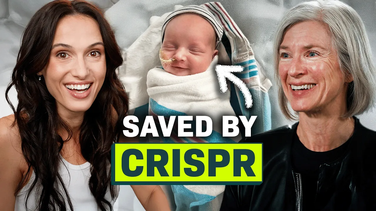
You’re Not Ready for What DNA Editing Will Do Next
826.6K
A critical distinction in gene editing is between somatic (non-reproductive) cells and germline (reproductive) cells. Somatic edits affect only the individual and do not get passed to future generations, while germline edits alter DNA in embryos and can be inherited by offspring. This distinction has profound ethical implications because germline changes are permanent, irreversible, and could potentially alter human populations over generations. The ethical concerns surrounding germline editing are significantly greater than those for somatic applications.
Click to watch →

Ethical, legal and social issues in epigenetics (ELSI): Session 3
419
Human germline gene editing became technically feasible with CRISPR-Cas9, yet remained ethically and legally contentious until Dr. He Jiankui's 2018 announcement of the first genome-edited babies, violating the 2015 international consensus against clinical use. Ethical frameworks emphasize that the human genome is humanity's heritage (Article 1, Universal Declaration) and that germline interventions impact future generations (Article 16, UNESCO). European legal frameworks provide strict bans: the Oviedo Convention prohibits germline modifications for procreation, while EU clinical trials regulation (Article 90) bans germline-modifying trials. Both frameworks contain amendment procedures requiring broad consensus across member states, reflecting tensions between enabling therapeutic benefits and protecting societal values. The path forward requires considerable agreement between different representatives of member states wearing different institutional hats, with neither framework set in stone.
Click to watch →

Bambine geneticamente modificate? Feat. Alessandro Tavecchio
23.2K
Editing the human germline fundamentally differs from other biomedical research because changes persist across generations and affect all descendants. Unlike somatic cell modifications that benefit only the individual, germline edits create permanent alterations in the human gene pool. This creates unprecedented ethical and practical challenges: (1) Future generations inherit modifications without consent; (2) Unintended consequences may emerge decades later; (3) Once released, modified genes cannot be recalled; (4) The modified population becomes a permanent branch of human evolution. These characteristics distinguish germline editing from any other form of medical intervention and require extraordinary caution and international consensus before proceeding.
Click to watch →

The Realities of Gene Editing with CRISPR I NOVA I PBS
239K
There are two fundamental categories of gene editing with distinct implications. Somatic gene editing modifies genes in body cells only, meaning changes do not get passed to future generations. This type is already being used in clinical trials to treat diseases like sickle cell disease and cancer. Germline gene editing, in contrast, modifies genes in reproductive cells (eggs, sperm, embryos), meaning changes would be inherited by all future offspring. The distinction is crucial because germline editing raises profound ethical questions about permanently altering the human gene pool and creating so-called 'designer babies.'
Click to watch →
Deep Dive
Current clinical breakthroughs, active clinical trials, and real-world therapeutic applications of CRISPR (e.g., sickle cell disease treatments).
![[다큐S프라임] 인류의 삶을 바꾸어 놓을 수 있는 유전자 가위 .. 불치병 치료를 넘어 생명 연장의 꿈 가능해지나.. / YTN 사이언스](https://i.ytimg.com/vi/z3Lmkhq39j4/maxresdefault.jpg)
[다큐S프라임] 인류의 삶을 바꾸어 놓을 수 있는 유전자 가위 .. 불치병 치료를 넘어 생명 연장의 꿈 가능해지나.. / YTN 사이언스
30.4K
Sickle cell anemia and beta thalassemia are single-gene disorders ideal for gene therapy. Clinical trials using CRISPR-Cas9 to reactivate fetal hemoglobin showed 42 of 44 beta thalassemia patients achieving transfusion independence. CRISPR Therapeutics' Casgevy therapy is expected to receive regulatory approval in 2023, potentially becoming the first CRISPR-based drug. UCLA researchers conducted trials using CRISPR-modified T-cells to target cancer-specific mutations, with 5 of 16 solid tumor patients showing tumor growth cessation. These clinical advances demonstrate translation from laboratory research to patient treatment, representing milestones in gene therapy development.
Click to watch →

The CRISPR Craze
1.2K
CRISPR has achieved significant clinical success in treating sickle cell disease. In late 2020, CRISPR Therapeutics in partnership with Vertex Pharmaceuticals reported remarkable results in patients treated with CRISPR-edited cells. Instead of correcting the mutated beta-globin gene, the therapy activated fetal hemoglobin production (normally turned off after birth), which compensates for the defective adult hemoglobin. Patients experienced elimination of pain episodes, hospitalizations, and blood transfusions, representing what physicians describe as a near-cure.
Click to watch →

CRISPR Tx is Done with Enrollment... (Ep. 291)
265
CRISPR-based gene therapy programs for sickle cell disease and thalassemia have achieved full enrollment in clinical trials (CLIMB 111 and CLIMB 121, each designed for 45 patients), with the company dosing over 70 patients and progressing toward Biologic License Application (BLA) filing by year-end, demonstrating the advancement of CRISPR technology from academic research to commercial therapeutic development.
Click to watch →
Cafe Scientifique - Gene Therapy in Sickle Cell Disease with Suchi Pandey, MD
497
Multiple CRISPR-based clinical trials for sickle cell disease are underway. The CTX001 trial (using CRISPR to disrupt BCL11A) enrolled 12 subjects aged 18-35 with severe sickle cell disease. The trial uses autologous CD34+ cells modified with CRISPR-Cas9 to disrupt BCL11A expression, aiming to increase fetal hemoglobin production. Fast-track designation from the FDA accelerates development for this and other gene-editing therapies targeting sickle cell disease, HIV, and leukemia.
Click to watch →

Las nuevas terapias génicas para tratar las enfermedades raras. Conferencia de Lluis Montoliu CNB
911
CRISPR enables gene deletion by cutting twice at the same location, removing intervening sequences. This approach was used to study albinism by deleting regulatory sequences in the tyrosinase gene. For Leber congenital amaurosis type 10, Editas Medicine removed cryptic sequences in the CEP290 gene, restoring vision in 11 patients. In cancer immunotherapy, CRISPR inactivates PD1 in extracted lymphocytes, making them more aggressive against tumors. For transthyretin amyloidosis, Intellia Therapeutics used lipid nanoparticles to deliver CRISPR components, reducing toxic protein levels for over a year. The breakthrough therapy for sickle cell disease uses CRISPR to inactivate BCL11A, restoring fetal hemoglobin production and changing red blood cell shape from sickle to normal. Approved in the UK (November 2023) and US (December 2023), it costs $2.2 billion per patient.
Click to watch →
Deep Dive
The mechanism and ecological consequences of 'gene drives' to control vector-borne diseases or invasive species.
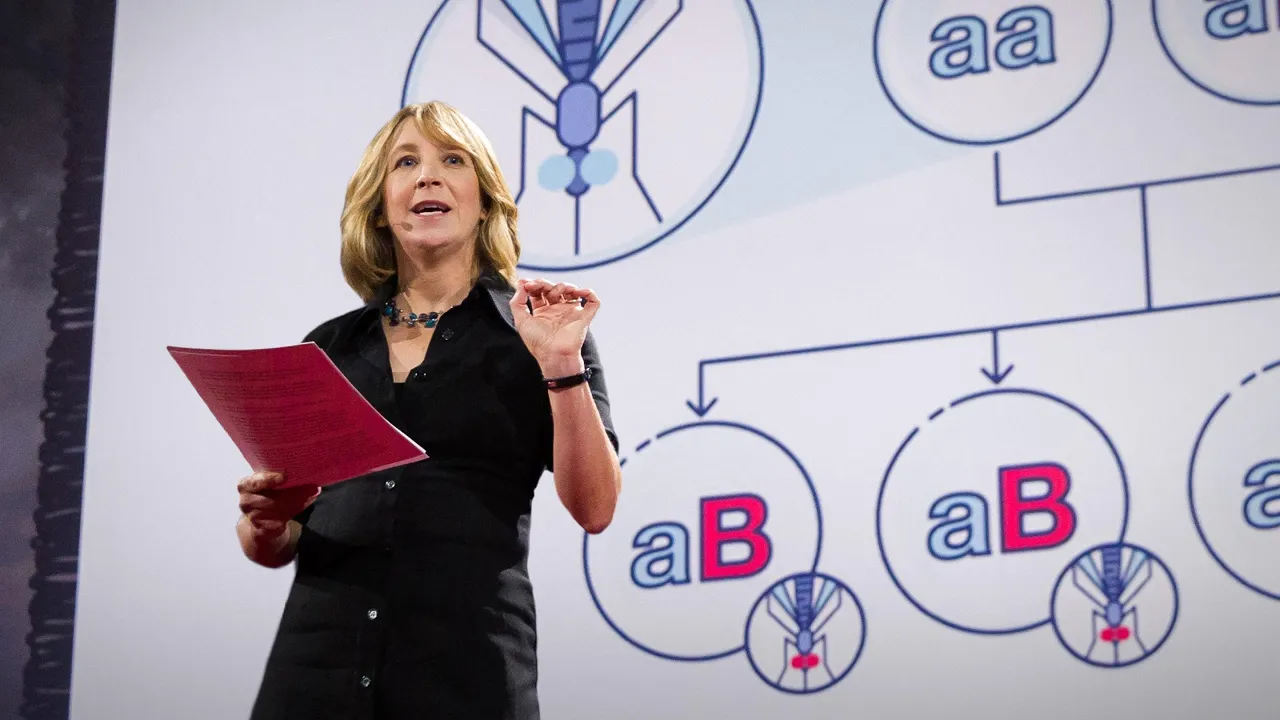
Gene editing can now change an entire species -- forever | Jennifer Kahn
511.9K
Gene drives offer transformative applications for public health and conservation. For disease control, introducing an anti-malarial gene drive into just 1% of Anopheles mosquitoes could spread to the entire population within a year, potentially eliminating malaria. Similar approaches could address dengue fever, chikungunya, and yellow fever. For invasive species management, gene drives could be used to suppress populations by releasing organisms that produce only male offspring, leading to population collapse within generations. This could help restore native species pushed to extinction by invasive species like Asian carp in the Great Lakes.
Click to watch →

Penser le vivant autrement (11) - Virginie Courtier-Orgogozo (2022-2023)
2K
Gene drive applications include: malaria eradication (Target Malaria project), invasive species control (Asian fruit fly), disease vector control (ticks, nematodes). Risks include: gene flow to related species, cryptic populations, uncontrollable spread, ecosystem impacts. Unlike pesticides, gene drives continue spreading once released, requiring complex 'break' mechanisms for control.
Click to watch →

Genetic Tools For Conservation and Health: What’s The Role of Gene Drives?
145
Gene drive technology enables the biased inheritance of genetic traits, allowing for rapid population modification in species like mosquitoes and invasive rodents; this emerging tool offers promising applications for controlling vector-borne diseases (such as malaria and dengue) and invasive alien species, though its deployment requires careful consideration of ecological impacts, regulatory frameworks, and community acceptance.
Click to watch →

Jonathan Latham PhD: The Dangers of Gene Editing and Gene Drives
155
Gene drives are the scariest approach because only one organism needs to be introduced into the environment, and the entire genetic construct spreads through that entire population. This is intended to overtake the population and change or send it extinct. If gene drives work as intended, they could potentially spread to other species through hybridization, potentially driving multiple species extinct through a chain reaction. Various mosquito population control methods exist, including Wolbachia bacteria, irradiation, and gene editing, but gene drives represent the most concerning approach. The regulatory system for these technologies is unclear because the set of questions that need to be answered is very large, and answers are not available for wild species that we know very little about.
Click to watch →

Can Gene Drive Mosquitoes Help Fight Malaria - Without Harming the Environment? – 4.12.2025
213
Gene drive is a genetic system that biases inheritance toward near 100% transmission instead of the typical 50% Mendelian rate, enabling population replacement (releasing disease-resistant mosquitoes) or suppression (crashing populations via fertility reduction or sex ratio manipulation). Due to its powerful nature, gene drive carries risks of irreversible genetic spread and unintended ecological consequences. Historical vector control studies show that conventional tools like bed nets and insecticides can alter species composition—bed nets decreased Anopheles gambiae but increased Anopheles arabiensis, while spraying reduced Anopheles but allowed replacement by other species. Effective gene drive monitoring requires detecting unintended spread beyond trial sites, identifying resistance mechanisms, and tracking non-functional effects within intervention areas.
Click to watch →
DNA & Genes
CRISPR Editing
Future Scope
DNA & Genes
0:01- 1
All cells contain a genome with 20,000 genes.
- 2
DNA pairing rules and genes influence health risks.
- 3
Sequencing advances identify disease-related genes.
Limitations of Double-Stranded Breaks and the Shift to Precision Editing Alternatives
While CRISPR-Cas9 is celebrated for its gene-cutting abilities, a major critical perspective in genomic science focuses on the biological risks of introducing double-stranded DNA breaks (DSBs). When CRISPR-Cas9 cleaves DNA, the cell's error-prone natural repair mechanisms can cause unintended large deletions, insertions, or chromosomal rearrangements. Furthermore, inducing DSBs can trigger a p53-mediated DNA damage response, which may kill healthy cells or inadvertently select for cancer-prone cells. Additionally, 'off-target' effects remain a persistent concern, where the system cuts unintended genomic sites. Because of these safety risks and unpredictable outcomes, many scientists advocate for newer alternatives like Base Editing and Prime Editing. These next-generation technologies allow for precise, single-nucleotide alterations or search-and-replace editing without severing the DNA backbone, offering a safer and more controlled pathway for therapeutic applications.
The structure of DNA and the rules of complementary base pairing (A-T, C-G).
The Central Dogma of Molecular Biology, specifically how DNA is transcribed into RNA and translated into proteins.
The nature of genetic mutations and how alterations in the genetic code can lead to inherited diseases.
The cell's endogenous DNA repair pathways, specifically Non-Homologous End Joining (NHEJ) and Homology-Directed Repair (HDR).
Advanced gene-editing technologies that build upon CRISPR-Cas9, such as base editors and prime editors.
In vivo and ex vivo delivery systems for CRISPR components, including viral vectors and lipid nanoparticles (LNPs).
The ethical, legal, and societal implications (ELSI) of germline versus somatic gene editing in humans.
Current clinical breakthroughs, active clinical trials, and real-world therapeutic applications of CRISPR (e.g., sickle cell disease treatments).
The mechanism and ecological consequences of 'gene drives' to control vector-borne diseases or invasive species.
4.4M views42.3Klikes4:13@McGovernInstituteOriginal Release: 2014-11-05
[Music] every cell in our body contains a copy of our genome over 20,000 genes 3 billion letters of DNA DNA consists of two strands twisted into a double helix and held together by a simple pairing rule a pairs with T and G pairs with C our genes shape who we are as individuals and as a species genes also have profound effects on health and thanks to advances in DNA sequencing researchers have identified thousands of genes that affect our risk of disease to understand how genes work researchers need ways to control them changing genes and living cells is not easy but recently a new method has been developed that promises to dramatically improve our ability to edit the DNA of any species including humans the CRISPR method is based on a natural system used by bacteria to protect themselves from infection by viruses when the bacterium detects the presence of virus DNA it produces two types of short RNA one of which contains a sequence that matches that of the invading virus these two RNAs form a complex with a protein called caste 9 caste 9 is a nucleus a type of enzyme that can cut DNA when the matching sequence known as a guide RNA finds its target within the viral genome the caste 9 cuts the target DNA disabling the virus over the past few years researchers studying the system realized that it could be engineered to cut not just viral DNA but any DNA sequence at a precisely chosen location by changing the guide RNA to match the target and this can be done not just in a test tube but also within the nucleus of a living cell once inside the nucleus the resulting complex will lock onto a short sequence known as the pan the cast nine will unzip the DNA and match it to its target RNA if the match is complete the cast 9 will use two tiny molecular scissors to cut the DNA when this happens the cell tries to repair the cut but the repair process is error-prone leading to mutations that can disable the gene allowing researchers to understand its function these mutations are random but sometimes researchers need to be more precise for example by replacing a mutant gene with a healthy copy this can be done by adding another piece of DNA that carries the desired sequence once the CRISPR system has made a cut this DNA template can pair up with the cut ends recombining and replacing the original sequence with the new version all this can be done in cultured cells including stem cells that can give rise to many different cell types it can also be done in a fertilized egg allowing the creation of transgenic animals with targeted mutations and unlike previous methods CRISPR can be used to target many genes at once a big advantage for studying complex human diseases that are caused not by a single mutation but by many genes acting together these methods are being improved rapidly and will have many applications in basic research in drug development in agriculture and perhaps eventually for treating human patients with genetic disease [Music] [Music]
Up Next

1:44
Non-Homologous End Joining (NHEJ) | DNA Double-Strand Break Repair Animation
@OUPAcademic
319.8K views•2014-04-11
14:12
Quantitative Real-Time PCR (qPCR): Principle, Method & Data Analysis
@animatedbiologywitharpan
129.2K views•2023-09-06
23:33
Microbial Degradation of Plastics: Biodegradation Pathways & Sustainability
@majeedhammad
2.9K views•2021-04-11
16:04
CRISPR and Genetic Engineering: How Gene Editing Works and Why It Matters
@kurzgesagt
30.5M views•2016-08-10
Related Study Plans & Knowledge Roadmaps
Structured learning paths in Biotechnology
biotechnology
AAV Production: Transfection, Prep & qPCR
01Introduction to Gene Therapy and AAV Biology
02Mammalian Cell Culture: Suspension HEK293 Cells
03Upstream Bioprocessing: Triple-Transfection Optimization
+ 2 more learning modules
18h5 modules
Explore Roadmap
biotechnology
Building Gut-on-a-Chip: PDMS, Cells & TEER
01Foundations of Microfluidics and Gut Anatomy
02Cleanroom Fabrication & PDMS Soft Lithography
03Epithelial Cell Culture and On-Chip Seeding
+ 2 more learning modules
26h5 modules
Explore Roadmap
biotechnology
Optimizing CFPS: E. coli Extract & Yield
01Fundamentals of Molecular Biology and Translation
02Introduction to Cell-Free Protein Synthesis (CFPS)
03Preparing E. coli Crude Lysate (The Extract)
+ 2 more learning modules
19h5 modules
Explore Roadmap
biotechnology
Haematococcus PBR: Design, Stress & Astaxanthin
01Biology and Life Cycle of Haematococcus pluvialis
02Bench-Scale Photobioreactor (PBR) Design and Engineering
03Culture Media Formulation and Inoculation
+ 2 more learning modules
15h5 modules
Explore Roadmap