

![AP Psychology Unit 1 Review [Everything You NEED to Know]](https://i.ytimg.com/vi_webp/4yvfd8aoUBc/maxresdefault.webp)














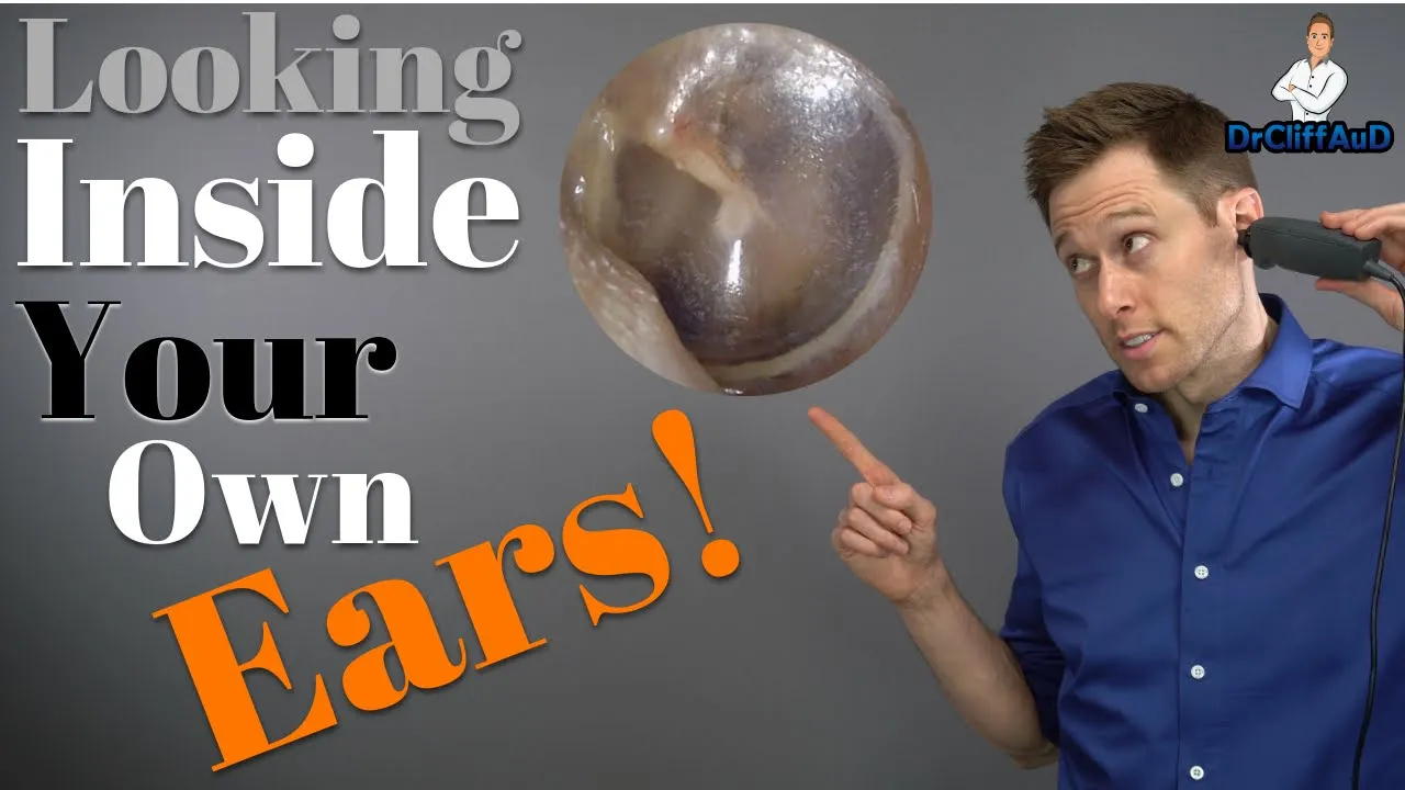
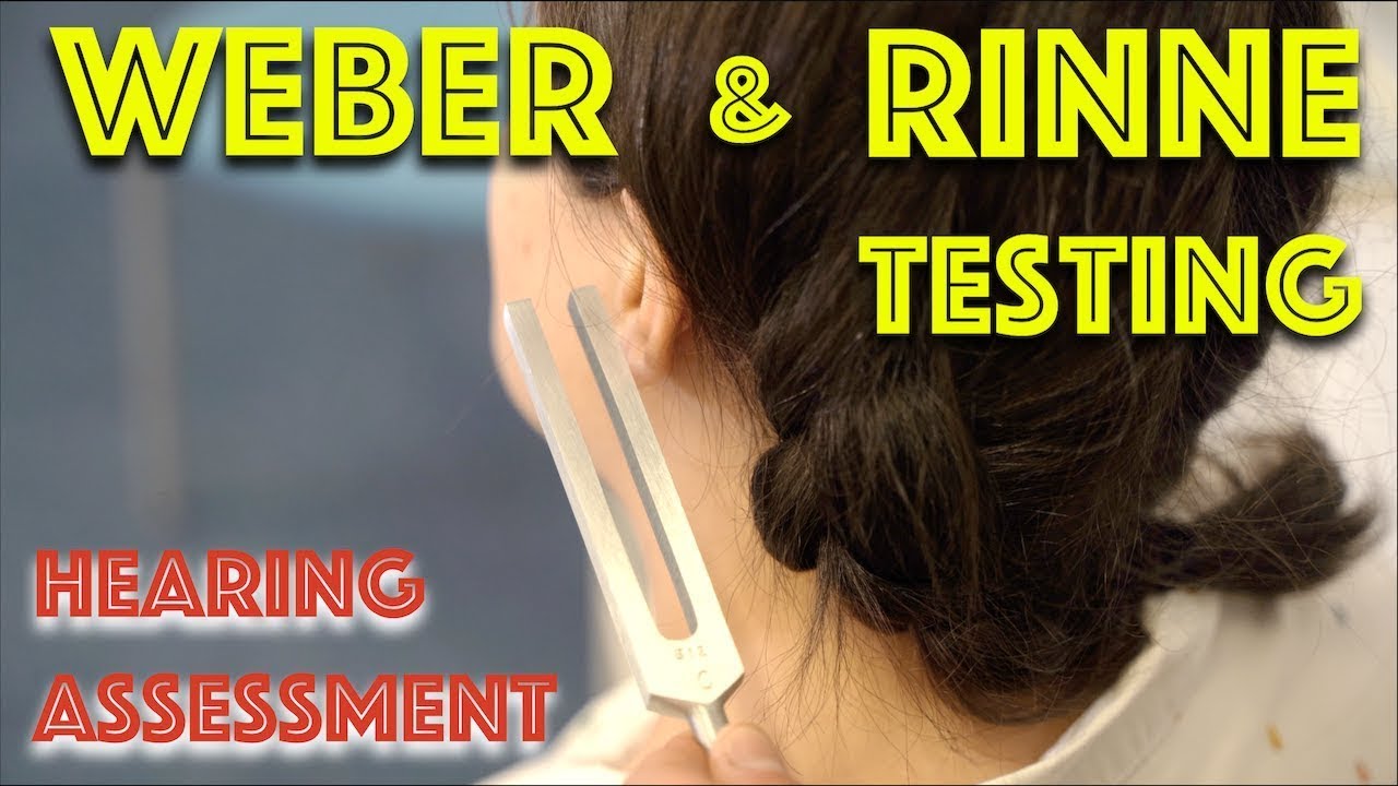


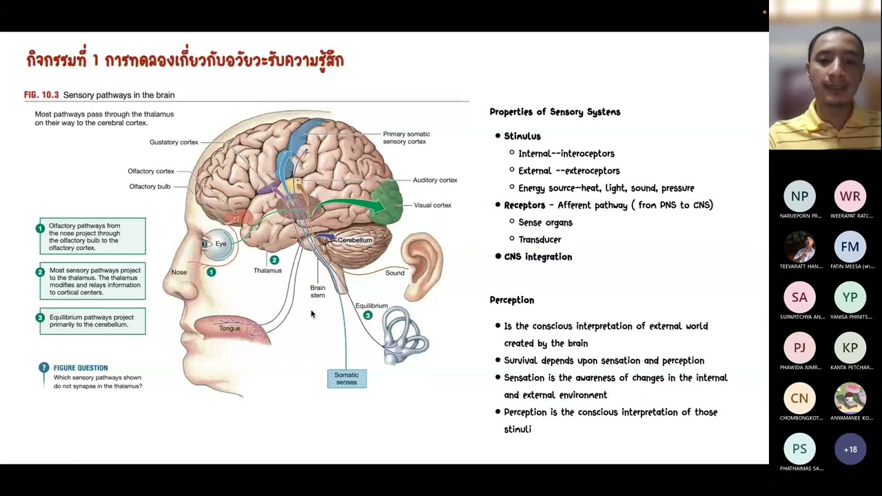
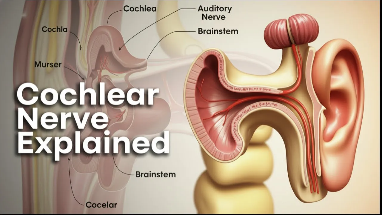




The ear is anatomically divided into three main parts: the outer ear (visible portion including the pinna, helix, lobe, ear canal, and eardrum), the middle ear (containing the three smallest bones called malleus, incus, and stapes, plus the Eustachian tube), and the inner ear (comprising the vestibule, semicircular canals for balance, and the cochlea for hearing); sound vibrations travel through these structures to be interpreted by the brain via the vestibulocochlear nerve.
Anatomy of the Ear: Outer, Middle, and Inner Ear Explained
Added:Basic physics of sound, including waves, frequency, amplitude, and how sound travels through different mediums.
Fundamental human anatomical terminology, particularly directional terms and the skeletal structure of the skull (specifically the temporal bone).
The general concept of sensory transduction—how the human nervous system converts physical stimuli into electrical signals.
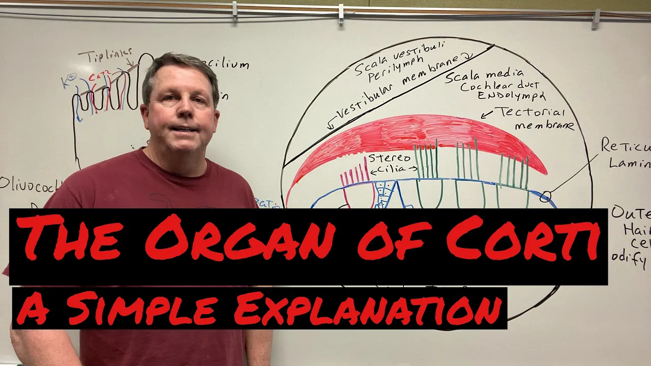
The physiological process of auditory transduction, specifically focusing on the organ of Corti and hair cell activation.
The vestibular system's role in balance, spatial orientation, and equilibrium managed by the semicircular canals.
Common clinical pathologies of the ear, such as conductive versus sensorineural hearing loss, otitis media, and tinnitus.
The central auditory pathway, detailing how nerve impulses travel from the vestibulocochlear nerve (CN VIII) to the auditory cortex in the brain.
Biomedical interventions for hearing impairment, including the mechanics of hearing aids and cochlear implants.
1.9K views23likes5:43Original Release: 2024-04-24
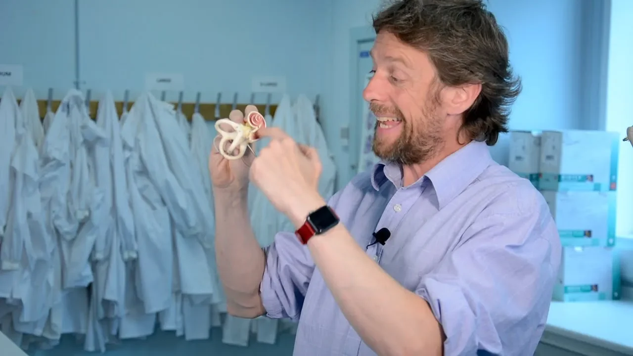
hi and welcome to my channel learning biology with Dr Vanessa I'm Dr Vanessa and I'm on a mission to make difficult Concepts easy to understand within the realm of anatomy and physiology and biology if that resonates with you please make sure to subscribe to my channel and click the notification Bell so that you never miss out on a new video in today's video we're going to take a look at the anatomy of the ear we're going to be using this all awesome 3D model to be able to show you the different parts of the ear I think that being able to see these big life-size models really helps to visualize the different parts in a way that is very different than being able to see it on a piece of paper so we're going to dive in and take a look at the anatomy of the ear now the ear is separated into three main parts the outer ear the middle ear and the inner ear so let's go through each different part of the ear and what we find there so the outer ear consists of the part of the ear that you can actually see okay the outer part of the ear and that portion is referred to as the Oracle or the PA this portion here is referred to as the Helix and this portion here is referred to as the lobe now if we go in through the ear this portion right here is called the ear canal it's it's also got a few other names um the external acoustic meatus or sometimes it's referred to as the external auditory canal so a lot of different names but this is the ear canal for to make it easy okay still part of the outer ear this is still considered part of the outer ear and then we go to the tanic membrane right here or easier to remember the eardrum so this is the tanic membrane also known as the eardrum so that all consists all is part of the outer ear so the Oracle or the ear canal tanic membrane all consist of um the outer ear now the ear itself is made of of elastic cartilage it is um mobile surrounded by skin and then you can kind of see some inner Anatomy here where you have um some fat tissue some more elastic cartilage and then you also have bone um in through this inner portion so external ear or outer ear outer ear and then we move to the middle ear the middle ear is behind the tanic membrane okay behind the eardrum and there are three little bones that make up the middle ear and so in the middle ear we have um the malus the incus and the stapes now these are the smallest bones in the the body they are actually tiny little bones that connect to the eardrum they also then connect to the inner ear and that's going to allow us to hear sound now we're just going through the anatomy in this video in um my next video I'm going to talk about how we hear because in order to really understand how we hear we first need to know how the ear is made up so that's the middle ear the middle ear also has this portion right here this is called the asuan tube and helps to drain any fluid uid from this area normally this area does not have any fluid in here when fluid starts to build up this can create an ear infection pressure pain okay so when that fluid starts to build up it creates pressure in this area it starts to hurt if the fluid stays there long enough bacteria can grow and create an infection so that's the middle ear which would connect to um the inner ear so here's the inner ear right here in this in this area right here if you look at the inner ear what you'll notice is that there's this portion right here between these two very distinct areas of the inner ear and that is where the um bones of the eardrum the bones right behind the eardrum or the bones of the middle ear are going to connect to the inner ear that portion right there is called the vestibule and then there's two other portions of the inner ear we have the semicircular canals which function in equilibrium and balance and then we have the ccka this snail like structure back here that is going to function in hearing so we'll delve more into that in my next video and talk about how we hear uh when this takes the signals that it gets from the bones those vibrations it's going to converge back here onto this nerve called the vestibulo clear nerve which is going to take those signals to the brain in order to interpret what we're hearing so those are all the different parts of the ear hopefully taking a look at this with a model really helps you to understand these different parts of the ear um let me know what you think about these videos that I'm using these models in is it helping you to really understand that Anatomy um put it down below let me know um stay tuned because I am going to make a video on how we hear and we'll go more into the science of how that happens looking really into um the inner part of the ear uh if you have any questions or comments put those in the comment box below and thank you so much for watching my video
Up Next
46:48
External and Middle Ear Anatomy: Special Senses | Ninja Nerd Lectures
@NinjaNerdOfficial
1.2M views•2017-08-12

50:12
Bacterial Communication: Quorum Sensing and Biofilm Formation Explained
@JHUAAP
12.6K views•2011-06-28

5:35
Enteric Nervous System Explained: The Gut's Brain | Neurobiology Lecture
@alumniu6029
438 views•2018-09-12

7:08
Bacteriophages: Earth's Deadliest Killers and Future Antibiotics
@kurzgesagt
34.6M views•2018-05-13
Related Study Plans & Knowledge Roadmaps
Structured learning paths in Biology
biology
RNAi: Biogenesis, RISC & Gene Silencing
01Molecular Biology Foundations: DNA to Protein
02Introduction to RNA Interference (RNAi)
03Molecular Biogenesis of miRNA and siRNA
+ 2 more learning modules
10h5 modules
Explore Roadmap
biology
Mycorrhizae: Signaling, Biophysics & Ecology
01Foundations of Mycorrhizal Symbiosis
02Molecular Signaling and Root Colonization
03Biophysics and Bioenergetics of Nutrient Exchange
+ 1 more learning modules
11h4 modules
Explore Roadmap
biology
Circadian Clocks: TTFL, SCN & Periphery
01Foundations of Molecular Biology & Gene Expression
02Introduction to Chronobiology and Actograms
03The Core TTFL: Molecular Architecture of the Clock
+ 3 more learning modules
14h6 modules
Explore Roadmap
biology
Population Genetics: Math, Drift & Selection
01Genetic & Probability Foundations
02The Hardy-Weinberg Principle and Baseline Models
03Mathematical Modeling of Natural Selection
+ 3 more learning modules
18h6 modules
Explore Roadmap