


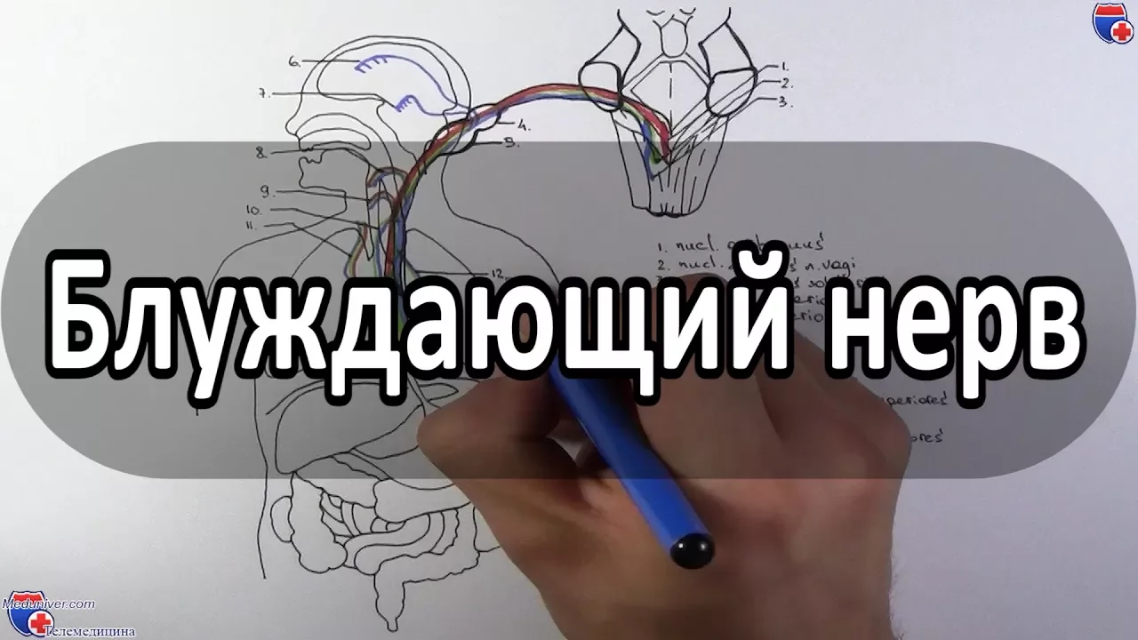

















The vagus nerve (cranial nerve X) is the longest and most widely distributed cranial nerve, serving as the primary parasympathetic conduit with sympathetic influences; it originates from four nuclei in the medulla oblongata (dorsal motor nucleus, nucleus ambiguus, solitary nucleus, and spinal trigeminal nucleus), contains two sensory ganglia (superior/jugular and inferior/nodosum), and carries general somatic efferent, special sensory, branchial motor, and visceral motor fibers, with its extracranial course divided into cervical, thoracic, and abdominal segments that innervate the pharynx, larynx, heart, lungs, esophagus, stomach, liver, and intestines.
Vagus Nerve Anatomy Animation: Origin, Course, Nuclei & Branches
Added:Basic organization of the 12 cranial nerves, including their naming, numbering, and general functional classifications.
Gross anatomy of the brainstem, with particular focus on the structure of the medulla oblongata where the vagus nerve nuclei reside.
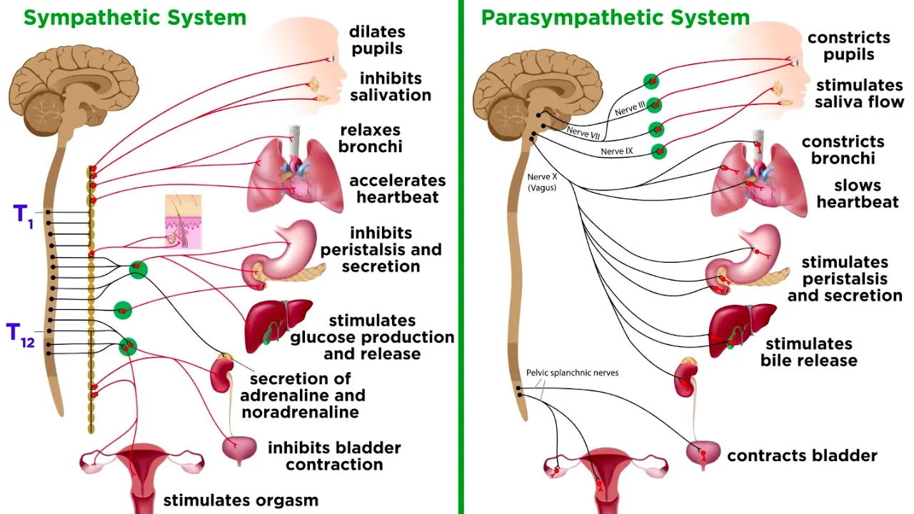
Fundamentals of the Autonomic Nervous System (ANS), specifically the physiological distinction between the sympathetic and parasympathetic divisions.
Basic neuroanatomical functional pathways, such as the difference between general visceral afferent/efferent and special visceral afferent/efferent fibers.
Clinical pathology of the vagus nerve, including diagnostic testing for lesions, vocal cord paralysis, dysphagia, and uvular deviation.
The physiological role of the vagus nerve in autonomic reflexes, such as the baroreceptor reflex and the mechanism behind vasovagal syncope.
The enteric nervous system and the gut-brain axis, exploring how the vagus nerve mediates complex biochemical signaling between the gut microbiota and the brain.
Clinical and therapeutic applications of Vagus Nerve Stimulation (VNS) in treating refractory epilepsy, treatment-resistant depression, and inflammatory diseases.
117.8K views2.1Klikes11:53Original Release: 2021-11-25
[Music] vegas nerf the tenth cranial nerve introduction the vagus nerve or the tenth cranial nerve is primarily associated with the parasympathetic division of the autonomic nervous system however it also has some sympathetic influence through peripheral chemoreceptors vagus is the longest and most widely distributed cranial nerve this is the only nerve that has the thoracic and abdominal distribution the vagus nerve is a mixed nerve as it contains both efferent or sensory and efferent or motor fibers this means it is responsible for not only carrying motor signals to the organs it innervates but it also carries sensory information from these organs back to the central nervous system it has two sensory ganglia superior or jugular and inferior or nodosum the vagus nerve contains general efferent or sensory fibers special sensory visceral efferent or sensory fibers branchial efferent or motor fibers visceral efferent or motor fibers general efferent fibers are responsible for perceiving touch pain temperature pressure vibration and proprioceptive sensation from the posterior ear external auditory meatus and posterior and the external surface of the tympanic membrane branchial efferent fibers innervate the muscles that develop from the branchial arches such as muscles of mastication the tensor tympani and the tensor villi palatini special sensory convey taste from palette and epiglottis visceral efferent fibers innervate the viscera including all smooth muscle and glands visceral efferent fibers are responsible for perceiving sensory input with the exception of pain from the viscera or internal organs of the body's main cavity nuclei of vagus nerve within the medulla oblongata of the brain stem there are four vagal nuclei onto which the axons of the vagus nerve emerge from or converge onto these include the dorsal motor nucleus the nucleus ambiguous the solitary nucleus and the spinal trigeminal nucleus the dorsal motor nucleus supplies parasympathetic inference primarily to the gastrointestinal tract and lungs the efferent fibers that arise from the nucleus ambiguous supply the muscles of the soft palate pharynx and larynx it also gives rise to branchial efferent fibers and preganglionic parasympathetic neurons for the heart the solitary nucleus receives primary efference from visceral organs as well as taste information finally the efferents that converge onto the spinal trigeminal nucleus relay sensory information regarding pain temperature and deep touch of the outer ear the dura of the posterior cranial fossa and the mucosa of the larynx intracranial course the vagus nerve arises from the posterior lateral sulcus between the 9th and tenth cranial nerves after its origin it passes along with the cranial accessory nerve towards the jugular for ramen to exit from the cranial cavity sensory vagal ganglia the superior ganglion or jugular is located in the jugular foramin contains unipolar first sensory neurons for general somatic sensations the inferior ganglion or nodosum is located below the jugular foramin and contains unipolar first sensory neurons for general visceral and special visceral or taste sensation branches of vegas in jugular for ramen auricular branch of vegas or alderman's nerve is distributed to skin off upper part of the auricle external acoustic miatas and posterior inferior part of the tympanic membrane extracranial course of vagus is divided into three parts cervical thoracic and abdominal let's take a look at the cervical cores the vagus nerve in the neck is present as a content of carotid sheath in between the internal carotid artery and internal jugular vein up to the level of superior margin of thyroid cartilage below this level it lies between common carotid artery and internal jugular rain the vagus nerve comes out of the carotid sheath at the root of the neck branches in the neck pharyngeal branch this branch carries cranial accessory fibers the 11th cranial nerve which takes part in the pharyngeal plexus through which it supplies the pharyngeal muscles except stylopharynges it also supplies muscles of the soft palate except the tensor palette superior laryngeal nerves arise just below the pharyngeal branch it immediately divides into internal and external laryngeal nerves the external laryngeal nerve supplies cricothyroid and inferior constrictor whereas the internal laryngeal nerve is a sensory nerve which supplies the mucosa of the larynx above the vocal cords recurrent laryngeal nerves the nerves of both sides supply all intrinsic muscles of larynx except cricothyroid muscles and the sensory fibers to mucosa of larynx below the vocal cords cardiac branches are two sets superior and inferior forms superficial and deep cardiac plexus to supply muscles off the heart vagus nerve in the thorax it enters into the thorax through the thoracic inlet the course of the right vegas is different from that of the left vegas in the thorax course of the right vegas the right vegas enters the thoracic cavity by crossing in front of the first part of the subclavian artery posterior to the internal jugular vein in the superior mediastinum right vegas runs posterior to the right brachiocephalic vein and superior vena cava in contact with the right surface of trachea in the posterior mediastinum the nerve passes posterior to right principal bronchus reaches the hilum of the right lung to form the pulmonary plexus at posterior surface of esophagus branches of right and left vagus forms the esophageal plexus let's take a look at the course of the left vegas it enters the thoracic cavity between the left subclavian and left common carotid artery posterior to brachiocephalic vein it is in relation to left surface of the arch of aorta near the hilum of the left lung it gives off the pulmonary plexus branches of right and left vegas in the thorax deep cardiac plexus supplies the heart pulmonary plexus to the trachea and bronchial tree esophageal plexus to the esophagus abdominal course during development of the foregut the stomach rotates 90 degrees clockwise shifting the left vagus to anterior and right vagus to posterior so in the abdomen anterior and posterior vagal trunks are present and here your vagal trunk gives rise to hepatic and gastric branches hepatic branches supply liver gallbladder and the extra biliary system gastric branches supply anterior and superior surfaces off the stomach posterior vagal trunk gives rise to gastric and celiac branches gastric branches the main gastric branch which is the nerve to let our jet runs in the lesser momentum to supply the posterior inferior part of the stomach celiac branches pass around the left gastric artery and join with celiac and superior mecentric plexus to supply the abdominal organs clinical correlation unilateral lesions of vagus nerve causes unilateral paralysis of soft palate except tensor palate due to this lowering of the palate on the affected side is observed during phonation the palate fails to elevate the uvula shifts to normal side results in nasal tongue of nasal regurgitation of food unilateral paralysis of pharyngeal muscles causing deviation of posterior pharyngeal wall loss of gag reflex on the same side of the lesion ipsilateral loss of sensations in the pharyngeal mucosa and paralysis of the laryngeal muscles
Up Next

111:21
Daily Gut Health Checks and How to Improve Microbiome
@doctors_kitchen
39.2K views•2025-02-25

48:47
Graphic Medicine: Comics for Collaborative Healthcare Communication
@nationalpatientadvocate
189 views•2023-12-04
22:48
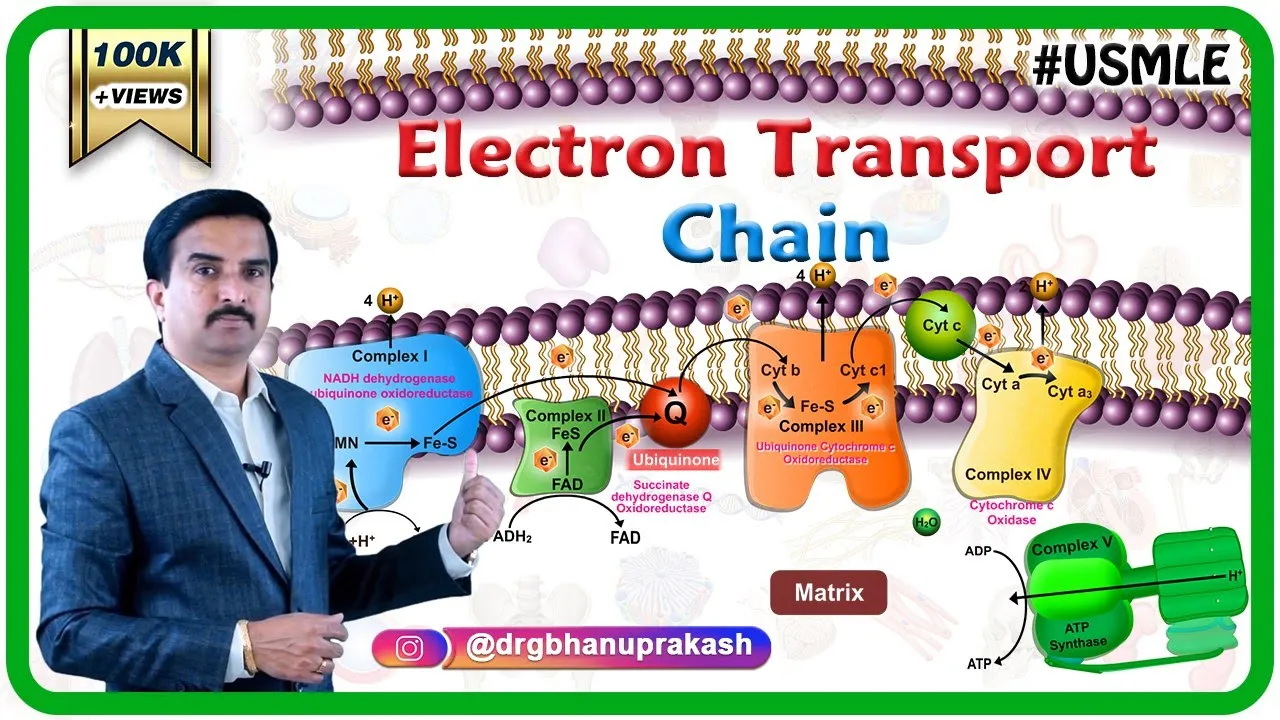
Electron Transport Chain & Oxidative Phosphorylation | Biochemistry Animation
@doctorbhanuprakash
186.8K views•2018-11-13

3:05
Stages of Labor and Vaginal Birth | Childbirth Animation
@nucleusmedicalmedia
52.1M views•2017-08-18
Related Study Plans & Knowledge Roadmaps
Structured learning paths in Medicine
medicine
Abnormal LFTs: Diagnostic Workup & Pathology
01Liver Anatomy and Basic Physiology
02Introduction to Liver Function Tests (LFTs)
03Hepatocellular vs. Cholestatic Patterns
+ 3 more learning modules
12h6 modules
Explore Roadmap
medicine
Analyzing Blood Smears: RBC, WBC & Platelets
01Introduction to Blood Components & Smear Preparation
02Normal Morphology of Blood Cells
03Erythrocyte Pathologies & Morphological Variants
+ 2 more learning modules
12h5 modules
Explore Roadmap
medicine
Mechanical Ventilation: Waveforms & Alarms
01Anatomy and Physiology of Breathing
02Introduction to Mechanical Ventilation & Core Modes
03Ventilator Settings and Waveform Interpretation
+ 2 more learning modules
12h5 modules
Explore Roadmap
medicine
Neuro Exam: Localization & Stroke Syndromes
01Foundations of Neuroanatomy and Vascular Supply
02The Neurological Examination: Step-by-Step
03Principles of Neurological Localization
+ 3 more learning modules
18h6 modules
Explore Roadmap