Long-term depression (LTD) is a process by which synaptic connections between neurons become weaker, serving as the opposing mechanism to long-term potentiation (LTP); the best-understood LTD mechanism involves NMDA and AMPA glutamate receptors, where prolonged low-intensity stimulation causes partial magnesium unblocking of NMDA receptors, allowing low-level calcium influx that triggers a cellular cascade removing AMPA receptors from the postsynaptic membrane, thereby weakening the synapse and potentially resetting previous synaptic changes to enable new memory formation via LTP.
Long-Term Depression (LTD): Mechanisms and Role in Memory
Added:Deep Dive
Fundamentals of synaptic transmission, including neurotransmitter release (specifically glutamate) and postsynaptic potential generation.
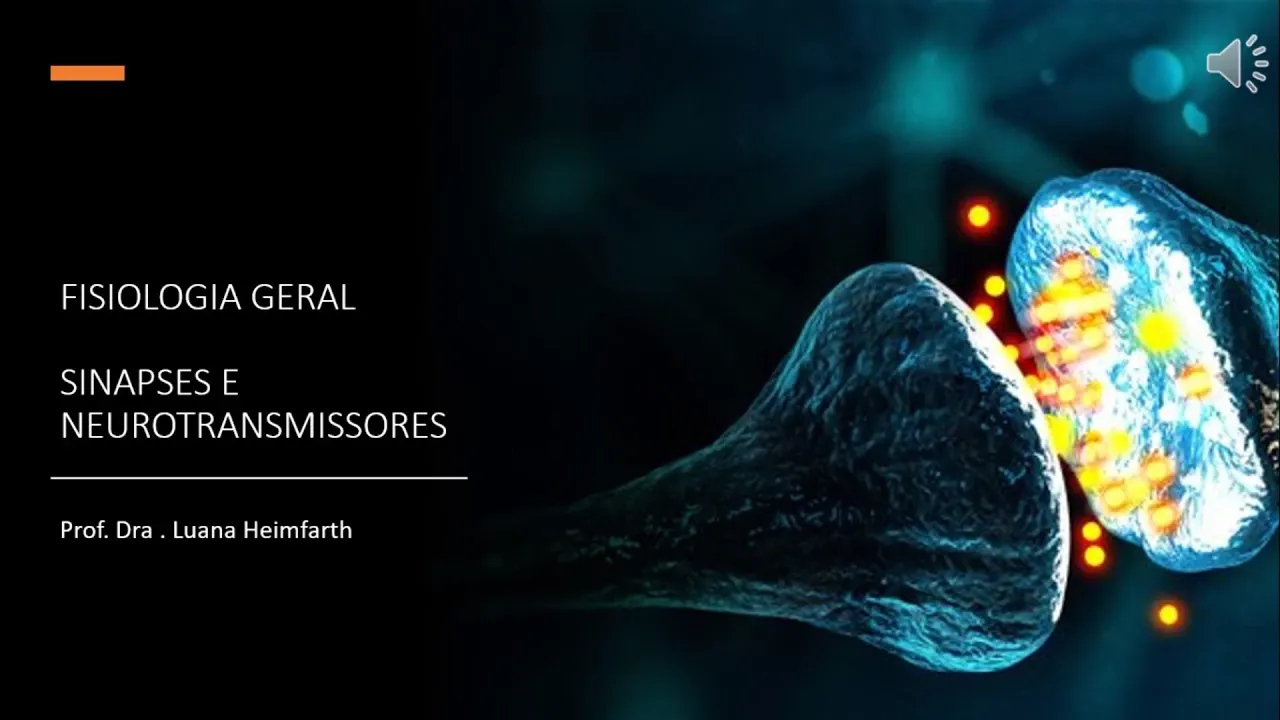
MÓDULO 1 - Aula 3: Sinapses e neurotransmissores
57.6K
After release, neurotransmitters diffuse across the synaptic cleft and bind to specific receptors on the postsynaptic membrane. Each neurotransmitter binds only to its specific receptor type. Binding can generate excitatory postsynaptic potentials (EPSPs) that depolarize the neuron, making it more likely to fire, or inhibitory postsynaptic potentials (IPSPs) that hyperpolarize the neuron, making it less likely to fire. Glutamate is the main excitatory neurotransmitter in the CNS, binding to ionotropic receptors (AMPA and NMDA) that allow sodium and calcium entry, causing depolarization.
Click to watch →
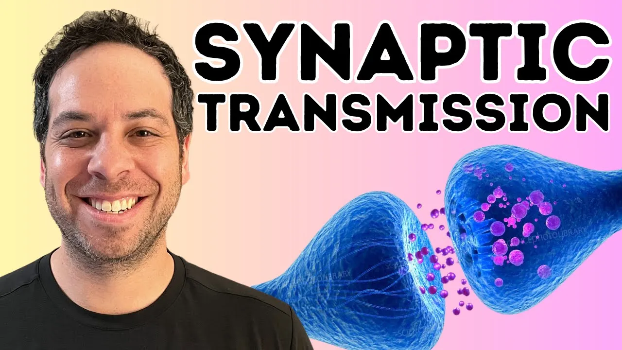
Synaptic Transmission Explained: How Neurons Communicate
147.6K
At the axon terminal, the action potential activates voltage-gated calcium channels, allowing calcium to enter the presynaptic neuron. Calcium binds to proteins on synaptic vesicles, causing them to fuse with the presynaptic membrane in a process called exocytosis, releasing neurotransmitters into the synaptic cleft. These neurotransmitters diffuse across the gap and bind to ligand-gated receptors on the postsynaptic neuron's membrane. When bound, these receptors open channels that allow ions (typically sodium) to enter, potentially depolarizing the postsynaptic neuron and triggering a new action potential if threshold is reached.
Click to watch →
ZIDD SERIES PART-I | INTRODUCTION OF AUTONOMIC NERVOUS SYSTEM
158K
Synaptic transmission involves: (1) Impulse conduction to presynaptic terminal, (2) Neurotransmitter release via calcium-triggered exocytosis, (3) Neurotransmitter binding to postsynaptic receptors, (4) Generation of postsynaptic potentials, (5) Termination through metabolism, reuptake, or diffusion. Excitatory neurotransmitters increase sodium permeability, causing depolarization and generating Excitatory Postsynaptic Potentials (EPSP). Inhibitory neurotransmitters increase chloride permeability, causing hyperpolarization and generating Inhibitory Postsynaptic Potentials (IPSP). The resting membrane potential is approximately -70 mV, with sodium higher outside and potassium higher inside.
Click to watch →
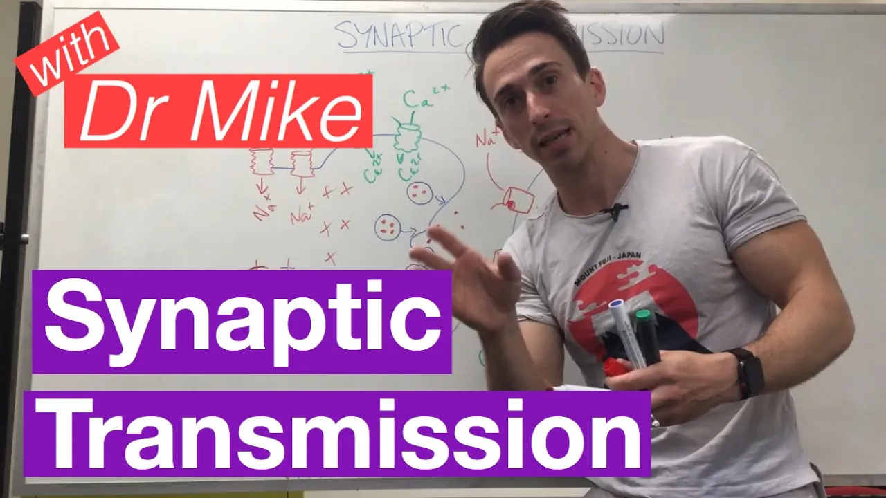
Synaptic Transmission | Neuron
258.7K
Calcium entry stimulates vesicles containing neurotransmitters to fuse with the presynaptic membrane, releasing their contents into the synaptic cleft. Excitatory neurotransmitters like glutamate diffuse across the synapse and bind to receptors on the postsynaptic neuron. This binding opens channels that allow sodium to enter the postsynaptic neuron, depolarizing it from its resting -70 mV toward the threshold of -55 mV, potentially triggering a new action potential.
Click to watch →
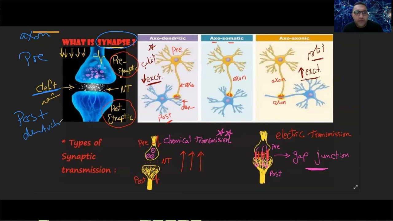
CNS Introduction crash course د/الشريف
34.5K
The mechanism of neurotransmitter release involves: action potential arrival at the presynaptic terminal, opening of voltage-gated calcium channels, calcium influx, calcium binding to synaptotagmin protein, binding to SNARE proteins, vesicle fusion with the presynaptic membrane, and exocytosis of neurotransmitters. Synaptic potentials can be excitatory (EPSP) or inhibitory (IPSP). EPSPs depolarize the postsynaptic membrane by opening sodium channels, bringing it closer to threshold. IPSPs hyperpolarize the membrane by opening chloride or potassium channels, moving it further from threshold. The type depends on which ions flow through opened channels and the membrane's resting potential.
Click to watch →
Deep Dive
The structure and function of ionotropic glutamate receptors, specifically AMPA and NMDA receptors, and their gating mechanisms.

Receptores Ionotrópicos.
886
Ionotropic receptors are membrane proteins that directly couple neurotransmitter binding to ion channel opening. They generate rapid ion flux (up to 10^8 ions/second) passively down electrochemical gradients without metabolic energy. Major families include: (1) Nicotinic acetylcholine receptors at neuromuscular junctions (5 subunits, Na+/K+ permeable); (2) GABA receptors as primary CNS inhibitors (5 subunits, Cl- permeable, causing hyperpolarization); (3) Glutamate receptors as primary excitatory receptors (4 subunits, Ca2+ permeable). Glutamate receptors include AMPA and NMDA subtypes, with NMDA receptors uniquely requiring both glutamate binding and membrane depolarization to overcome magnesium block. This voltage-dependent gating mechanism is fundamental to synaptic plasticity and information storage in synapses.
Click to watch →
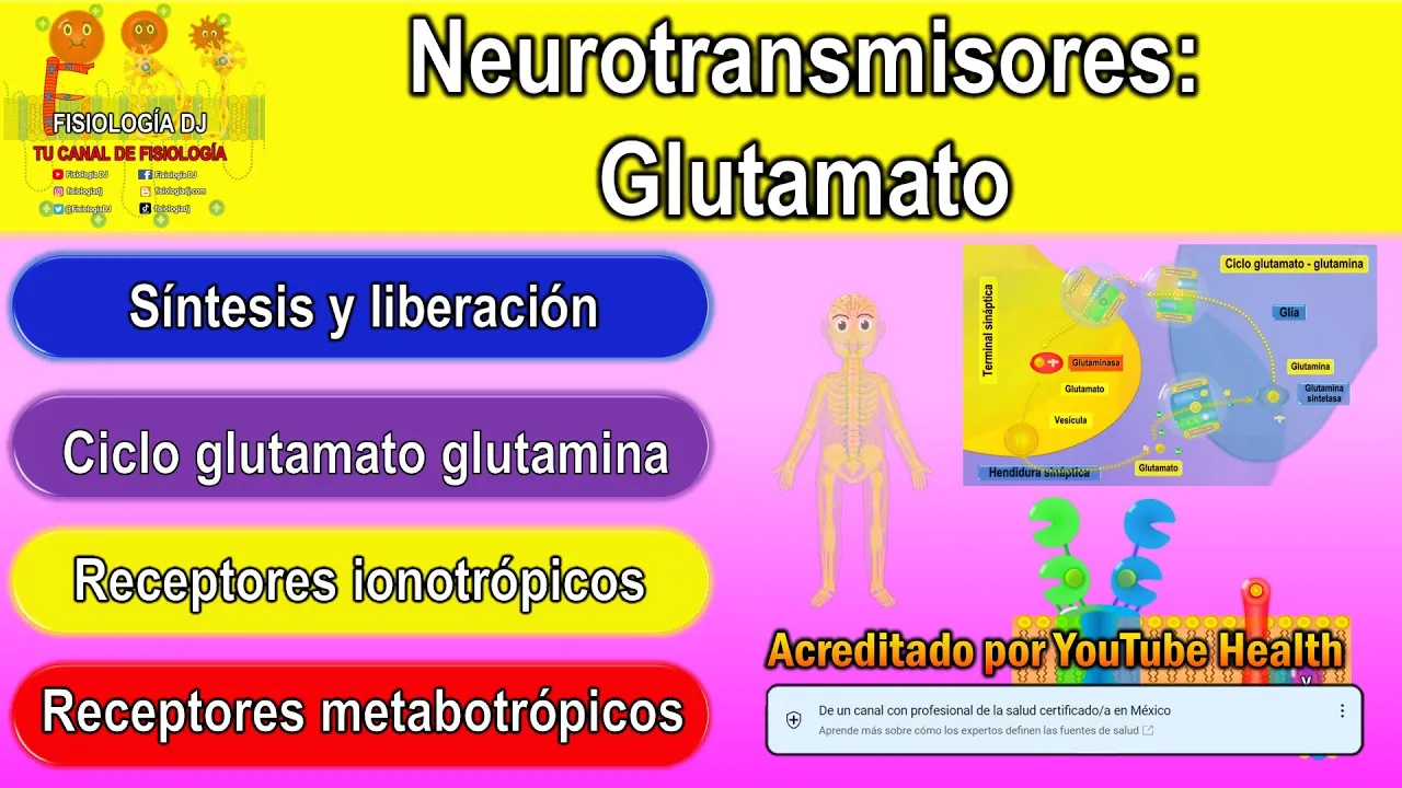
Neurotransmisores | Sistema nervioso central | Glutamato neurotransmisor | Glutamato función
21.6K
Ionotropic glutamate receptors are tetrameric structures with four subunits containing amino-terminal, ligand-binding, transmembrane, and carboxy-terminal domains. AMPA receptors have four subunits (GluA1-4), while kainate receptors have five (GluK1-5). NMDA receptors require both glutamate binding (GluN1) and glycine binding (GluN2/3) for activation. At rest, magnesium blocks the pore; depolarization removes this block, enabling calcium influx that serves as a second messenger.
Click to watch →
GLUTAMATO - NEUROTRANSMISSOR (FISIOLOGIA DE GUYTON) - FISIOLOGIA DO GLUTAMATO - NEUROFISIOLOGIA
22.1K
Ionotropic glutamate receptors function as ligand-gated ion channels that directly control ion flow upon neurotransmitter binding. Three main subtypes exist: AMPA receptors permit sodium and potassium influx; NMDA receptors allow calcium influx but require membrane depolarization to remove magnesium block; and kainate receptors have affinity for sodium and potassium. These receptors mediate rapid synaptic transmission essential for neural processing and plasticity.
Click to watch →
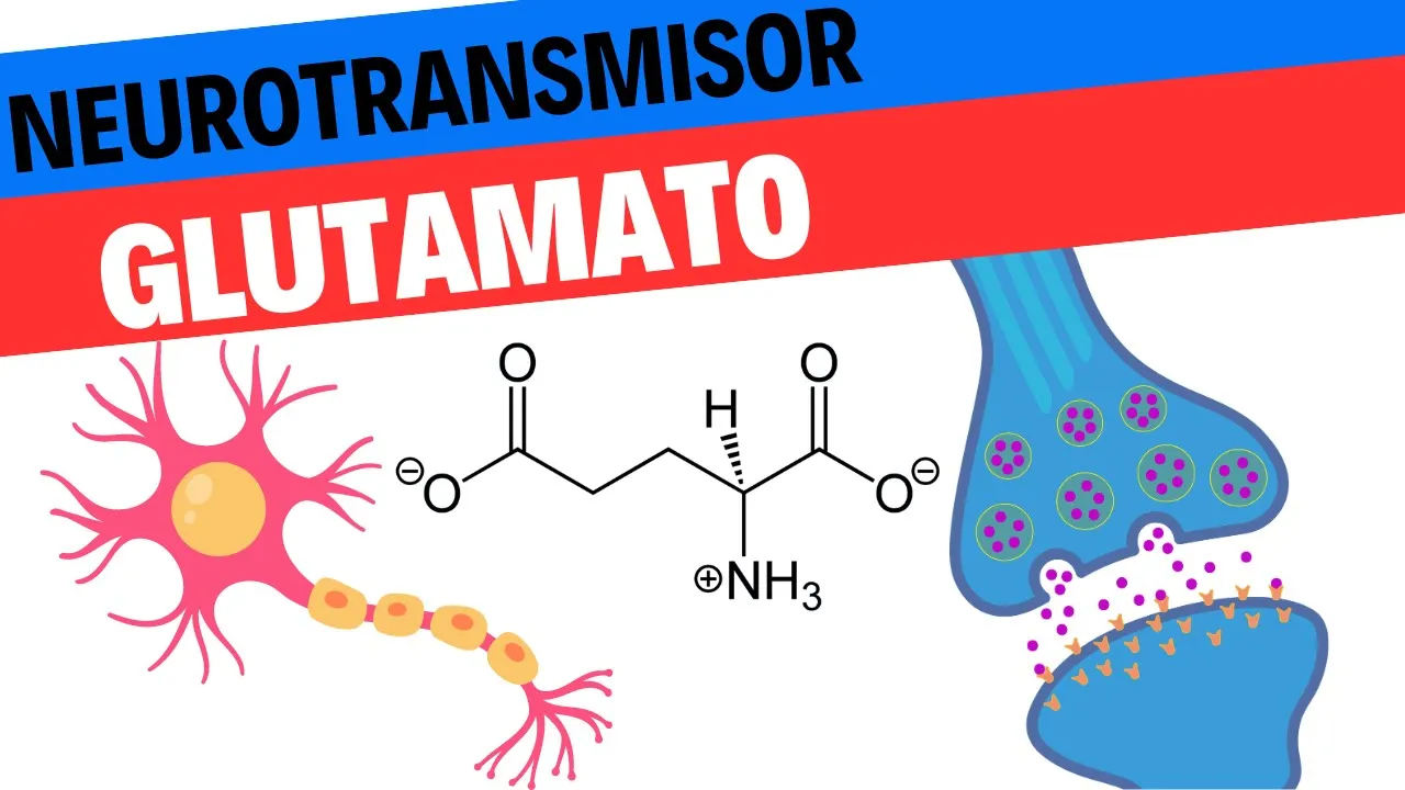
Fisiología del Glutamato. Neurociencias 8
5.5K
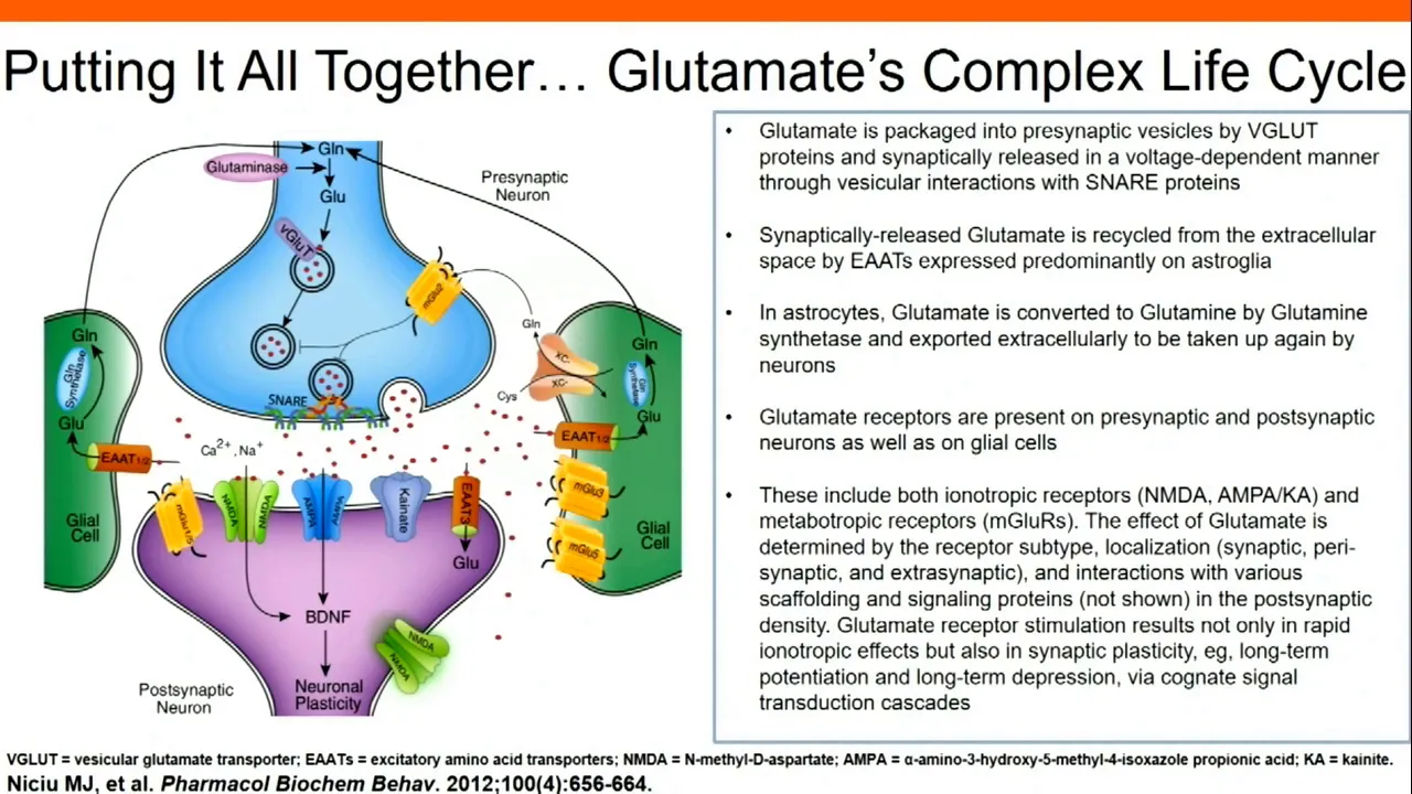
Glutamate binds to ionotropic receptors including AMPA and NMDA. AMPA receptors are highly permeable to sodium, generating rapid excitatory postsynaptic potentials that activate and deactivate quickly. NMDA receptors are blocked by magnesium ions until sufficient depolarization occurs through AMPA receptor activation, then allow calcium and sodium influx. NMDA receptors generate prolonged activation and are primarily responsible for long-term potentiation (LTP), which strengthens synaptic connections through calcium-dependent signaling cascades that increase AMPA receptor expression and promote dendritic spine growth.
Click to watch →
The Role of Glutamatergic Signaling in Major Depressive Disorder
86.3K
Glutamate receptors are classified into ionotropic (fast) and metabotropic (slow) types. Ionotropic receptors like NMDA and AMPA allow rapid ion flow producing effects within seconds to minutes, while metabotropic receptors trigger slower metabolic changes over hours to days. The NMDA receptor requires co-activation by both glutamate and glycine to open, functioning like a safety-critical system requiring dual authorization. AMPA receptors work in concert with NMDA receptors to produce synaptic plasticity and neurogenesis within 15 minutes. This receptor diversity creates multiple therapeutic targets for psychiatric intervention.
Click to watch →
Deep Dive
The concept of synaptic plasticity, particularly Long-Term Potentiation (LTP) as the opposing mechanism of synaptic strengthening.

Clase 6, Plasticidad Sináptica parte 1
3.6K
Long-Term Potentiation (LTP) is the primary mechanism of synaptic plasticity in the central nervous system, where glutamate binding to AMPA receptors causes sodium influx and depolarization, which removes the magnesium block from NMDA receptors, allowing calcium influx that activates CaMKII kinase to increase AMPA receptor conductance and insert additional AMPA receptors into the postsynaptic membrane, thereby strengthening synaptic connections and serving as the cellular basis for learning and memory.
Click to watch →

Learning and Memory: How It Works and When It Fails
1.4M
Long-term potentiation (LTP), discovered in 1973, is the fundamental cellular mechanism underlying learning and memory formation. LTP involves strengthening synaptic connections between neurons through repeated stimulation, where repeated activation causes receiving neurons to respond with increasingly larger electrical signals. This strengthening persists for hours, days, or even weeks after initial stimulation. The molecular mechanism involves glutamate binding to NMDA receptors, triggering calcium influx that activates signaling cascades leading to gene expression and increased receptor production. Associative long-term potentiation extends this principle: when a weak synaptic input coincides with the firing of a strong synapse (such as emotional input from the amygdala), the weak synapse becomes strengthened. This explains why emotionally charged events are remembered more vividly. Memory formation fundamentally depends on changing the strength of synaptic connections—a process called synaptic plasticity—where transient changes become stable modifications to neural networks through hippocampal coordination.
Click to watch →

Molecular Mechanisms of Long-Term Potentiation in Synaptic Plasticity and Memory Formation
609.3K
Long-term potentiation (LTP) is a long-lasting enhancement in signal transmission between neurons that serves as the molecular foundation for learning and memory; it occurs through a process where repeated high-frequency stimulation of the Schaefer collateral axons of CA3 neurons causes calcium influx via NMDA receptors, triggering two phases of synaptic strengthening: the early phase involves immediate insertion of new AMPA receptors onto the postsynaptic membrane, while the late phase involves gene expression and protein synthesis that creates new synaptic connections, ultimately strengthening the connection between neurons and enabling memory retention.
Click to watch →

Brain and Behavior - Learning and Memory: Neural Mechanisms
39.8K
Long-term potentiation (LTP) is a stable, enduring increase in synaptic effectiveness discovered in the 1970s in rabbit hippocampus. High-frequency tetanus stimulation induces lasting enhancement of synaptic responses persisting for 90+ minutes. LTP occurs at multiple hippocampal sites (mossy fiber-CA3 and Schaffer collateral-CA1 synapses). This phenomenon demonstrates Hebbian plasticity at the cellular level, where coordinated pre- and postsynaptic activity strengthens synapses. LTP provides the cellular mechanism for how the hippocampus might implement learning and memory, bridging behavioral observations with molecular understanding.
Click to watch →

뇌의 구조와 작동 방식을 바꾼다 - 신경가소성
265K
Long-term potentiation (LTP) is a process where repeated stimulation of a synapse leads to enhanced synaptic transmission. Through repeated training, new synaptic branches form and existing connections strengthen. This involves gene expression and protein synthesis that creates additional synaptic contacts, allowing information to be transmitted more strongly with the same stimulus. LTP represents the molecular basis of learning and memory formation.
Click to watch →
Deep Dive
The role of calcium ions (Ca2+) as intracellular second messengers and how different concentration thresholds trigger different cellular pathways.

G Protein Signalling
242K
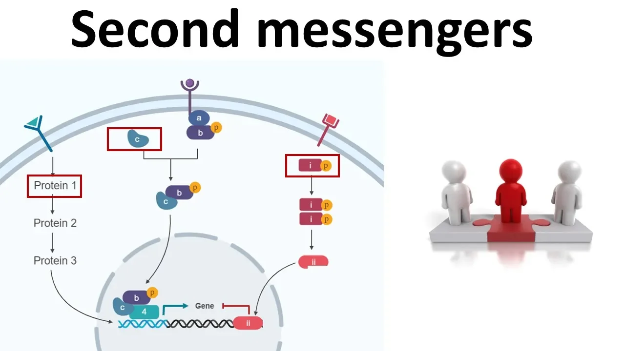
Calcium ions themselves act as second messengers. A rising concentration of calcium ions in the cytoplasm triggers many types of cellular events including muscle contraction and apoptosis (programmed cell death). This demonstrates how calcium ions integrate multiple signaling pathways.
Click to watch →

Біохімія. Лекція. Роль Ca+2 в активації сигнальних шляхів. Фосфоінозитидна система. Гормони ІІ типу
240
Calcium ions are evolutionarily ancient intracellular messengers regulating cell growth, division, muscle contraction, blood clotting, hormone secretion, and neurotransmitter release. Intracellular calcium concentration increases from 10^-8 M to 10^-6 M (100-1000 fold) during activation. This massive change serves as a powerful signal for cellular responses.
Click to watch →

Signal Transduction (PART 5)
293
Calcium ions (Ca2+) themselves function as second messengers in cellular signaling. The cytoplasmic calcium concentration is normally very low (approximately 100 nM), but signaling events can cause rapid increases in calcium concentration. These calcium signals activate calcium-dependent enzymes such as calmodulin-dependent kinases, protein kinase C, and phospholipase C. Calcium signals are transient and are terminated by calcium pumps that return calcium to the endoplasmic reticulum or extracellular space.
Click to watch →
Second messengers: cAMP, cGMP, IP3 & DAG ,Calcium
160.4K
Calcium signaling occurs through specific intracellular pathways. In Gq signaling, ligand binding activates Gq proteins which stimulate phospholipase C to break down membrane phospholipids, producing IP3. IP3 diffuses to the endoplasmic reticulum where it binds to IP3 receptors, releasing stored calcium into the cytosol. Increased cytosolic calcium is detected by calmodulin, which has four calcium-binding domains. Calmodulin can activate various effectors including phosphatases that mobilize nuclear transcription factors. This pathway demonstrates how calcium acts as a versatile second messenger capable of producing diverse cellular responses depending on context and cell type.
Click to watch →

CALCIUM SIGNALLING PATHWAY
848
Calcium serves as a critical second messenger in cellular signaling due to two key properties: (1) intrinsically low cytoplasmic calcium concentration (~100 nM) allows detection of even small concentration changes, and (2) calcium ions (Ca²⁺) strongly interact with negatively charged protein regions, including side chains of glutamate/aspartate and oxygen atoms of carbonyl groups, inducing conformational changes that activate target proteins. When cytoplasmic calcium exceeds 500 nM, calmodulin (a 17 kDa protein with four calcium-binding sites) undergoes conformational changes and activates calmodulin-dependent kinases, which phosphorylate proteins to regulate metabolism, ionic permeability, and neurotransmitter release.
Click to watch →
Prerequisite Knowledge
- Concept 01Fundamentals of synaptic transmission, including neurotransmitter release (specifically glutamate) and postsynaptic potential generation.
- Concept 02The structure and function of ionotropic glutamate receptors, specifically AMPA and NMDA receptors, and their gating mechanisms.
- Concept 03The concept of synaptic plasticity, particularly Long-Term Potentiation (LTP) as the opposing mechanism of synaptic strengthening.
- Concept 04The role of calcium ions (Ca2+) as intracellular second messengers and how different concentration thresholds trigger different cellular pathways.
Subsequent Learning
- Step 01The molecular mechanisms of receptor endocytosis, specifically how AMPA receptors are internalized during LTD.
- Step 02The distinct characteristics of cerebellar LTD (occurring at Purkinje cells) and its role in motor learning and coordination.
- Step 03The connection between aberrant LTD mechanisms and neurodegenerative or developmental disorders, such as Alzheimer's disease and Fragile X syndrome.
- Step 04The concepts of homeostatic plasticity and metaplasticity, which keep neural networks stable despite continuous LTD and LTP changes.
- Step 05Current scientific debates regarding the 'forgetting' hypothesis and the necessity of LTD for behavioral flexibility and spatial memory reversal.
LTD basics
Core mechanisms
Stimulation types
Calcium cascade
Other changes
LTD basics
0:05Playing Section
- 1
LTD weakens synaptic connections between neurons.
- 2
It is the opposite process to long-term potentiation.
- 3
May help reset synaptic changes for new memory formation.
The Dissociation of LTD and Learning: Redundant and Non-Synaptic Mechanisms
While Long-Term Depression (LTD) is traditionally viewed as essential for memory formation and circuit pruning, significant research challenges its necessity. This counterpoint is highlighted by studies in cerebellar motor learning—long considered the classic model for LTD-dependent memory. Researchers have generated mutant mice completely lacking parallel fiber-Purkinje cell LTD that still exhibit normal motor learning and adaptation. This 'dissociation' suggests that LTD may not be the primary or exclusive mechanism for memory storage. Critics argue that learning relies on highly redundant systems, pointing instead to changes in intrinsic neuronal excitability (non-synaptic plasticity) and homeostatic synaptic scaling as the true drivers of network modification. Furthermore, some information-theoretic models propose that synapses are too unstable for long-term storage, suggesting that memories are instead encoded intracellularly via molecular mechanisms rather than synaptic strength adjustments.
Deep Dive
The molecular mechanisms of receptor endocytosis, specifically how AMPA receptors are internalized during LTD.
Long term synaptic depression (LTD )
9.2K
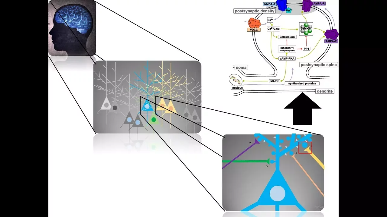
Long-term depression (LTD) is a long-lasting weakening of synaptic transmission efficiency that occurs when low-frequency stimulation (1 Hz) is applied for approximately 10 minutes, causing calcium levels in the postsynaptic region to rise from 100 nM to 400 nM; unlike LTP which requires high calcium levels (1 μM), LTD depends on NMDA receptor-mediated calcium entry and calcineurin activation, which triggers AMPA receptor internalization and structural changes in the postsynaptic region to reduce synaptic strength.
Click to watch →
LTP vs LTD
14.7K
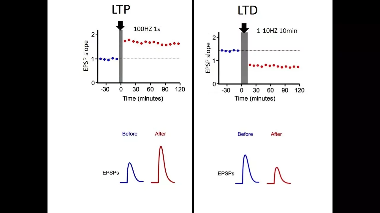
LTP involves AMPA receptor insertion into the postsynaptic membrane, increasing synaptic strength. LTD involves AMPA receptor endocytosis, reducing postsynaptic current. CAMKII acts as a calcium decoder requiring high calcium and repetitive stimulation (40-50 pulses) for optimal activation. Single pulses fail to induce changes. CAMKII activation involves competition with phosphatases—brief high calcium outcompetes phosphatases, enabling structural changes like spine volume increase. This molecular machinery enables precise control of synaptic strength.
Click to watch →

MoM by the Cerebellum Closer Look at Circuits part 2
578
LTD in the cerebellum involves AMPA receptor internalization triggered by metabotropic glutamate receptors. Unlike the cortex where high calcium causes long-term potentiation, cerebellar high calcium causes long-term depression. Climbing fiber activation provides massive depolarization that opens voltage-gated calcium channels, allowing calcium influx that engages second messenger systems (phospholipase C) leading to AMPA receptor internalization and synaptic weakening.
Click to watch →

"Механизмы клеточной рецепции", Дурыманов М. О. 12.02.2021г.
705
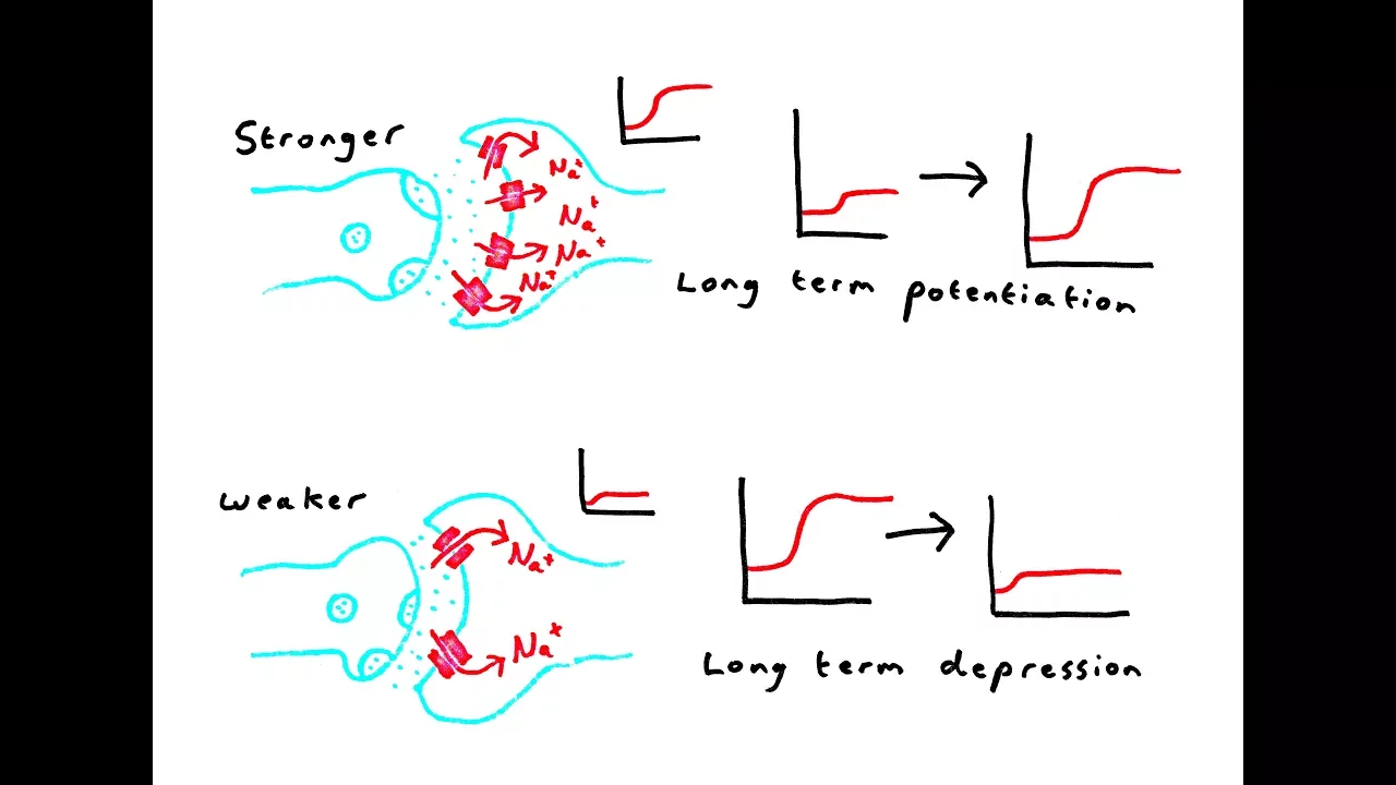
Long-term depression (LTD) weakens synapses in response to low-frequency stimulation. Low-frequency stimulation causes modest glutamate release, activating AMPA receptors but not enough to remove the magnesium block from NMDA receptors. The small calcium influx binds calmodulin, but instead of activating CaMKII, it activates calcineurin (a phosphatase). Calcineurin dephosphorylates and inactivates CaMKII, preventing synaptic strengthening. It also activates endocytosis of AMPA receptors and transcription factors that reduce synaptic protein expression.
Click to watch →
Synaptic Plasticity: How the Brain Strengthens Neural Connections
219K
The molecular machinery underlying synaptic plasticity centers on glutamate receptors and calcium signaling. Glutamate activates AMPA receptors (causing depolarization) and NMDA receptors (blocked by magnesium at resting voltage). Only when depolarization displaces magnesium do NMDA receptors allow calcium influx. Large calcium influx activates kinases that increase AMPA receptor exocytosis and phosphorylation, strengthening synapses. Small calcium influx activates phosphatases that promote receptor endocytosis, weakening connections. These processes occur locally on individual dendrites, enabling precise, independent modulation of neural circuits.
Click to watch →
Deep Dive
The distinct characteristics of cerebellar LTD (occurring at Purkinje cells) and its role in motor learning and coordination.

Fundamentos das Neurociências: Como o Cérebro Aprende?
15.9K
In the cerebellum, long-term depression (LTD) occurs when climbing cells and granule cells simultaneously release excitatory glutamate neurotransmitters onto Purkinje cells. This simultaneous stimulation causes a reduction in the amplitude of action potentials in Purkinje cells, unlike the increase seen in hippocampal LTP. This LTD mechanism is hypothesized to be important for motor learning, allowing the cerebellum to refine and adjust motor responses based on experience.
Click to watch →

Lecture 2. The Cerebellar System Structure, Function and Dysfunction
11.8K
The cerebellum enables motor learning through synaptic plasticity mechanisms. Long-term depression (LTD) occurs at parallel fiber-Purkinje cell synapses during associative learning. Using eyeblink conditioning as a model, repeated pairing of conditioned stimulus (tone) and unconditioned stimulus (shock) induces LTD. This requires simultaneous activation of metabotropic glutamate receptors (by parallel fibers) and voltage-gated calcium channels (by climbing fibers), triggering protein kinase C. This enzyme phosphorylates glutamate receptor 2, causing its internalization and making Purkinje cells less excitable. Reduced Purkinje cell inhibition allows appropriate motor responses to develop. This molecular mechanism demonstrates how the cerebellum stores learned motor skills through experience-dependent changes in synaptic strength, providing the cellular basis for motor learning and adaptation.
Click to watch →

The Cerebellum and Motor Learning Part 2
6.6K
Long-term depression (LTD) in cerebellar Purkinje cells occurs when climbing fiber activation coincides with granule cell-Purkinje cell synaptic activity, triggering a molecular cascade: glutamate binding to type 1 metabotropic glutamate receptors activates Gq proteins, which stimulate phospholipase Cβ to produce IP3; IP3 then binds to IP3 receptors on the endoplasmic reticulum, releasing calcium that opens these receptors as a coincidence detector, ultimately weakening the synaptic connection to correct motor calibration errors.
Click to watch →

The Cerebellum and Motor Learning Part 3
4.2K
Long-term depression (LTD) in cerebellar Purkinje cells is a form of synaptic plasticity essential for motor learning, occurring when granule cell and climbing fiber inputs coincide. The mechanism involves the IP3 receptor acting as a coincidence detector: granule cell activation produces IP3 that primes the receptor, while climbing fiber activation provides calcium influx that opens the receptor. This calcium release from intracellular stores triggers synaptic weakening at the granule cell-Purkinje cell synapse, which may occur presynaptically (reduced glutamate release) or postsynaptically (reduced receptor expression).
Click to watch →

Вячеслав Дубынин: "Мозг и движение"
814.5K
The motor cortex is located in the frontal lobe and contains a motor homunculus - a body map where different body parts are represented in specific areas. Body parts requiring fine control (hands, face, tongue) occupy disproportionately large areas. The motor cortex sends commands to spinal cord motor neurons through the corticospinal tract. Motor learning involves the transformation of voluntary movements into automated skills through repetition, with the cerebellum and basal ganglia playing key roles. The cerebellum is located at the base of the brain and has three evolutionary divisions: the vestibulocerebellum (balance and eye movements), the spinocerebellum (motor control and posture), and the cerebrocerebellum (motor learning and coordination). The cerebellum contains more than half of all neurons in the human brain (60-70 billion). Purkinje cells are large inhibitory neurons in the cerebellar cortex that receive input from many sources and send output to deep cerebellar nuclei. They fire at high frequencies (up to 60 impulses per second) and provide powerful inhibition to deep cerebellar nuclei, creating a 'brake' on motor output. During motor learning, Purkinje cells adapt their firing patterns based on sensory feedback, reducing their inhibitory output as movements become more accurate.
Click to watch →
Deep Dive
The connection between aberrant LTD mechanisms and neurodegenerative or developmental disorders, such as Alzheimer's disease and Fragile X syndrome.

Nahum Sonenberg - Translational control of Autism Spectrum Disorder and Fragile-X syndrome
851
Long-term depression (LTD) is an electrophysiological measurement of synaptic plasticity that is impaired in fragile X syndrome. In wild-type mice, LTD induction causes a moderate decrease in synaptic strength that recovers over time. However, in fragile X knockout mice, LTD is exaggerated (the synaptic response decreases much more dramatically) and fails to recover. This provides a cellular model for understanding the synaptic abnormalities underlying cognitive deficits in fragile X syndrome.
Click to watch →

Prof. Mark Bear of Massachusetts Institute of Technology on Pathophysiology of amblyopia
835
Long-term depression (LTD) refers to activity-induced decrease in synaptic transmission strength, weakening synaptic connections. Research on LTD mechanisms led to discoveries about how activity regulates protein synthesis at synapses. This basic research question about protein synthesis regulation became crucial for understanding fragile X syndrome and other neurodevelopmental disorders.
Click to watch →

SCSB Colloquium Series: André Fenton, Ph.D.
293
In hippocampal slices from fmr1 null mice, long-term potentiation (LTP) expression and induction appear normal, but long-term depression (LTD) induced by group I mGluR agonists like DHPG shows greater magnitude and is more persistent. This dysregulation of mGluR-dependent translation pathways has led to the 'angular theory' proposing that dysregulated translation causes various phenotypic problems in fragile X syndrome.
Click to watch →

Picower Institute Inaugural Symposium 2005: Future of the Brain (1/4) S.Tonegawa, S.Brenner, R.Axel
300
Long-term depression (LTD), a form of synaptic weakening induced by sensory stimuli, plays crucial roles in developmental plasticity. Research on LTD mechanisms led to discoveries about fragile X syndrome, the best-known inherited cause of mental retardation and autism. This basic research finding suggests that malfunctioning LTD pathways may be correctable by drugs, potentially leading to clinical trials within a few years. Additionally, prefrontal cortex and basal ganglia interactions in habit formation show that while basal ganglia initiate rule-following faster, the prefrontal cortex acts as the final arbiter of behavior.
Click to watch →

Multiple Roles for Oligophrenin at the Synapse
669
The importance of mGluR-LTD in cognitive impairment has been demonstrated by findings that this form of plasticity is altered in mouse models of fragile X syndrome. This has led to the development of new therapeutics for fragile X syndrome that act on mGluR-LTD pathways, demonstrating the clinical relevance of understanding this plasticity mechanism.
Click to watch →
Deep Dive
The concepts of homeostatic plasticity and metaplasticity, which keep neural networks stable despite continuous LTD and LTP changes.

Using Neurobiology to Overcome Genetic and Environmental Adversity
1.6K
Excitatory synapses use glutamate, activating AMPA receptors for fast transmission and NMDA receptors for plasticity modification. Strong NMDA activation induces long-term potentiation (strengthening synapses), while modest activation causes long-term depression (weakening). These processes depend on correlated pre- and post-synaptic activity. Metaplasticity describes how plasticity rules themselves change based on prior activity levels—prolonged darkness shifts the function to make potentiation easier and depression harder. This homeostatic mechanism ensures neural networks maintain appropriate dynamic ranges.
Click to watch →
46b. Plasticité synaptique
5.4K
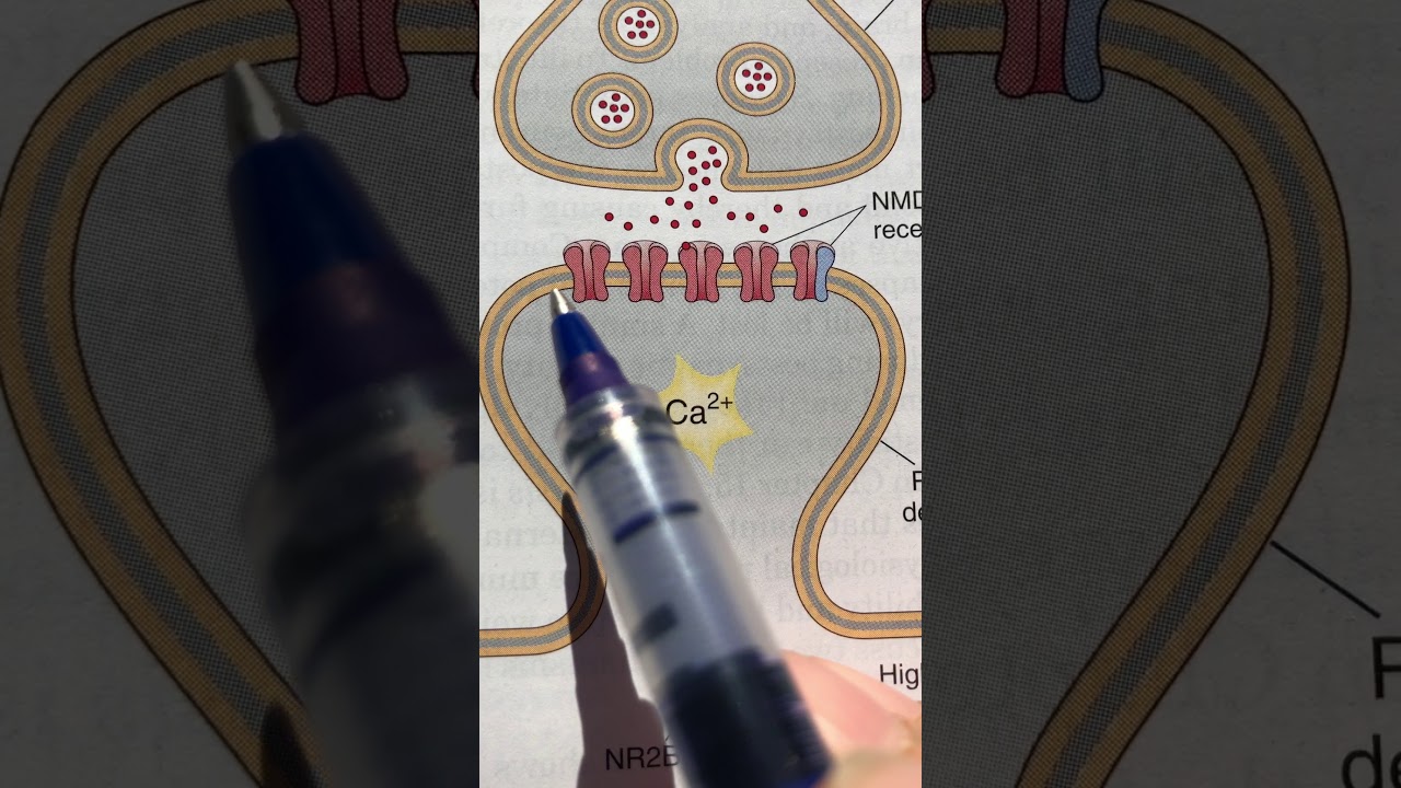
Metaplasticity is the phenomenon where prior synaptic plasticity (LTP or LTD) alters the threshold for future plasticity, such that inducing LTP makes subsequent LTP easier but LTD more likely, while inducing LTD makes subsequent LTD easier but LTP more likely; this is regulated by changes in NMDA receptor subunit composition (NR2A vs NR2B), where high activity shifts receptors toward NR2B subunits that allow more calcium entry, facilitating LTP, while low activity shifts toward NR2A subunits that allow less calcium entry, facilitating LTD.
Click to watch →

Jennifer Raymond: Cerebellar metaplasticity
695
Metaplasticity encompasses two fundamental phenomena: threshold metaplasticity (experience-dependent changes in how easy it is to induce plasticity) and temporal metaplasticity (experience-dependent changes in timing requirements for inducing associative plasticity). This research investigates how neural circuits implement learning algorithms at the algorithmic level by studying the cerebellum's simple architecture. The textbook model proposes climbing fiber-driven LTD at parallel fiber-Purkinje cell synapses, but evidence shows LTD selectively supports VOR increase learning but not VOR decrease learning. Paradoxically, enhanced LTD (in MHC class I and fragile X knockouts) causes selective impairment rather than enhancement, challenging simple views of plasticity mechanisms.
Click to watch →

Plasticidad cerebral: Reconfigura tus vías neuronales | aprendizaje, memoria y hábitos sostenibles.
32.5K
Brain plasticity is the nervous system's fundamental capacity to modify structure and function in response to activity and environment, supporting learning, memory, injury recovery, and adaptation. Neurons communicate through synapses using chemical messengers, with Long-Term Potentiation (LTP) strengthening synapses following intense activation and Long-Term Depression (LTD) weakening them during scarce activity. Structural plasticity involves dendritic spine changes, while functional plasticity adjusts synaptic strength and neuronal excitability. Myelination by oligodendrocytes adjusts conduction velocity, and the excitatory-inhibitory balance regulated by GABA prevents circuit saturation. Neuromodulators like dopamine, acetylcholine, and norepinephrine influence plasticity through attention, motivation, and context-dependent learning. Neural ensembles activate together to represent elements, with the hippocampus forming associations and coordinating with cortex for memory consolidation. Homeostatic plasticity maintains activity within functional ranges, while metaplasticity adjusts the brain's capacity to change based on activation history.
Click to watch →

"Synapses: From Vesicles to Circuits" by Dr. Venkatesh Murthy
437
Homeostatic plasticity is a mechanism by which neural networks maintain stable activity levels despite perturbations. When a network is made inactive (e.g., by blocking activity), synapses strengthen to restore normal activity. Conversely, when neurons become hyperactive (like in epilepsy), synapses weaken to reduce excessive connectivity. This auto-regulatory mechanism helps maintain network stability over timescales of days.
Click to watch →
Deep Dive
Current scientific debates regarding the 'forgetting' hypothesis and the necessity of LTD for behavioral flexibility and spatial memory reversal.

working memory model, Long term memory, Long term potentiation|Attention perception learning memory
597
Long-Term Depression (LTD) is the weakening of synaptic connections between neurons. It is the opposite of Long-Term Potentiation. LTD occurs when synaptic connections are not activated frequently enough. It involves the removal of AMPA receptors and reduction in dendritic spine size. LTD is important for memory refinement and forgetting. It allows for the elimination of weak or irrelevant memories. LTD is the reverse process of LTP.
Click to watch →

Why Does Your Brain Forget On Purpose? Psychology for Sleep
292
A growing body of work points to forgetting as a regulated process the brain runs rather than a decay it suffers. Researchers such as Ryan and Franklin have argued that forgetting is best understood as a form of learning that keeps memory flexible in a world that keeps changing. In animal studies, specific biological signals can speed forgetting up or slow it down without any damage to the machinery that stores memory in the first place. That means there is a rate and the rate can be adjusted.
Click to watch →

Как работает память: Нейронаука памяти простыми словами
2.5K
There is a molecular mechanism for forgetting called long-term depression (LTD), the opposite of long-term potentiation (LTD). Certain proteins in synapses change, receptors are removed from the membrane, and connections weaken. This is an active process as much as strengthening. Understanding both mechanisms gives us two tools: one for remembering and one for forgetting.
Click to watch →
Plasticidad cerebral: Reconfigura tus vías neuronales | aprendizaje, memoria y hábitos sostenibles.
32.5K
Long-term depression erases or attenuates connections no longer used. Synaptic pruning eliminates redundant routes. Reactivation during sleep selects what is maintained according to coherence with current goals. A translator who stops working in a language pair notes how certain associations fade while what is practiced daily maintains firmness. Plasticity is not endless accumulation but selection and update. Memory recovery is not perfect reproduction but reconstruction guided by cues and schemas.
Click to watch →

CNS Physiology lecture-3 Kasr Alainy 2025 by dr. Elsherif
8.6K
LTP involves glutamate release binding to NMDA receptors, allowing calcium entry when the postsynaptic neuron is depolarized. Calcium activates CaMKII, which increases AMPA receptor number and function. LTD involves weak stimulation that activates protein phosphatase 1, removing AMPA receptors. These mechanisms allow the brain to strengthen or weaken synaptic connections based on the pattern of stimulation. Memory formation and forgetting involve a balance between LTP and LTD. The hippocampus coordinates this balance during memory consolidation. When memories are not rehearsed or are emotionally neutral, synapses may undergo LTD, making memories more likely to be forgotten. This balance allows the brain to efficiently encode important information while maintaining efficiency by pruning unused connections.
Click to watch →
LTD basics
Core mechanisms
Stimulation types
Calcium cascade
Other changes
LTD basics
0:05- 1
LTD weakens synaptic connections between neurons.
- 2
It is the opposite process to long-term potentiation.
- 3
May help reset synaptic changes for new memory formation.
The Dissociation of LTD and Learning: Redundant and Non-Synaptic Mechanisms
While Long-Term Depression (LTD) is traditionally viewed as essential for memory formation and circuit pruning, significant research challenges its necessity. This counterpoint is highlighted by studies in cerebellar motor learning—long considered the classic model for LTD-dependent memory. Researchers have generated mutant mice completely lacking parallel fiber-Purkinje cell LTD that still exhibit normal motor learning and adaptation. This 'dissociation' suggests that LTD may not be the primary or exclusive mechanism for memory storage. Critics argue that learning relies on highly redundant systems, pointing instead to changes in intrinsic neuronal excitability (non-synaptic plasticity) and homeostatic synaptic scaling as the true drivers of network modification. Furthermore, some information-theoretic models propose that synapses are too unstable for long-term storage, suggesting that memories are instead encoded intracellularly via molecular mechanisms rather than synaptic strength adjustments.
Fundamentals of synaptic transmission, including neurotransmitter release (specifically glutamate) and postsynaptic potential generation.
The structure and function of ionotropic glutamate receptors, specifically AMPA and NMDA receptors, and their gating mechanisms.
The concept of synaptic plasticity, particularly Long-Term Potentiation (LTP) as the opposing mechanism of synaptic strengthening.
The role of calcium ions (Ca2+) as intracellular second messengers and how different concentration thresholds trigger different cellular pathways.
The molecular mechanisms of receptor endocytosis, specifically how AMPA receptors are internalized during LTD.
The distinct characteristics of cerebellar LTD (occurring at Purkinje cells) and its role in motor learning and coordination.
The connection between aberrant LTD mechanisms and neurodegenerative or developmental disorders, such as Alzheimer's disease and Fragile X syndrome.
The concepts of homeostatic plasticity and metaplasticity, which keep neural networks stable despite continuous LTD and LTP changes.
Current scientific debates regarding the 'forgetting' hypothesis and the necessity of LTD for behavioral flexibility and spatial memory reversal.
212.8K views2.7Klikes2:00@neurochallengedOriginal Release: 2017-08-03
Welcome to 2 minute neuroscience, where I simplistically explain neuroscience topics in 2 minutes or less.
In this installment I will discuss long-term depression, or LTD.
LTD is a process by which synaptic connections between neurons become weaker.
It is the opposing process to long-term potentiation.
Although the functions of LTD are not completely understood, it’s thought to be important to memory formation, perhaps by resetting previous synaptic changes to allow for new memories to be formed via long-term potentiation.
There are several different mechanisms by which LTD may occur, but the best understood of them involves the same glutamate receptors involved in long-term potentiation: NMDA and AMPA receptors.
NMDA receptors are typically blocked by a magnesium ion, which is only removed if the postsynaptic neuron becomes sufficiently depolarized as can occur through activation of the AMPA receptor; when the block is removed, calcium is able to flow into the neuron, causing further depolarization.
While long-term potentiation typically occurs after brief but high-intensity stimulation of a post-synaptic neuron, LTD can be caused by prolonged low-intensity stimulation or stimulation that occurs after the firing of an action potential.
With the type of modest stimulation that results in LTD, there is not enough depolarization to cause widespread removal of the magnesium blockage of the NMDA receptor.
However, there is enough to cause some NMDA receptors to allow calcium into the cell.
This low level of calcium is insufficient to activate the enzymes that facilitate long-term potentiation, but it is thought to activate a cellular cascade that causes the removal of AMPA receptors.
This reduces the number of glutamate receptors on the postsynaptic neuron and weakens the synapse.
LTD may also result in other changes that decrease the strength of synapses, like a decrease in the amount of glutamate released from the presynaptic neuron, and it also can involve other receptors like metabotropic glutamate receptors or other neurotransmitter receptors altogether.
Up Next
2:02
Corticospinal Tract | Upper Motor Neuron Pathway Explained
@neurochallenged
592K views•2015-12-06

21:55
Deciphering Neural Circuits Controlling Anorexia in Mice | Richard Palmiter
@AllenInstitute
2.3K views•2011-10-10
1:59
How Smell Works: Olfaction Explained in 2 Minutes | Neuroscience
@neurochallenged
303.7K views•2018-01-31

13:03
How Exercise Benefits Your Brain: Science Explained
@TED
11.4M views•2018-03-21
Related Study Plans & Knowledge Roadmaps
Structured learning paths in Neuroscience
neuroscience
Appetite Regulation: Brain Circuits & Hormones
01Foundations of Endocrinology and Brain Anatomy
02Introduction to Energy Homeostasis
03Peripheral Signals: Leptin, Ghrelin, and Insulin
+ 2 more learning modules
14h5 modules
Explore Roadmap
neuroscience
Neuroeconomics: Value, Risk & Brain Mechanisms
01Foundations of Neuroscience and Brain Anatomy
02Introduction to Neuroeconomics and Subjective Value
03Dopaminergic Prediction Errors & Reinforcement Learning
+ 3 more learning modules
18h6 modules
Explore Roadmap
neuroscience
Spatial Navigation: Cells, Grids & Brain Maps
01Foundations of Brain Anatomy and Memory
02Place Cells and Head-Direction Cells
03Grid Cells: The Brain's Coordinate System
+ 2 more learning modules
12h5 modules
Explore Roadmap
neuroscience
BCIs: Neural Decoding & Prosthetic Control
01Neuroscience Foundations: How the Brain Signals Intent
02Signal Acquisition: Recording the Brain's Activity
03Neural Decoding: Translating Brain Waves into Data
+ 2 more learning modules
25h5 modules
Explore Roadmap