


![Anatomy of the Heart: Structures and Blood Flow [Cardiology Made Easy]](https://i.ytimg.com/vi/1b4V09HzhBw/maxresdefault.jpg)







![مصطلحات طبيه || الجهاز الدوري [ 13 ] Medical terminology of the cardiovascular system](https://i.ytimg.com/vi/wGsYfIR9Skg/maxresdefault.jpg)









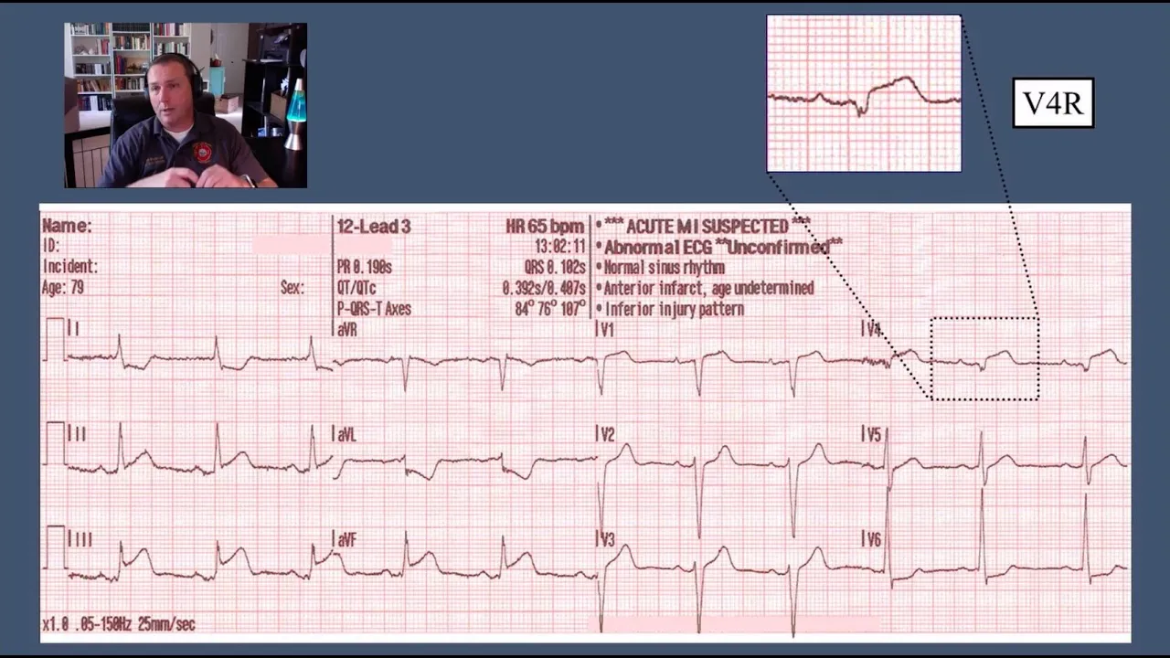







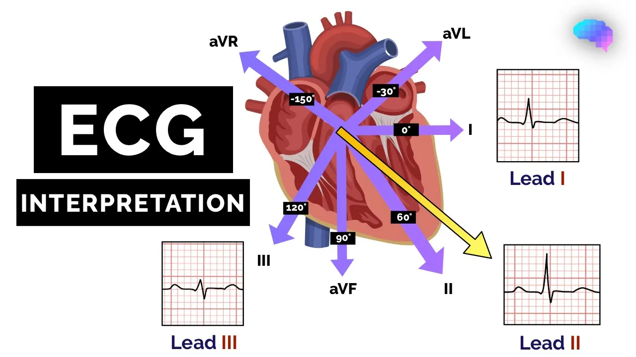


A 12-lead ECG captures the heart's electrical activity from 12 different angles using hexaxial leads (I, II, III, aVR, aVL, aVF) and precordial leads (V1-V6), where hexaxial leads view the heart from the sides and precordial leads view it from the front; the normal ECG waveform includes P wave (atrial depolarization), QRS complex (ventricular depolarization), and T wave (ventricular repolarization), with key measurements being: small box = 1mm² and 0.04 seconds, large box = 5mm² and 0.20 seconds, PR interval = 0.12-0.20 seconds, QRS <3 small boxes, ST segment at isoelectric line, and QT interval = 0.33-0.44 seconds.
ECG Interpretation Made Easy: 12-Lead EKG Basics
Added:Basic anatomy and physiology of the heart, including the four chambers, valves, and systemic/pulmonary circulation.
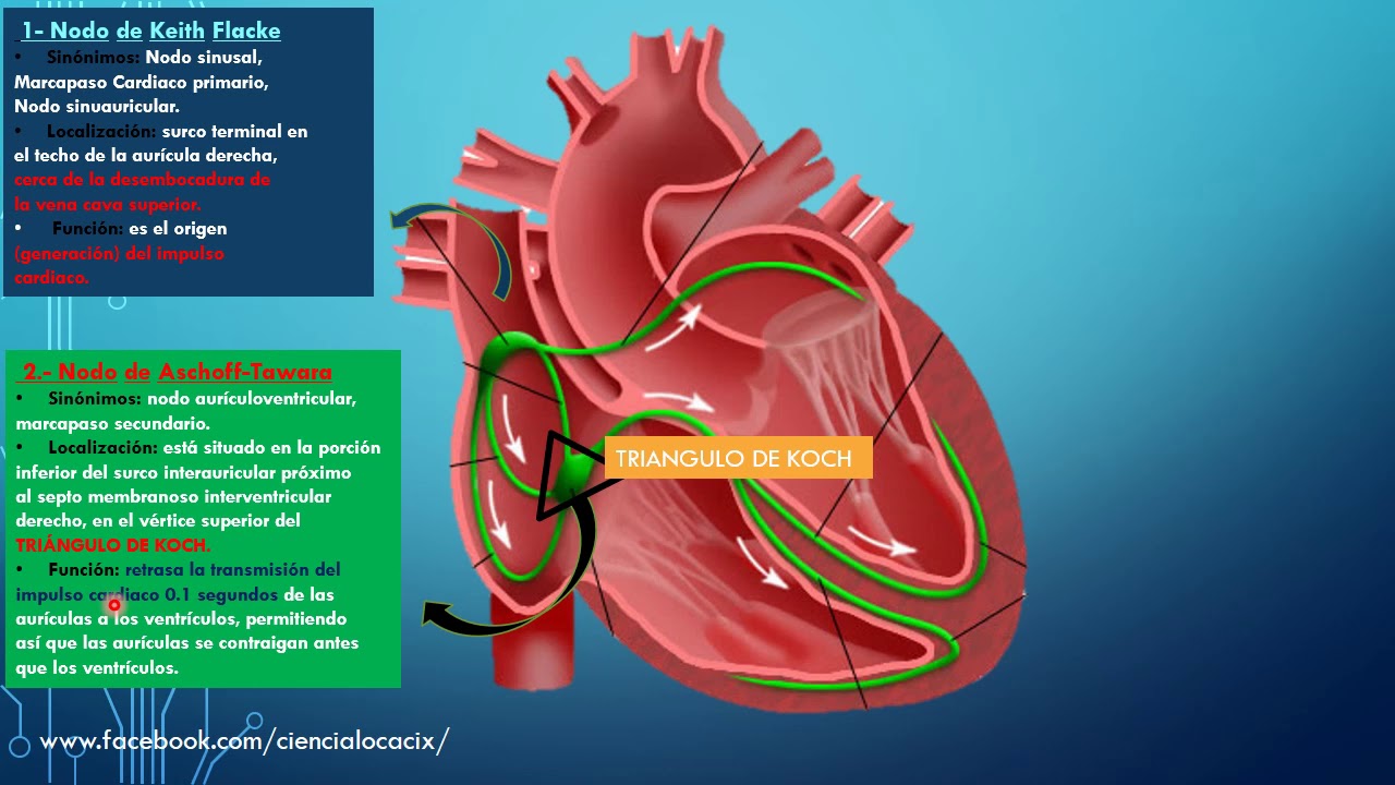
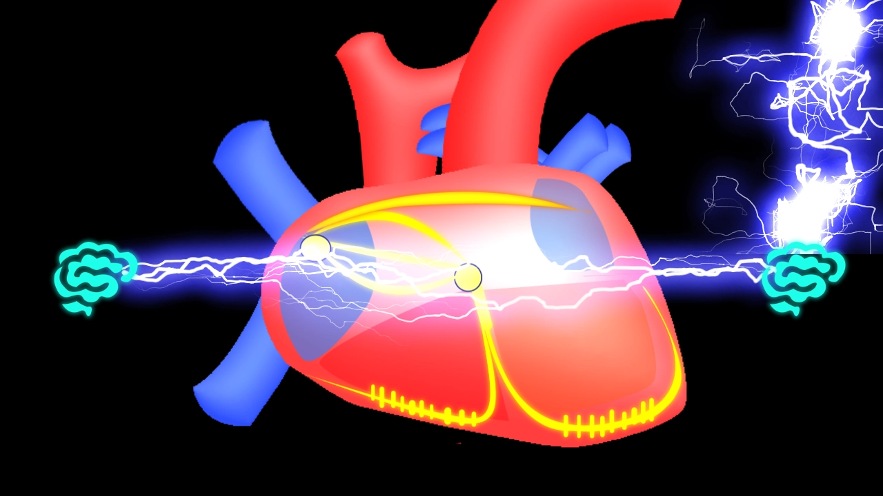
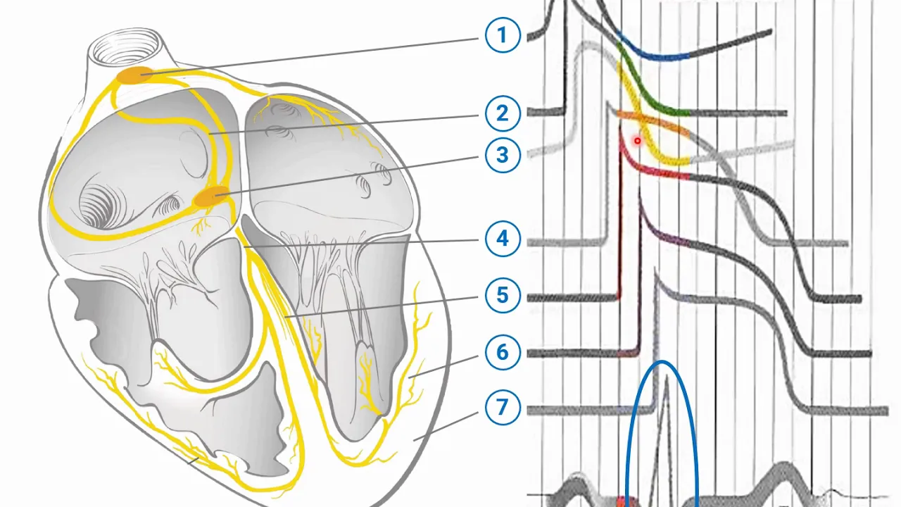
The cardiac conduction system, specifically the pathways of electrical impulses from the SA node to the Purkinje fibers.
The cellular concepts of cardiac depolarization and repolarization.
Standard medical terminology related to the cardiovascular system, such as bradycardia, tachycardia, and ischemia.
Identification and classification of common cardiac arrhythmias, such as atrial fibrillation, atrial flutter, and ventricular tachycardia.
Recognition of myocardial infarction (heart attack) patterns, including distinguishing between STEMI and NSTEMI on a 12-lead ECG.
Analysis of advanced ECG findings, including bundle branch blocks, hemiblocks, and axis deviations.
Understanding the impact of electrolyte imbalances (like hyperkalemia or hypokalemia) and specific medications on ECG intervals and waves.
Clinical integration of ECG findings with patient history, physical examination, and emergency cardiac protocols (ACLS).
1.1M views26.9Klikes26:57Original Release: 2023-02-04
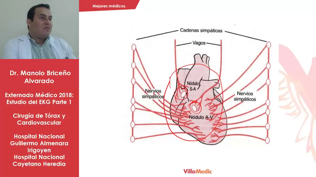
okay welcome to the first video of ECG interpretation Made Easy video series by the end of this video series I'll make sure that you have mastered all the important ecgl all the high yield ECG rhythms and their abnormalities we will solve ECGs together we will learn ECGs together we will interpret different 12 lead ECGs first of all what is the CG ECG is basically electrocardiogram in which you place certain sensors on the human body and you detect the electrical activity of the heart that electrical activity of the heart is recorded in the in a piece of paper that is called as ECG basically in our heart there is essay node and that SA node produces electrical signals and those electrical signals cause contraction of the Atria those electrical signals travel from SA node towards AV nodes and AV node sends those signals toward the ventricles and causes contraction of the ventricles it sends signals via right bundle branch and left bundle branch these bundle branches further divide up into small fibers called as purkinje fibers and they when the electrical signal goes through these fibers they cause contraction of the ventricles Whenever there is any abnormality in this electrical circuit of the heart abnormalities as in myocardial infarction abnormalities as in hypertrophies abnormalities as in ventricular tachycardias ventricular fibrillations we will learn all these ECGs now in your words you would have seen an ECG strip like this with all different leads capturing the activity of the heart that is called as a 12 lead ECG what does 12 lead ECG means 12 lead ECG means that we are capturing the activity of heart from 12 different angles we are looking at the heart electrical activity from 12 different angle that is called as a 12 lead ECG now where does these 12 leads come from these leads are basically divided into hex axial leads and precordial leads hex axial leads include lead one two three AVR AVL avf precordial leads include V One V two V three V four V five V6 now first we will discuss the hex axial leads now to get the hex axial leads we place certain electrodes on the human body parts and these electrodes capture the electrical activity of the heart to get lead one we place positive electrode on the left arm of the patient and we place the negative electrode on the right arm of the patient that is the lead one now remember this positive electrode acts as a camera and this positive electrodes is basically the electrode that is looking at the heart from the positive direction to the negative Direction so the positive electrode always acts as a camera and it is looking towards the negative side and it is capturing the electrical activity of the heart now same applies for lead 2 in lead 2 we place the positive electrode on the left foot of the patient and you place the negative electrode on the right arm of the patient and this positive electrode is acting as a camera and it is looking at the heart from the left foot towards the right side so it is looking at the heart from beneath so we are capturing another angle of the heart another view of the heart coming to lead 3 in lead 3 we place the positive electrode on the left put over the patient and negative electrode is placed on the left arm of the patient so we are capturing another angle to look at the heart from positive to negative the positive electrode acts as a camera so this is how lead one two three are placed and these are placed at different locations and they are looking at the heart from different angles now this is another picture showing lead one two three and these leads are looking at the heart from three different angles now what mathematicians and physicists did is that they performed certain calculations and they derived three more leads from these very leads and those leads are called as augmented leads augmented leads include lead AVR AVL and avf and for AVR AVL avf you do not need to put any more electrodes on the patients these three leads are basically derived from lead one two three from the vectors of lead one two three these three leads are derived these mathematicians and these physicists performed calculations and they derived three more leads these are leads AVR AVL avf and they actually gave us three more angles to look at the heart these three leads are actually acting as a camera and heart is acting as their negative electrode and these leads are also looking at the heart from three different angles so this is so amazing that we put three different leads on the patient and from those three very leads we are actually getting six different views of the hard three views from the leads that we place and three views from the math of those leads we get another three angles those leads are called as augmented leads or Elite AVR AVL avf these leads are actually called augmented leads because when they were deriving these leads from the vectors of the main leads that we placed their voltage was actually low so what ECG machine did was that they increased the voltage from ECG machine so that these we get a proper tracing on ECG paper so these AVR AVL avf leads are basically derived from these leads and their voltage is low so they increase the voltage a little bit so that we get a proper beautiful tracing on the ECG strip and we get a different angle to look at the heart that is called as hex axial leads in HEX axial leads we have read one two three AVR AVL avf now what we will do is that we will move these vectors and we will make them pass through the heart and then you will get an idea that where these leads are actually looking at if you pick up lead one and you place it over the heart it will look like this if you take lead two and you place it over the heart it looks like this you do the same with lead three you pick it up you put it over the heart and we get an excess plane like this you do the same with these leads the augmented leads you take the lead avf and you let it you place it over this axis you take lead AVL and AVL looks like this you take lead AVR and AVR is looking like this now all these leads are looking at the heart in different direction lead one is looking in this direction lead 3 is looking in this direction lead 2 is looking in this direction lead AVL is looking at the heart from beneath lead AVL is looking from this direction so we are capturing the electrical activity of heart from all these sides now coming to precordial leads precordial leads also has six lead V1 V2 V3 V4 V5 V6 now if you would have noticed we were looking at heart in these hex axial lead in just one plane we were looking at the heart from this side or this side or we were looking at the heart from above or below but we did not have any lead that was looking at the heart from the front in HEX axial plane we had all the leads that were looking at the heart in this plane in this plane but we did not have any lead that was looking directly at the heart from the front so the precordial leads are the leads that we place on chest and these chest leads these precordial leads actually look at the heart from the front and hexagonal leads look at the heart from the sides so this is the concept now we are getting six more angles to look at the heart we place chest leads also called as precordial leads these precordial leads are placed on chest and you capture the electrical activity of the heart and these precordial leads are actually looking at the heart from the front now how will you place these precordial leads basically you palpate the sternal notch or jugular Notch and you keep going downwards you will feel that there is a small Ridge called as Angle of Louis also called as sternal angle where manubrium joins the sternum and when you go on the right side you will feel that there is a rip present beneath that rib there is second intercostal space then you palpate the third intercostal space and you palpate the fourth intercostal space and on the fourth intercostal space beside the sternal body you place the V1 lead so you place V1 in the fourth intercostal space beside the sternal body in the same way you place V2 in the fourth intercostal space on the left side beside the sternal body you place it beside the sternal body because the fat content over here is less and we can capture the electrical activity very well then you don't Place V3 now what you do is that you move to the fifth intercostal space you were in the fourth intercostal space on the left side where you place the V2 what you do is that you palpate the fifth intercostal space and what you do is that you place the V4 in fifth intercostal space then between these V2 and V4 when you have already placed V2 and V4 between these V2 and V4 you put the lead V3 now what you do is that in the direction of V4 you put V5 and you put V6 the V6 is placed in the mid auxiliary line and most of the time you will have to ask the patient to move their arm away to put V6 if you do not ask the patient to move their arm away most likely you are putting V6 in the wrong space so V 1 V 2 in the fourth intercostal spaces V one on right side V2 on the left side V4 in four fifth intercostal space and V3 is placed between V2 and V4 V5 and V6 are placed in the same line of V4 V5 and V6 V6 is placed in the mid axillary line these are the locations of precordial leads now when you have placed these precordial leads these leads are looking at the heart in this direction they are capturing the electrical activity from the front execil leads are looking from the sides precordial leads are looking from the front V1 and V2 are called as septal leads because they look at the septum of the heart lead V2 V3 V4 are called as anterior leads because they are looking at the interior part of the heart lead with with 4 V5 V6 are called as lateral leads because they look at the left lateral side of the heart so whenever there will be an MI in the left lateral side leads you will capture it in lead V4 V5 V6 now whenever there is MI inceptal leads there will be changes in lead V1 and V2 so these are more six angles to look at the heart now on an ECG strip these leads are placed in this direction bleed one two three AVR AVL avf these are hex axially after which precordial leads are placed One V One V two V three V four V five v six this is an ECG paper in which you can see lead one two three and all these leads placed in the same way now if you have noticed beneath these leads you have another strip called as a rhythm strip in this Rhythm strip one lead is picked up from here and that lead is spread out beneath these leads that is called as a rhythm strip what is the use of Rhythm strips we will discuss it in our video in abnormal rhythms of ECG you can use lead 2 as a rhythm strip you can also use lead V1 as a rhythm strip now if you have understood these leads you will be able to answer these simple questions now if I ask you which are the leads that are looking at the inferior surface of the heart what are the leads that are looking at the inferior surface of the heart pause the video and think over it leads that are looking at the inferior surface of the hearts are lead to 3 and avf and if there is myocardial infarction in the inferior surface if it is an inferior wall myocardial infarction lead to 3 and avf will show changes lead two three and a VF show us the inferior wall of the heart now if I ask you what are the leads that are looking at the septum these are lead V1 V2 what are the leads that are looking interior aspect of the heart these include lead V2 V3 V4 so V1 V2 looks at the septal part V2 V3 V4 looks at the interior aspect of the heart if there are changes in these leads it's most likely an anteroceptile MI now if I ask you what are the leads looking at the lateral side of the heart these are lead V4 V5 V6 and in the hex axial lead what are the leads that are looking at the lateral side of the heart from positive to negative side these are basically lead one looking at the lateral aspect of the heart from positive to negative lead AVL looking at the lateral aspect of the heart from positive to negative so lead one lead AVL are looking at the left lateral aspect of the heart lead V five V6 are also looking at the lateral aspect of the heart so if there is a lateral wall Mi you will see changes in lead one lead AVL lead V5 lead B6 now this is the importance of understanding the different leads of PCG because when you know about what lead represents which part of the heart you can easily tell where the problem in the heart is if there is problem with lead 2 3 avf it means that there is inferior wall problem if there is if there are changes in lead V One V two it means its a septal wall problem if there are problem with lead V3 V4 it's an interior wall problem lead one AVL V5 V6 if they show changes it means that the problem is with the left lateral part of the heart now this is another picture to see how these hex axial leads are looking at the heart in this plane and how lead V1 V2 V3 are looking from the front now if we look at the normal flow of current normally current flows from SA node to AV node to bundle branches to per kanji fiber and if we take a vector of flow of this current that Vector will be in the left lateral direction that is the normal flow of current and if if there is an electrode present directly in front of this current flow that electrode that positive electrode that camera will show a positive deflection if there is a lead that is present in front of this current flow that lead will show a positive deflection and if a lead is present at the back if a lead is present behind the current flow that lead will capture a negative deflection because the current is moving away from this lead that will show a negative deflection current is Flowing towards the lead that shows a positive deflection now if you see in this picture you will see that the current is flowing in this direction the normal Vector of the heart in the left lateral Direction which lead is lying just in front of this current flow it is the lead 2 lead 2 will capture the most prominent positive deflections because it is present directly in front of the current flow and which is the lead that is present behind this Vector that's the AVR AVR is presented at the back of the current flow so it will show the most prominent negative changes the AVR will have negative deflections the lead 2 will have positive deflections now if you look at this ECG there is p q r s and t wave and it is very prominent and beautiful in lead two and if you look at AVR AVR is lead to but upside down AVR is fully negative AVR is just the copy of lead 2 but it is fully upside down now what's the importance of understanding this thing now if in any ECG if AVR has a positive deflection it's abnormal it means that the excess of heart has changed the current flow of the heart has changed current flow has changed its path therefore area is showing a positive deflection so a VR tells us about the direction of current flow if AVR is fully upside down if the AVR is fully negatively deflected it is normally CG and if AVR is showing any positive deflection in it it means that there is a problem with the heart axis the current is not flowing in the right direction if you zoom in you see the lead AVR is fully upside down and it's a normal finding a positive deflection in AVR shows that the current is not flowing in the right direction the current is flowing in the wrong direction the axis has changed now on ECG we have an isoelectric line isoelectric line means that there is no electrical activity and whenever current flows in the direction there will be positive deflection when the current flows in the opposite direction there will be negative deflection and you see a classical P Q R S T wave where does this P Q R S T wave come from Peak wave shows atrial depolarization when the current flows from SA node towards AV node it causes contraction of the Atria at the spread out of current in the Atria represents atrial depolarization and it appears as P wave on ECG now the current flows from SA node towards the AV node and from AV node the current goes towards the bundle branches and in the bundle branches remember the left bundle branch depolarizes first and the right one depolarizes later what the left side does is that left side gives current to the right bundle branch block and forth a small period of time for a short period of time current flows from left side towards the right side current is Flowing from left side to right side to deep polarized the right side because right side has thicker fibers the depolarize force they depolarize rapidly and when they depolarize they send signals towards the right side and when they send signals towards the light side you will see that the current moves in this direction that is captured as small Q wave on ECG a small negative deflection that appears as Q wave because the current is flowing in the opposite direction not in the downward Direction that's where that's why we see a negative deflection the after that the current flows forward in the direction of lead 2 and you will see a beautiful QRS complex a beautiful tall QRS complex that shows ventricular depolarization after that there is ventricular repolarization and ventricular repolarization Vector has also the same direction towards the lead to therefore you see a positive deflection that is called as a ventricular repolarization P wave shows atrial depolarization QRS shows ventricular depolarization T wave is ventricular repolarization now on ECG paper you see an ECG strip like this and on ECG paper you will appreciate large boxes and in these large boxes you will see these small boxes each large box is a square composed of five small squares in width and five small squares in height each small box is equal to one millimeter square it means that one small box has one millimeter height one millimeter width one millimeter square very important and that one small box in time is equal to 0.04 seconds very important must memorize this one small box is equal to one millimeter square and one small box is equal to zero point zero four seconds now each large box is composed of five small boxes so we multiply these numbers with five five multiplied by one five millimeter squares so the size of large box is five millimeter square five multiplied by zero point zero four seconds is 0.20 seconds so one large box represents 0.20 seconds these remember the small box measurements and it will be very easier for you because this is where we will detect the PR interval prolongation Q T prolongation QRS prolongation wide narrow QRS this thing is very important must memorize this now I'll briefly go through the P wave P wave should be smooth a normal P wave should be round and upright with a duration of 0.10 seconds or less that's why I was stressing on the last slide that you should memorize the size of small boxes now coming to PR interval PR interval starts from the start of P wave to the start of R wave this is PR interval PR interval is very important PR interval should be of 0.12 to 0.20 seconds which is equal to three to five small boxes so that's why I was stressing on the previous slide memorize those numbers PR segment remember PR interval is all all the waves and lines and PR segment is the line remember PR segment is the line PR segment is depressed in pericarditis now coming to QRS complex a normal QRS complex should be narrow narrow means that it's the uh the small boxes which should be less than three a normal QRS is narrow it has steep angles and sharp points that is a normal QRS a wide QRS is abnormal we will discuss we attack in those abnormal rhythms there will be wide QRS complexes now a QRS complex can present in different forms a QR wave where the Q wave is present our wave is present and the S Wave is absent that is called as a QR wave another form of QRS is R S Wave where the Q wave is absent another form is R S Wave where the Q wave is absent and there is huge S Wave that is written as small R and S Wave so these are all the different forms of QRS complexes we will go into more details in the subsequent videos now coming to ST segment this is ST segment remember segment is the line interval has all the squiggles and waves in it segment is only the line PR segment ST segment ST segment should be at isoelectric line one millimeter variance in one lid is normal one millimeter is one small box and its length is 0.0 or two zero seconds coming to T wave remember T wave should be asymmetrical a normal T wave is asymmetrical where there is slow rise and a fast fall a symmetrical T wave is not normal a symmetrical T wave can be seen in ischemia a symmetrical T wave can be seen in head injury in electrolyte abnormalities symmetrical T wave is abnormal Q T interval Q T interval starts from the start of Q to the end of T wave and it shows all of the ventricular activity ventricular depolarization and depolarization and its rate its size is 0.33 to 0.44 Second it is also heart rate dependent where the heart slows down this will prolong where the heart is rapidly beating this will shorten U wave is sometimes seen after the T wave it has no clinical significance and it is sometimes seen in patients with hypokalemia but sometimes even presence with the of the normal potassium these wave can be seen and it has no clinical significance now before going into the summary please click on the Subscribe button we talked about the normal flow of current 12 lead ECG hex axial leads what these does these hexagonal leads represent where does these AVR AVL avf derive from and how what do they show the precordial leads and their importance what do the leads represent and then we discuss about the pqrs where are these derived from we discussed about the graph the big boxes small boxes normal P wave PR interval PR segment QRS complex ST segment QT sex interval U wave if you like my video please click on the Subscribe button and in the next video we'll discuss the abnormal rhythms the normal rhythms the the rate determination the Rhythm determination the link of those videos is given in the description below thank you very much
Up Next

8:16
ECG Axis Tutorial Part 2: Hexaxial Reference System Explained
@Meducator01
46.9K views•2012-10-30

48:47
Graphic Medicine: Comics for Collaborative Healthcare Communication
@nationalpatientadvocate
189 views•2023-12-04

10:04
Right Ventricular Hypertrophy RVH ECG Interpretation Made Easy
@MedNerdDrWaqasFazal
81K views•2023-06-18

3:05
Stages of Labor and Vaginal Birth | Childbirth Animation
@nucleusmedicalmedia
52.1M views•2017-08-18
Related Study Plans & Knowledge Roadmaps
Structured learning paths in Medicine
medicine
Abnormal LFTs: Diagnostic Workup & Pathology
01Liver Anatomy and Basic Physiology
02Introduction to Liver Function Tests (LFTs)
03Hepatocellular vs. Cholestatic Patterns
+ 3 more learning modules
12h6 modules
Explore Roadmap
medicine
Analyzing Blood Smears: RBC, WBC & Platelets
01Introduction to Blood Components & Smear Preparation
02Normal Morphology of Blood Cells
03Erythrocyte Pathologies & Morphological Variants
+ 2 more learning modules
12h5 modules
Explore Roadmap
medicine
Mechanical Ventilation: Waveforms & Alarms
01Anatomy and Physiology of Breathing
02Introduction to Mechanical Ventilation & Core Modes
03Ventilator Settings and Waveform Interpretation
+ 2 more learning modules
12h5 modules
Explore Roadmap
medicine
Neuro Exam: Localization & Stroke Syndromes
01Foundations of Neuroanatomy and Vascular Supply
02The Neurological Examination: Step-by-Step
03Principles of Neurological Localization
+ 3 more learning modules
18h6 modules
Explore Roadmap