

![[#2] INTRODUÇÃO À FISIOLOGIA DO SISTEMA NERVOSO: CÉLULAS DA GLIA E NEURÔNIOS | MK Fisiologia](https://i.ytimg.com/vi_webp/AlWtsInlvMY/maxresdefault.webp)

![Bioplayoffs [Nervous System] EP.1 Animal nervous system | กิจกรรมติวฟรีชีววิทยาเพื่อแข่งขันและสอวน.](https://i.ytimg.com/vi/svz0uK1ht8A/maxresdefault.jpg)









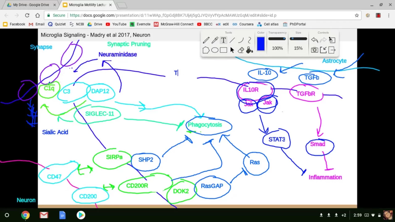
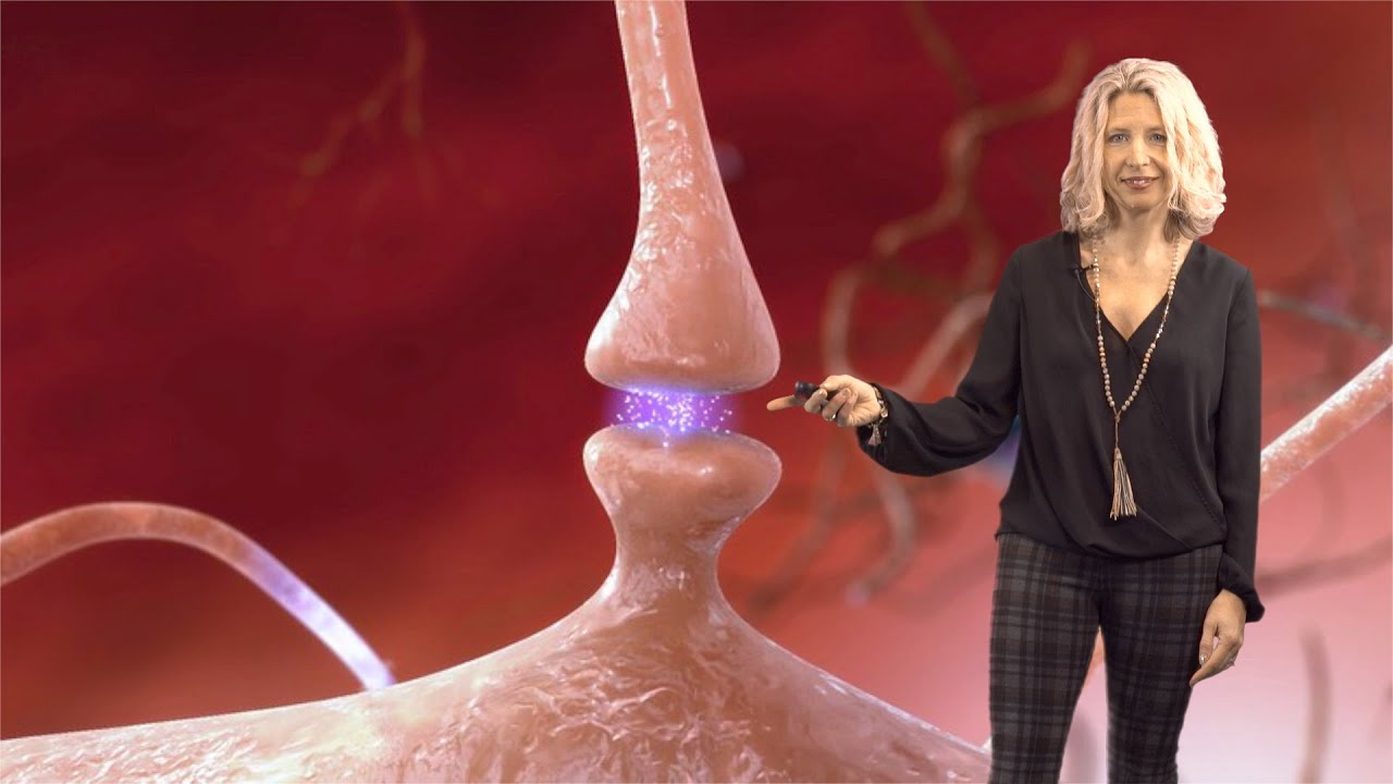












Microglia, the brain's resident immune cells comprising 7-10% of brain cells, play diverse roles in brain development, function, and disease through dynamic interactions with neurons and synapses; they are essential for synaptic pruning during development via complement-mediated mechanisms, but their heterogeneity across different states and contexts makes understanding their beneficial versus detrimental roles in conditions like Alzheimer's disease challenging.
Microglia in Brain Development & Disease | Beth Stevens | iBiology
Added:Fundamental brain anatomy and the primary cell types of the central nervous system, specifically distinguishing between neurons and glial cells.
The basics of synaptic transmission and neural circuit formation, including how neurons communicate across synapses.
Basic immunology concepts, particularly the function of macrophages, phagocytosis, and how immune cells protect host tissues.
The general concept of neurodevelopment and brain plasticity, including the idea that the brain overproduces connections that must later be refined.
The specific molecular pathways of synaptic pruning, such as how the classical complement cascade (e.g., C1q and C3) tags synapses for elimination by microglia.
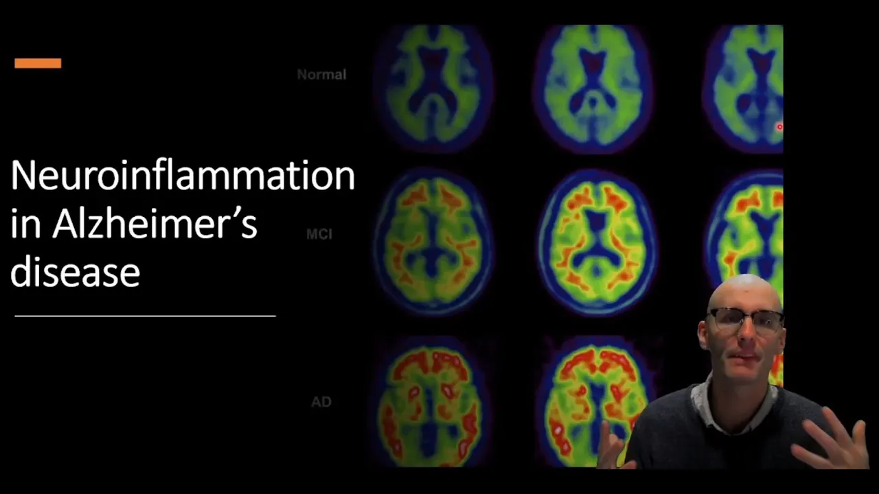
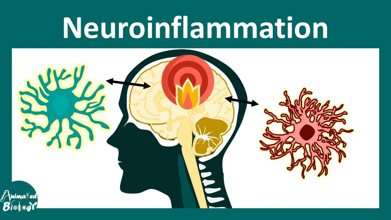
The role of aberrant microglial activation and chronic neuroinflammation in the pathogenesis of neurodegenerative conditions like Alzheimer's disease.
How disruptions in microglial-mediated synaptic pruning during development are linked to neurodevelopmental and psychiatric disorders like autism and schizophrenia.
Current therapeutic approaches and clinical trials aimed at targeting microglial receptors or pathways to prevent synapse loss in disease.
33.4K views762likes21:41Original Release: 2020-09-09
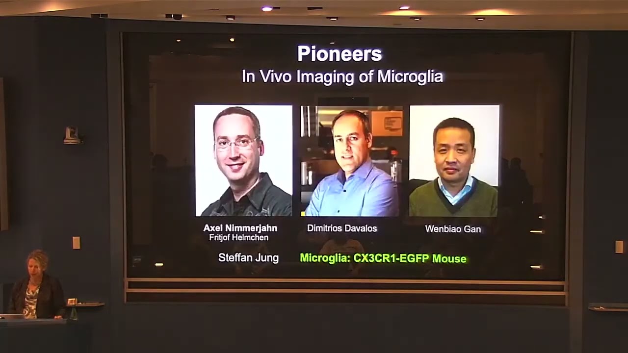
hello my name is beth stevens and i'm an hhmi investigator at boston children's hospital in the broad institute and my lab is largely interested in understanding how the immune system helps sculpt developing neural circuits now from the time i was a graduate student i've been fascinated by glial cells the non-neuronal cells of the brain but even as a card-carrying glial biologist i've largely ignored the cell i'm going to tell you about today microglia our residential immune cells now there are many reasons for that but it largely was in part because we really lack the tools in which to study these cells until fairly recently we now know that microglia our resident phagocytes and immune cells play central roles in both brain development function and dysfunction and disease they make up about seven to ten percent of the cells in our brain and despite being described over a hundred years ago by rio hertega we're only now beginning to understand their dynamic functions in the brain now we've known for quite some time they play a key role when the brain is injured or into context of disease microglia changed dramatically in the context of even local perturbations in the brain they largely are these beautiful cells that have these dynamic processes that you can't quite appreciate from this image but under the context of injury and disease they can dramatically change shape they become more phagocytic in many cases and they are known to play key roles in neuroinflammation by releasing molecules like cytokines into the brain they're also known as our resident phagocytes to be very good at clearing pathogens and debris and moreover they can remove local and toxic proteins in the brain now these functions which have been described for many years are both detrimental and beneficial depending on the context but what we're now appreciating is the fact that these cells play far more diverse roles than we previously appreciated and this again is because until recently we lacked tools to study them in detail one of the things that really has cracked open the field is new reporter transgenic lines like this one that i'm showing you that was developed by stefan young a number of years ago this is a mouse that's been genetically engineered to express egfp this fluorescent green protein in all the microglia and myeloid cells and using this tool researchers can look at microglia in the developing or adult or even diseased brain and watch these cells in action and this is in large part due to the fact that we can do two photon imaging by making these thin cranial windows and put a microscope over these fluorescent green mice and using these tools a number of pioneers in the field really were the first to start to make some of these really fundamental new observations about microglia that certainly changed the way i've been thinking about them so axel nimrod and demetrius davolos and wenbialgan were some of these pioneers that started to do imaging experiments like i just described so as i mentioned microglia are really good at eating or engulfing things they can rapidly clear debris or dead cells in the brain and they respond very dramatically to injury and this is illustrated by an experiment that axel nimrod did a number of years ago and this was enabled by the fact that again using this reporter line this green gfp fractal kind receptor mouse that was crossed to a neuronal reporter line where the neurons are labeled in red what axel did is in the purkinje cells in the cerebellum he made a little laser injury to that red neuron and what you can see from this movie is that the microglia were dramatically responding to that local injury and engulfing or pulling up the parts of that that neuron after the injury now this is a very dramatic example but this just illustrates their ability to phagocytose through the expression of a lot of different phagocytic receptors now in addition to phagocytosis as i mentioned microglia are incredibly dynamic the processes in particular are constantly surveying the brain environment on the brain parenchyma and these experiments again that were first done by these pioneers showed that by just looking at microglia under normal conditions in the brain you can see their fine processes moving but if you were to make an injury in this case with a laser the microglia dramatically move and extend their processes toward the injury as shown here now this is really rapidly happening although it's quite sped up in this movie but you can see the fine processes chemo attracting to the injury site and it's been shown now that one of the signals released that actually brings the microglia to the cidr injury are atp or pure they have pure energy receptors that recognize the signal but this is just one example of under under a real acute injury what microglial look like okay so microglia not only respond when things go wrong but two photon imaging like this has revealed that microglia are constantly surveying the brain environment with their fine processes so the question of course is what is it that they're surveying what are the signals that are regulating their motility and although you can't see the cells underneath there what signals are they using to communicate with neurons and other cells to regulate synapse and brain development so microglia are indeed integral parts of brain circuitry over the last many years more and more evidence is suggesting that microglia actively signal with neurons and synapses but also with other glial cells where they play critical roles in brain development and plasticity and function now what we also appreciate is that microglia are the only cell not born in the brain a recent work um bait mapping study by miriam murad's lab in florentino a number of years ago did some really important fate mapping studies and showed that microglia actually derived from the yolk sac as myeloid progenitor cells they enter the brain quite early in development and then once they're in the brain they're exposed to brain signals they become known as what we call resident microbial cells and they differentiate into these cells that i just mentioned these surveying microglia that mature over the first several weeks of development now we know that these microglia have many different functions uh many more that i'm probably going to tell you about today that we're just beginning to on earth but in particular even before birth there's been evidence to suggest that microglia are critically important in neurogenesis for example as resident phagocytes they actually help to clear and phagocytoses newborn neurons and there's actually even some evidence that they can engulf neurons without clear evidence of apoptosis so some of those signals have been identified there's also evidence to suggest that microglia release signals such as chemokines and cytokines that are also important in regulating not just neurogenesis but different aspects of neuronal differentiation during development and there's also evidence to suggest that axons that microglia are also dynamically regulating the refinement and pruning back of these uh developing axon tracks before birth and that they can also regulate the fate and the outcome of certain subtypes of inner neurons in the adult brain so this is all happening during our embryonic development but of course after birth more and more evidence points to the fact that these cells are playing heroes in myelination so they can regulate the process of myelination but also the uh the the process of oligodendrocyte differentiation they're involved in synaptic development synaptic maturation and synaptic plasticity and they're also key regulators in synaptic remodeling or synaptic pruning which i'll tell you more about today so this suggests that microglia are existing in specialized states and we as i mentioned before these cells are obviously dynamically associating with synapses and this is now illustrated in this movie where we can overlay a microglia in green with neurons which are labeled in red and this led to the observation that these microglia are dynamically interacting with subsets of synapses spines and axons especially during developmental critical periods these cells are incredibly dynamic and their as you'll tell you are particularly phagocytic in times and places of synaptic remodeling which is a normal developmental process that's happening over the first weeks of life um and and so the question is could microglia be involved in this process of synaptic refinement or synaptic pruning now synaptic pruning is as i mentioned a normal developmental process in which excess synapses or connections are permanently removed while others get strengthened and maintained they happen over this process happens over different critical periods um so there are parts of your brain in like sensory systems that are that are pruned relatively early in development but there are some parts of our brain like the frontal cortex that continue to prune and remodel into early adulthood and so the question is you know could microglia play a role in this process and when you look at microglia morphology and you start to characterize some of the markers they express we noticed pretty early on that during this developmental critical period of pruning microglia were particularly phagocytic meaning they expressed a lot of phagocytic receptors they had more lysosomal activity at these periods where they're associating with synapses and in the most visual system the model system that we've been using we provided evidence that microglia are in fact engulfing some of these axons or inputs during development this is a 3d surface rendering of a microglia from the visual thalamus for example during its critical period of remodeling and dorothy shaffer in my lab showed that most of the microglia at this time of eye segregation and the visual thalamus were engulfing large numbers of these presynaptic inputs which could also be shown by em and it was happening again during this window of development and they largely decreased their ability to engulf synapses after that critical period raising the question about what's the signals that are instructing microglia to engulf synapses and are there's even more specifically signals that are telling microglia to engulf specific synapses for example the less active inputs during development and as i mentioned you know this is an activity dependent process how do microglia know which synapse to prune and which synapse to leave alone raising the question that they may be some sort of cues or instructive signals that are recruiting microglia to certain synapses or inputs and there may also be signals that are telling microglia not to engulf certain synapses or inputs so one of the signals that we have been focused on and we identified a number of years ago was a group of molecules called complement which was a group of innate immune molecules well studied in the innate immune system in the periphery these are eat me signals that are known to tag apoptotic cells or debris and that they're a way that circulating macrophages in the periphery recognize these apoptotic cells so essentially these these proteins bind or opsinize the surface of these the surface of membrane of these cells and and tell the the macrophages to engulf them now in the brain what we discovered when i was a postdoc in ben barris's lab is that a large number of these secreted complement molecules were actually expressed in the healthy brain by neurons and glial cells especially the microglia were making a lot of c1q for example that initiating protein of the cascade and that many of these molecules were actually binding to subsets of these developing synapses and importantly microglia are the only resident cell to express the receptors for these complement molecules or cr3 for example so in the immune system as i mentioned complement is tagging a bacterial cell for elimination that's recruiting macrophages which express complement receptors and what we discovered is that the brain in the brain especially in the healthy brain complement molecules are binding to or localizing to synapses and axons and this is mediating the engulfment of those inputs through complement receptors and so what we went on to show was that if you knock out either the molecules complement the secreted molecules or the receptor on microglia there were defects in synapse elimination and that microglia at least in the visual system were only about half as good at engulfing these synapses now this is just one example of a group of molecules that we that are instructive in synaptic pruning in this circuit but work over the last many years by many labs have identified more and more signals some secreted some surface-bound molecules both by microglia and the neurons and other glial cells that are contributing to different aspects of of both brain development and synaptic refinement i just illustrated a few here and more importantly which i want to touch on briefly it's it's it raises also the question of of whether or not in the context of disease where there's aberrant synaptic loss and an activation of microglia and other immune related pathways could some of these same pathways and mechanisms that normally function to regulate brain development could they go awry and become dysregulated in the context of brain injury and disease and indeed many of these molecules including trem2 apoe these are actually molecules and proteins that are also risk genes for diseases like alzheimer's disease and we now appreciate from emerging genetics that a large number of the late onset alzheimer's risk genes are expressed or enriched in microglia or myeloid cells and that really does change the way we're thinking about these cells in the context of these diseases not just as playing a secondary role but actually may even be uh driving some of the the disease in the context of at least alzheimer's disease for example trem2 and some of the complement genes as well so this raises the question how do microglia contribute to disease when do they contribute and these are really challenging questions to address obviously in human disease because we really do lack the tools to be able to both manipulate and label and track microglia changes over the course of disease especially non-invasively but even in fixed tissue we don't have very good markers that denote all these different functional states that i just told you about so imagine if we had a signature that denoted a microglia that was undergoing phagocytosis during neurogenesis or synaptic pruning and differentiating it from a microglia that was pruning a plaque versus aberrantly pruning synapses right now when these microglia change state we don't really have markers that denote the beneficial versus the detrimental states in the brain even inflammation we call these inflammatory states but more than likely this represents many different sub-states or sub-populations and even then inflammation can be both beneficial and detrimental depending upon the context and the cues so this really is raising the both the challenge but also the fact that we now are in a position to start to develop new tools to be able to start to track these different states how heterogeneous are microglia how diverse are they how many states really even exist before we can understand their role in alzheimer's disease we thought maybe we should step back and start to ask how many states exist under normal development across the lifespan of a mouse and what can we infer about their function and how do these these microglia states change in the context of disease so to address this question we've been applying a new technology that many in the field of course are using called single cell sequencing initially developed by steve mccarroll my collaborator and vivergev and evan mccosko now it's a technology that many are using routinely but we applied this a number of years ago and i should say tim hammond and connor in my lab in collaboration with steve's lab decided to use single cell sequencing to assess microglia diversity across the lifespan of a mouse and to do that and and you may recall that microglia only represent between seven and ten percent of the cells in the brain so that also involved having to develop new methods to both dissociate and purify and and isolate microglia only from the brains from many many mice in fact they ended up profiling 78 000 microglia across 41 mice um and using these um approaches that tim and conor developed to purify the microglia we put them through a 10x or single cell sequencing and in this way we can start to ask how diverse are they and the take-home of that this is a summary of of tim's paper but also a very very um consistent results were observed in a study by ben barris's lab which is that there's a lot of diversity during development by about one month at least in a mouse they're largely homogeneous one big population but then in the context of aging pathology there's a shift in some of these states and this is the data just to illustrate an example of that where you can actually we looked at all of the the data put together in these tsne plots and we started looking at what the different clusters or the different populations of microglia were what were the genes in those different clusters and in particular cluster four emerged only during development during this postnatal period and this particular cluster or state of microglia expressed genes like osteopontin or spp1 igf-1 lpl and others and these are interesting because they're the same signatures that are coming up in some of these disease-associated states in alzheimer's and other diseases yet we were seeing them in a small population of microglia only during development and using and interestingly these and other genes in this cluster suggested immune recognition lysosomal activity phagocytosis and so to would ask where do these microglia exist in the brain um connor and tim did spatial mapping and multiplex and situ hybridization they made probes for these markers and then found quite excitingly that the um these microglia and cluster four were largely localized to developing white matter tracks and nowhere else and that of course opened up lots of interesting questions um could these special axon associated microglia be important in myelination axonal refinement other aspects of refinement of these white matter tracks that we were not yet um thinking about and so using some of these tools we're now in a position to start to manipulate and deplete and knock out just these specific cluster four microglia to start to interrogate function of these cells and we're taking a similar approach and as our other in the field to start to use the single cell data to try to try to link changes in transcription to specific functions of microglia i gave you one example these axon tract associated microglia i don't think we necessarily would have found this this particular population had we not done the single cell sequencing but this is now giving us signature and markers to start to use as tools to start to manipulate specific states of microglia and i think this approach by combining single cell and spatial mapping and functional studies are going to enable the field to ask a number of fundamental questions what are the functions of these different populations in different contexts can we start to model faithfully model microglia can we how do the genetic mutations and variants influence microglia states how do we link genetics to function and ultimately can we use this information to target and track microglia into across disease progression and i think this really is sort of where i want to wrap up is that i do think that we're in a exciting time in the field but again without understanding the normal functions of these cells it's going to be really challenging to try to understand how they go awry in these complex disorders especially given the diversity and and heterogeneity of these microglia in the brain so we want to be able to develop new tools to identify and track microglia populations we want to be able to use this information to target the detrimental states for example of microglia and different diseases and we ultimately hope that maybe someday digging deeply into the biology could unveil new biomarkers or maybe even new therapeutic targets that we could we could use to start to um to manipulate microglia so i'll end there by thanking the human microglia in my lab that made a lot of the work i told you about today possible and also a lot of really terrific collaborators and lab members both past and present so thanks very much you
Up Next

21:23
Cell Surface Markers Explained: CD3, CD4, CD8, CD19, CD28, CD16, CD56
@ScienceAnswers101
79.4K views•2013-05-31

50:12
Bacterial Communication: Quorum Sensing and Biofilm Formation Explained
@JHUAAP
12.6K views•2011-06-28

42:45
MicroRNAs: Gene Regulation Explained by David Bartel | iBiology
@scicommlab
233.1K views•2014-03-24

7:08
Bacteriophages: Earth's Deadliest Killers and Future Antibiotics
@kurzgesagt
34.6M views•2018-05-13
Related Study Plans & Knowledge Roadmaps
Structured learning paths in Biology
biology
RNAi: Biogenesis, RISC & Gene Silencing
01Molecular Biology Foundations: DNA to Protein
02Introduction to RNA Interference (RNAi)
03Molecular Biogenesis of miRNA and siRNA
+ 2 more learning modules
10h5 modules
Explore Roadmap
biology
Mycorrhizae: Signaling, Biophysics & Ecology
01Foundations of Mycorrhizal Symbiosis
02Molecular Signaling and Root Colonization
03Biophysics and Bioenergetics of Nutrient Exchange
+ 1 more learning modules
11h4 modules
Explore Roadmap
biology
Circadian Clocks: TTFL, SCN & Periphery
01Foundations of Molecular Biology & Gene Expression
02Introduction to Chronobiology and Actograms
03The Core TTFL: Molecular Architecture of the Clock
+ 3 more learning modules
14h6 modules
Explore Roadmap
biology
Population Genetics: Math, Drift & Selection
01Genetic & Probability Foundations
02The Hardy-Weinberg Principle and Baseline Models
03Mathematical Modeling of Natural Selection
+ 3 more learning modules
18h6 modules
Explore Roadmap