The FAST (Focused Assessment with Sonography in Trauma) exam for female pelvis uses a phased array transducer to scan the pelvic region, starting with a sagittal view above the symphysis pubis to identify the anechoic bladder, pear-shaped uterus, and the Cul de Sac—a potential space where free fluid can be detected; the examination then transitions to a transverse view by rotating the transducer toward the patient's right, scanning from inferior to superior to visualize the bladder, uterus, and possibly the ovary while carefully searching for any evidence of free fluid.
How to Perform a FAST Exam: Female Pelvis Ultrasound
Added:Deep Dive
Basic principles of medical ultrasound physics, including transducer selection, echogenicity (anechoic vs. hyperechoic), and image optimization controls like gain and depth.
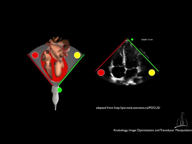
02 Knobology, Image Optimization and Transducer Manipulation.
15.4K
This section covers essential ultrasound image optimization controls. Depth control extends 1-2 cm beyond structures of interest to ensure complete visualization without sacrificing temporal resolution. Receiver gain adjusts overall image brightness by amplifying all signals equally, though it doesn't improve signal-to-noise ratio. Time gain compensation compensates for tissue attenuation by selectively amplifying signals at different depths to achieve uniform brightness. Focusing narrows the ultrasound beam at selected depths to improve lateral resolution. Transducer frequency creates a fundamental trade-off: higher frequencies provide better resolution but less penetration, while lower frequencies penetrate deeper but resolve less clearly. For adult transthoracic echocardiography, 2-4 MHz transducers are standard, with frequency options typically displayed as P (penetration), G (general), and R (resolution).
Click to watch →

Webinar gratis - Principios básicos de la ecografía - M.V.Z. Eduardo Moore
12.1K
Proper ultrasound interpretation requires understanding equipment controls and echogenicity patterns. Gain control amplifies or reduces echoes—excessive gain obscures details while insufficient gain makes images too dark. Depth control adjusts the field of view, with proper settings allowing organs to occupy about half the screen. Zoom control allows magnification of specific areas. Echogenicity terminology differs from X-ray: hyperechoic (white) indicates mineralized structures like bones and calculi; anechoic (black) indicates fluid; isoechoic matches surrounding tissue; heterogeneous suggests tumors or abscesses. Artifacts like posterior acoustic enhancement confirm fluid, while shadowing confirms calcifications. Gas causes significant artifacts, requiring fasting before abdominal ultrasound.
Click to watch →
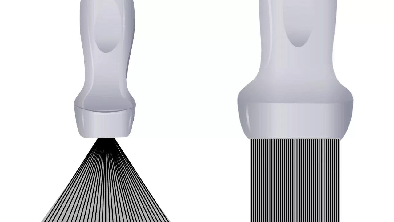
Ultrasound Physics for Emergency Medicine: Modes, Probes, Artifacts, Orientation
359K
Ultrasound is a mechanical pressure wave measured in Hertz, with diagnostic ultrasound operating above 20,000 Hz; key principles include the inverse relationship between wavelength and frequency, the reverse piezoelectric effect for sound generation, and the pulse-echo principle for imaging. The speed of sound varies through tissues (1,040 m/s in soft tissue, 330 m/s in air, 4,030 m/s in bone), affecting penetration and resolution. Higher frequencies provide better resolution but less penetration, while lower frequencies offer deeper penetration but reduced detail. Common imaging modes include B-mode (grayscale), M-mode (motion), and Doppler (color and spectral) for detecting blood flow. Transducers use piezoelectric crystals with matching layers to optimize sound transmission. Key artifacts include shadowing (high acoustic impedance interfaces), posterior acoustic enhancement (fluid-filled structures), lateral cystic shadowing (refraction at fluid interfaces), mirror images (strong reflectors), and reverberations (multiple internal reflections). Proper probe orientation follows conventions where the indicator points toward the patient's right or head, with sagittal, coronal, and transverse planes defined accordingly. Echogenicity describes tissue brightness relative to surroundings, classified as hypoechoic, isoechoic, hyperechoic, or anechoic.
Click to watch →

Clinical Ultrasound-Physics and Knobology.
19K
This video teaches the fundamental physics principles and machine controls essential for clinical ultrasound imaging, including how ultrasound waves interact with tissues based on density (echogenicity), the trade-off between transducer frequency and penetration depth, proper orientation using standard indicators, and how to optimize image quality by adjusting depth, gain, and frequency settings while understanding common artifacts like acoustic shadowing, enhancement, and reverberation.
Click to watch →
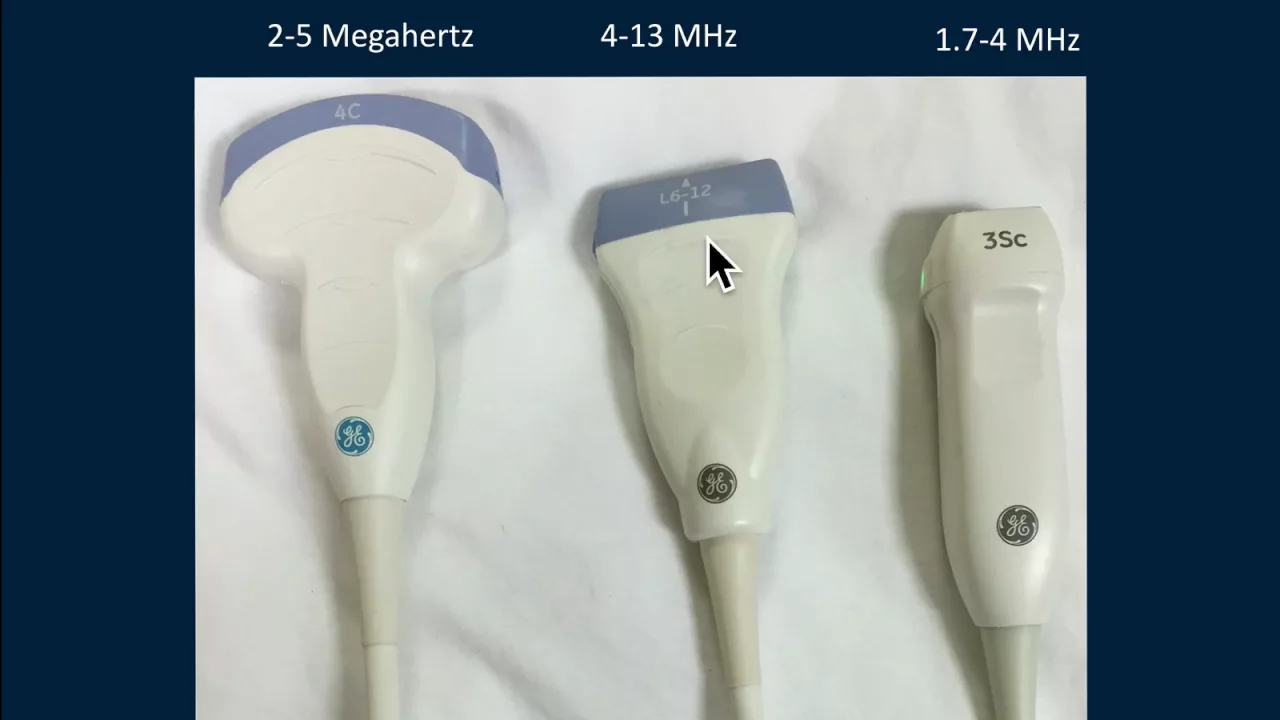
Ultrasound Physics and Instrumentation
135.9K
Ultrasound imaging works by emitting sound waves at 1540 m/s through tissue and plotting echoes based on their return time, with deeper structures appearing lower on the screen; clinicians must balance transducer frequency (higher for resolution, lower for penetration), use appropriate transducers (convex for deep structures, linear for superficial), and understand key controls including depth, gain, and Doppler settings to produce diagnostic-quality images while recognizing artifacts like posterior shadowing and mirror-image reflections that aid in clinical interpretation.
Click to watch →
Deep Dive
Gross anatomy of the female pelvis, specifically identifying the urinary bladder, uterus, rectouterine pouch (Pouch of Douglas), and vesicouterine pouch.
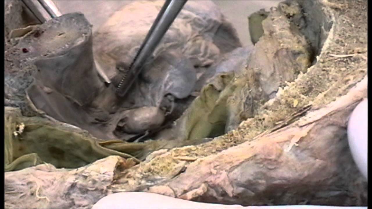
Anatomy of female genital organs - dissection
548.5K
This video demonstrates the anatomy of female genital organs through dissection, showing the urinary bladder, uterus (with its fundus, cornu, and isthmus), vagina with fornices, urethra, rectum, and anal canal. Key structures include the uterine tubes attached to the broad ligament, the ovary attached to the posterior leaf via mesovarium, and the mesosalpinx connecting ovary to tube. The round ligament of the ovary extends from the uterine pole to the cornu, while the round ligament of the uterus extends to the deep inguinal ring. Critical anatomical relationships include the uterine artery crossing the ureter (water under the bridge), and the cardinal ligament providing pelvic support. The peritoneal reflections create the uterovesical pouch and rectouterine pouch of Douglas.
Click to watch →
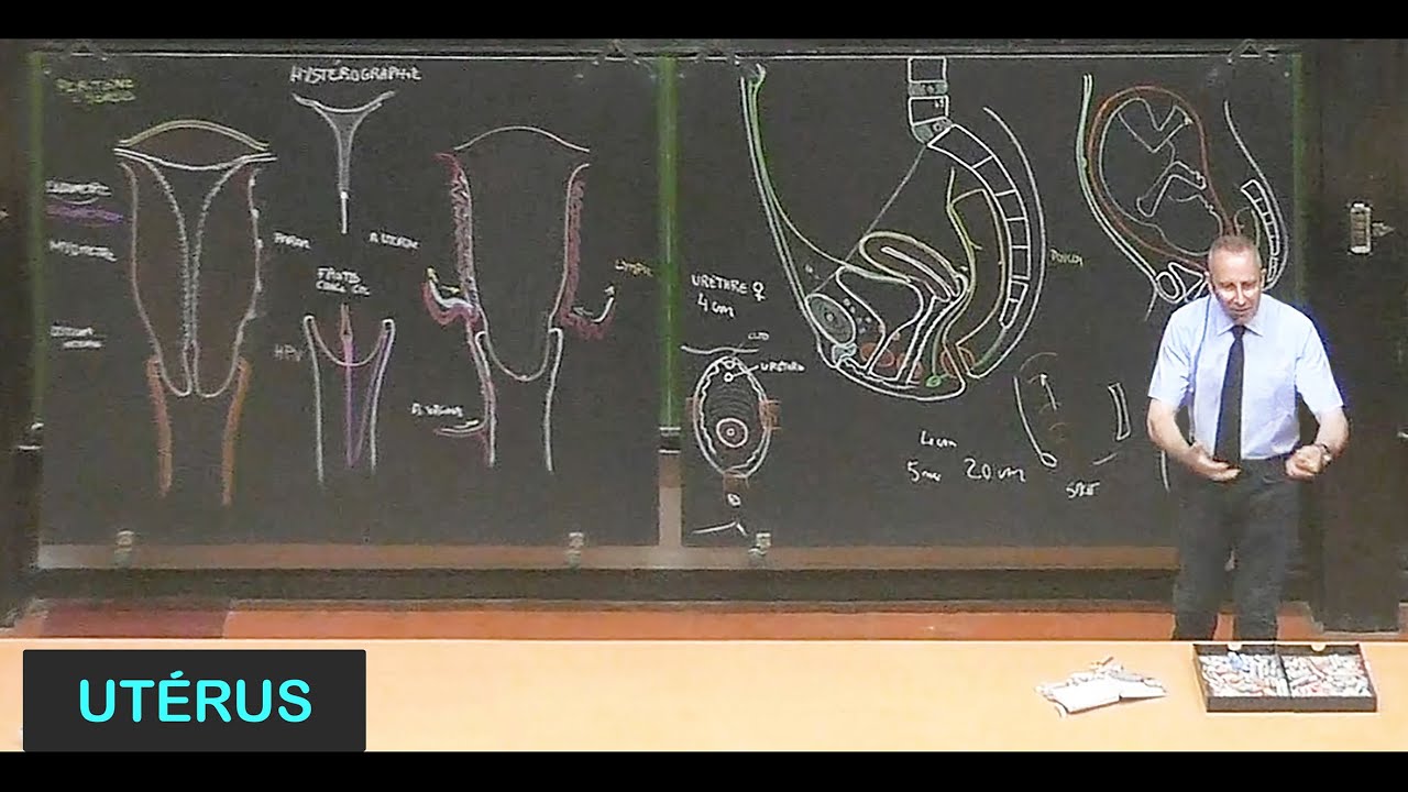
Anatomie de l'utérus (Pelvis 13)
21.7K
The peritoneum creates several pouches in the female pelvis: vesicouterine pouch (between bladder and uterus) and rectouterine pouch (pouch of Douglas, between uterus and rectum). The rectouterine pouch is the lowest point of the peritoneal cavity in females. This pouch allows for vaginal exploration of the uterine cavity, assessment of uterine size during pregnancy, and peritoneal fluid aspiration. The pouch of Douglas is also the site of the 'Douglas' sign in peritonitis.
Click to watch →

What is the pouch of Douglas?
52.7K
The female pelvis contains three peritoneal pouches formed by peritoneal folds between organs. The recto-uterine pouch (pouch of Douglas/cul-de-sac) lies between the uterus and rectum. The vesico-uterine pouch exists between the bladder and uterus. The recto-vaginal pouch is found between the vagina and rectum. These pouches are potential spaces containing minimal fluid. The pouch of Douglas is clinically significant as it is the most inferior and posterior peritoneal point, making it the primary site for fluid accumulation in conditions like peritonitis, abscesses, hemorrhage, and ascites.
Click to watch →

CHO Special MCQs Series #5 | CHO | Staff Nurse | RRB Nursing Preparation | Dr. Shailendra Sir
8.2K
External female genitalia include the clitoris, mons pubis, labia majora, labia minora, and vestibules. Internal organs include ovaries, fallopian tubes, uterus, and vagina. The rectouterine pouch (Pouch of Douglas) is between the uterus and rectum. The vesicouterine pouch is between the uterus and bladder. The Space of Retzius is between the pubic symphysis and bladder.
Click to watch →

Sistema genital femenino: generalidades
320.4K
The uterus has intimate relations with surrounding structures: the vagina is in direct contact with the cervix; the fallopian tubes extend posteriorly and laterally from the uterine cornua; the urinary bladder is related anteriorly to the uterine body and cervix; the rectum is related posteriorly; the rectouterine pouch (pouch of Douglas) is located posteriorly; and the vesicouterine pouch is located anteriorly.
Click to watch →
Deep Dive
The clinical indications and primary objectives of the Focused Assessment with Sonography for Trauma (FAST) protocol in emergency medicine.

Focused Assessment with Sonography for Trauma - FAST
6.1K
FAST (Focused Assessment with Sonography for Trauma) is a focused ultrasound examination for trauma assessment, coined in 1994 with standard windows established in 1999. FAST Extended, developed in 2004, adds pulmonary windows. This methodology evolved from focused abdominal ultrasound in 1971 to trauma-specific applications, replacing invasive peritoneal lavage with non-invasive bedside ultrasound. FAST is part of POCUS (Point-of-Care Ultrasound), performed dynamically at the bedside. Sensitivity for free abdominal fluid is 76%, while hemopericardium detection ranges from 56-90%. Sensitivity varies by trauma type: blunt torso injury (63-99%) versus penetrating trauma (28-100%). The primary objective is identifying free fluid enabling immediate surgical transfer. FAST is validated for hemodynamically unstable abdominal trauma patients and can follow stable patients under observation.
Click to watch →
Focused Assessment with Sonography in Trauma (FAST)
1.6K
The FAST (Focused Assessment with Sonography in Trauma) exam should be performed on any patient with trauma, whether blunt or penetrating, regardless of age (adult, pediatric, or pregnant). The primary goal is to identify blood in the peritoneal cavity and blood in the pericardial space. It is not intended to identify all intra-abdominal pathology or rare retroperitoneal trauma. The extended FAST exam includes assessment for hemothorax, pneumothorax, hemodynamic status, solid organ injury, and pelvic hemorrhage.
Click to watch →
Diagnostico por Imagen en Emergencias
186
FAST (Focused Assessment with Sonography for Trauma) is an ultrasound imaging technique used in emergency medicine to evaluate closed abdominal trauma. It can also be applied for thoracic trauma, though it is most frequently used for abdominal trauma assessment. The primary purpose is to rapidly determine the presence of fluid accumulation in the abdominal cavity, which is critical for patient management in trauma situations. FAST exam is indicated for patients with closed abdominal trauma, commonly occurring in motor vehicle accidents, motorcycle accidents, and various other circumstances that can cause blunt abdominal trauma. The technique is particularly valuable in emergency situations where rapid assessment is needed to identify life-threatening conditions requiring immediate intervention.
Click to watch →

III JORNADA NACIONAL LAUEC URGÊNCIA E EMERGÊNCIA 1º DIA
18K
The FAST (Focused Assessment with Sonography for Trauma) protocol is performed during the circulation assessment phase of trauma evaluation. It detects free fluid (blood) in the peritoneal cavity, pericardium, and pelvis. The primary indication is hemodynamic instability with suspected intra-abdominal or cardiac injury. FAST has >80% sensitivity for detecting significant fluid collections. It is particularly valuable in pregnant patients (avoiding radiation) and when CT is unavailable. A positive FAST in an unstable patient requires immediate laparotomy.
Click to watch →

Focused Assessment With Sonography in Trauma (FAST)
2.4K
FAST (Focused Assessment Sonography for Trauma) is a rapid bedside ultrasound examination performed in emergency settings to detect intraperitoneal fluid in trauma patients. The primary clinical purpose is to identify significant intra-abdominal injuries, which helps determine the need for surgical intervention. The examination is designed to be completed in 3-4 minutes, making it efficient for emergency assessment. The probe is positioned at four specific locations: epigastric region (for pericardial tamponade), right hypochondriac region (for liver fluid), left hypochondriac region (for spleen fluid), and pelvic region (for pelvic fluid). Unlike comprehensive abdominal ultrasound, FAST focuses solely on detecting intraperitoneal fluid accumulation.
Click to watch →
Deep Dive
Standard probe orientation and scanning planes (sagittal and transverse) used in transabdominal pelvic imaging.

Understanding Pelvic anatomy and orientation of ultrasound image video 2
5.9K
Pelvic ultrasound utilizes two primary scanning planes: sagittal (longitudinal/vertical) and transverse. In sagittal plane, the probe notch faces upward, with image top representing anterior body, bottom posterior, left side toward patient's head, and right side toward feet. In transverse plane, the probe notch faces the patient's right side, with image top showing anterior, bottom posterior, left side displaying patient's right side, and right side displaying patient's left side. The vagina lies between bladder and rectum, indicated by a bright line separating anterior and posterior walls. The cervix has internal os (uterine cavity-endocervical canal junction) and external os (endocervical canal-vagina junction). Uterine anatomy includes fundus (top), body, anterior wall (facing bladder), posterior wall, and pouch of Douglas (area behind posterior wall).
Click to watch →

How to perform a Transvaginal Ultrasound
212.3K
Proper probe orientation is fundamental to accurate ultrasound interpretation. In the transverse plane, hold the probe with the marker on your left (patient's right) and set the machine so the marker appears at the top left of the screen. The sound beam extends from the marker side to the opposite side. In the longitudinal plane, rotate the probe 90 degrees clockwise so the marker is uppermost at the head end, creating an image as if the patient lies face down beneath the screen. These principles apply identically to both transabdominal and transvaginal scanning, though the gray strip is not visible on the transvaginal probe tip.
Click to watch →

Basic Applications : Female Pelvis - Scanning Techniques
583.9K
Transabdominal scanning employs longitudinal and transverse planes with probe placement at the symphysis. Longitudinal sweeps involve pinning the probe and slow directional movements to visualize the entire uterus. The transverse plane provides cross-sectional views where the uterus appears round versus the vagina's oblong shape with V-stripe. The highly mobile uterus may be anteverted, retroverted, or tilted, requiring adaptation. Transvaginal/endocavitary scanning uses higher-frequency probes (5-8 MHz) with wide fields of view (>90°). Preparation involves sterile gel application, condom covers, and air bubble removal. The probe is introduced gently with marker anteriorly, then rotated to find the bladder as the sonographic landmark. Both approaches require understanding probe orientation: probe marker direction correlates with patient anatomy, with anterior structures appearing in the near field and posterior structures in the far field.
Click to watch →
![Essential First Trimester Pelvic Ultrasound for physician - HD [Basic Radiology]](https://i.ytimg.com/vi/WJO0dCez6Vw/sddefault.jpg)
Essential First Trimester Pelvic Ultrasound for physician - HD [Basic Radiology]
409
Use curved low-frequency transducer for transabdominal imaging. A full bladder provides an acoustic window and pushes the uterus posteriorly for better visualization. Obtain sagittal and transverse views, fanning through the entire area of interest. For sagittal views, slide probe down above pubic symphysis then tip into pelvic brim (common error: scanning too high toward head). For transverse views, rotate probe 90 degrees with marker toward patient's right. The bladder appears rectangular in transverse view, uterus circular. Evaluate pouch of Douglas for free fluid in both views.
Click to watch →
Ultrasound Physics and Instrumentation
135.9K
Standard conventions exist for orienting ultrasound probes relative to the patient's anatomy. In the sagittal plane (longitudinal view from anterior surface), the probe indicator points toward the patient's head, with superior structures above and inferior structures below. In the transverse plane (short-axis view), the probe indicator points toward the patient's right side, with structures to the right appearing on the right side of the screen. In the coronal plane, the probe indicator still points toward the patient's head, but the sound beam is directed medially into the body. Understanding these conventions ensures consistent image interpretation and communication among healthcare providers.
Click to watch →
Prerequisite Knowledge
- Concept 01Basic principles of medical ultrasound physics, including transducer selection, echogenicity (anechoic vs. hyperechoic), and image optimization controls like gain and depth.
- Concept 02Gross anatomy of the female pelvis, specifically identifying the urinary bladder, uterus, rectouterine pouch (Pouch of Douglas), and vesicouterine pouch.
- Concept 03The clinical indications and primary objectives of the Focused Assessment with Sonography for Trauma (FAST) protocol in emergency medicine.
- Concept 04Standard probe orientation and scanning planes (sagittal and transverse) used in transabdominal pelvic imaging.
Subsequent Learning
- Step 01Clinical differentiation between physiological pelvic fluid (e.g., normal menstrual cycle/ovulation) and pathological fluid (e.g., hemoperitoneum, ruptured ectopic pregnancy).
- Step 02Integration of FAST pelvic ultrasound findings into clinical decision-making pathways for unstable trauma patients (e.g., immediate laparotomy vs. CT imaging).
- Step 03Mastery of the remaining views of the Extended FAST (eFAST) protocol, including hepatic, splenic, cardiac, and thoracic windows.
- Step 04Introduction to transvaginal point-of-care ultrasound (POCUS) for advanced assessment of gynecological emergencies such as ovarian torsion or pelvic inflammatory disease.
FAST Pelvic Setup
Sagittal Bladder Scan
Uterus and Cul-de-sac
Sweep for Free Fluid
Transverse Probe Rotation
Inferior to Superior Sweep
Uterus and Ovary Check
FAST Pelvic Setup
0:15Playing Section
- 1
Begin FAST exam focused on female pelvic view.
- 2
Use phased array transducer with proper probe marking.
Diagnostic Limitations and False Positives in Female Pelvic FAST Exams
While the FAST (Focused Assessment with Sonography for Trauma) exam is a standard emergency protocol, critics and clinical studies highlight significant limitations in its application to the female pelvis. A primary concern is the high rate of false positives in reproductive-age females, who often have physiological free fluid (due to ovulation or menstruation) in the pouch of Douglas, which can be mistaken for traumatic hemoperitoneum. Conversely, the pelvic FAST exam has low sensitivity for detecting retroperitoneal bleeding, pelvic fractures, or specific uterine and ovarian injuries. Consequently, many trauma specialists argue against over-relying on pelvic ultrasound in stable patients, advocating instead for Computed Tomography (CT) as the diagnostic gold standard. They caution that a negative FAST exam can provide a false sense of security, potentially delaying critical interventions for internal injuries that ultrasound fails to detect.
Deep Dive
Clinical differentiation between physiological pelvic fluid (e.g., normal menstrual cycle/ovulation) and pathological fluid (e.g., hemoperitoneum, ruptured ectopic pregnancy).

УЗД новообразований яичников с помощью стратификационной системы O-RADS (версия 2023 г.)
13.1K
O-RADS provides standardized descriptors for common benign findings: simple cyst (anechoic, thin-walled, smooth, with acoustic enhancement), dermoid cyst (echogenic components including dermoid plug with shadowing, hair, fat, bone, teeth), endometrioid cyst (ground glass pattern, sometimes with hyperechoic aggregates), and hydrosalpinx (elongated, tubular, with incomplete septations). Free fluid in the pelvis is classified as physiological or pathological. Physiological fluid is expected in premenopausal women, particularly after ovulation. Pathological fluid (ascites) is defined as fluid above the uterine fundus. In postmenopausal women, any free fluid should be considered pathological and warrants follow-up.
Click to watch →

Ultrasound Evaulatio of Ectopic Pregnancy
548
The most common finding indicating ectopic pregnancy is a heterogeneous adnexal mass (blood clot) with 92% PPV. Free fluid with echogenic clots in the cul-de-sac is highly suggestive, with 15% showing only free fluid and 50-70% showing free fluid with other signs. Differentiating pathologic from physiologic free fluid is crucial: pathologic fluid contains echogenic irregular objects (clots), while physiologic fluid is small and non-echogenic. These findings help distinguish ectopic pregnancy from normal intrauterine pregnancy.
Click to watch →
1st Trimester OB US 2018 CUSE
140
Free fluid in pelvis can be physiological (pouch of Douglas) or pathological (ectopic, trauma). Simple fluid is anechoic; complex fluid has internal echoes. Rule of thirds: <1/3 posterior wall = small, 1/3-2/3 = moderate, >2/3 = large. Sonographic findings concerning for ectopic (least to most concerning): absence of intrauterine fetal parts, pseudogestational sac, fluid in cul-de-sac, complex fluid, adnexal mass, extrauterine fetal pole/yolk sac, extrauterine fetal heart rate (most definitive). The main goal is risk estimation, not definitive identification.
Click to watch →

УЗИ при неотложных и критических состояниях
4.3K
Free fluid in the pelvis appears as anechoic areas with sharp, angular margins, unlike bowel loops which have more rounded contours. In females, the rectouterine pouch (Pouch of Douglas) is the most dependent area for fluid accumulation. Fluid volumes less than 30 mm are considered physiological and related to the menstrual cycle. Fluid volumes greater than 30 mm are considered pathological and may indicate conditions such as ectopic pregnancy, ovarian cyst rupture, or other gynecological pathology. The probe should be positioned to visualize the bladder and surrounding structures, with the scanning surface directed toward the feet.
Click to watch →

Ultrasound of Normal and Abnormal early pregnancy YouTube
387
Echogenic fluid indicates hemoperitoneum until proven otherwise. The right upper quadrant window (8th-11th intercostal space) visualizes peritoneal fluid. Large volume complex fluid with no IUP is consistent with ruptured ectopic until proven otherwise. Clinical pearls: stable IUP with benign exam can be discharged; assisted reproduction patients have 1 in 150 heterotopic risk; no IUP with free fluid requires emergent consultation; no IUP with hCG less than 1,000 needs comprehensive scan.
Click to watch →
Deep Dive
Integration of FAST pelvic ultrasound findings into clinical decision-making pathways for unstable trauma patients (e.g., immediate laparotomy vs. CT imaging).

25 Most Important Surgery Questions | Dr. Vineet Gupta| MIST FMGE
90.8K
For blunt abdominal trauma, management differs based on hemodynamic stability (shock index = pulse/systolic BP). Stable patients (SI <1): admit for observation, perform FAST ultrasound first, then CECT if indicated. Unstable patients (SI >1): start IV fluids aggressively, avoid CECT initially, perform FAST to assess pericardial, liver, spleen, and pelvic injuries. If not stabilizing, proceed to urgent exploratory laparotomy with Pringle maneuver for hemorrhage control.
Click to watch →
![[WSFP DONOTHON DAY 9] Medical Education: Major traumatic injuries](https://i.ytimg.com/vi/Z1KjnuN5e1o/maxresdefault.jpg)
[WSFP DONOTHON DAY 9] Medical Education: Major traumatic injuries
236
FAST (Focused Assessment with Sonography for Trauma) is a bedside ultrasound used to detect fluid in trauma patients. It examines four areas: pericardial view (heart) for cardiac tamponade, hepatorenal space (right upper quadrant) for liver bleeding, splenorenal space (left upper quadrant) for spleen bleeding, and suprapubic view (bladder) for pelvic bleeding. FAST results guide management: stable patients with positive FAST get CT scan, unstable patients with positive FAST need immediate surgery, and unstable patients with negative FAST can get CT scan.
Click to watch →

Surgery - Basics of Trauma || Head trauma, Abdominal trauma, Thoracic trauma
104.4K
Most commonly injured organ in abdominal trauma is the spleen. For stable blunt abdominal trauma patients, FAST ultrasound is initial investigation; contrast-enhanced CT scan is definitive. For unstable patients, FAST is preferred and midline laparotomy is performed if peritonitis is present. Penetrating abdominal injuries require careful object removal only in operating theater to avoid losing tamponade effect.
Click to watch →

FAST ABDOMEN ULTRASOUND SCAN
354
FAST is the definitive imaging modality for unstable trauma patients due to its speed and bedside availability. CT scan is the gold standard for stable patients but requires patient transport and time. For unstable patients, FAST-positive results require immediate surgical intervention; FAST-negative results do not exclude injury and may require surgical exploration. Delaying treatment for CT in unstable patients can result in patient deterioration or death. FAST scoring is reported as positive, negative, or intermediate based on fluid detection in the four examination windows. Repeat FAST should be performed in unstable patients with negative initial scan, during secondary survey if patient deteriorates, or for conservative management patients to monitor for accumulating fluid.
Click to watch →

I Congresso Nacional de Trauma e Medicina de Emergência
19K
FAST (Focused Assessment with Sonography for Trauma) is an ultrasound examination used to detect free fluid (blood) in the abdomen and pelvis. It is performed in patients with trauma and hemodynamic instability, including pericardial window, right upper quadrant, left upper quadrant, and pelvic views. Pelvic fractures can cause massive hemorrhage due to venous plexus injury. Management includes pelvic binder application for temporary stabilization, pelvic external fixation for definitive stabilization, preperitoneal packing for tamponade, and angioembolization for controlled bleeding. Pelvic instability assessment is critical because pelvic hemorrhage is a leading cause of preventable death in trauma patients.
Click to watch →
Deep Dive
Mastery of the remaining views of the Extended FAST (eFAST) protocol, including hepatic, splenic, cardiac, and thoracic windows.

MOST EXPECTED SURGERY MCQ's- Part 1 l FMGE DEC 25- 17 Jan
11.4K
FAST (Focused Assessment with Sonography for Trauma) uses four views: subxiphoid (pericardial), right hypochondrium (perihepatic), left hypochondrium (perisplenic), and suprapubic (pelvic). EFAST extends to include thoracic views. For penetrating chest trauma with hemodynamic instability, EFAST is the next step. CT scan is contraindicated in hemodynamically unstable patients.
Click to watch →
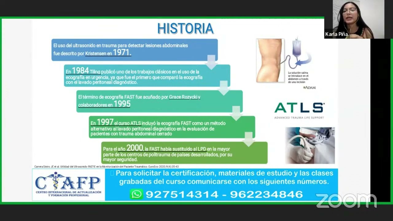
CURSO: ECOGRAFÍA FAST Y E-FAST EN EL PACIENTE POLITRAUMATIZADO
131
FAST requires a basic ultrasound machine with convex transducer (2.5-6 MHz) for abdominal evaluation, image recording system, ultrasound gel, and personal protective equipment. The protocol requires systematic evaluation of four 'P's windows: pericardial, perihepatic, perisplenic, and pelvic. For pericardial window, transducer is placed transversally at epigastrium directed toward cardiac apex. For perihepatic window, transducer is placed in right upper quadrant between 7th-9th intercostal spaces at midaxillary line, evaluating Space of Morrison. For perisplenic window, transducer is placed in left upper quadrant with spleen as acoustic window. For pelvic window, transducer is placed above pubic symphysis. In polytrauma context, free fluid is considered blood until proven otherwise. Minimum 200cc fluid is required for detection. E-FAST (Extended FAST) appeared in 2004, extending to thoracic region with 90% sensitivity and 99.7% specificity, superior to chest X-ray (requires 150cc vs 15cc). For E-FAST, transducer changes to linear (7.5-20 MHz). Anatomical landmarks include midclavicular line and axillary lines. Transducer is always placed in intercostal space between ribs.
Click to watch →
III JORNADA NACIONAL LAUEC URGÊNCIA E EMERGÊNCIA 1º DIA
18K
The FAST (Focused Assessment with Sonography for Trauma) protocol is performed during circulation assessment to detect free fluid. Four primary windows are used: subxiphoid (cardiac), right upper quadrant (Morison's pouch), left upper quadrant (splenorenal recess), and pelvic window. The probe marker faces cranially with patient supine. eFAST adds lung windows for pneumothorax detection. Interpretation: anechoic fluid around liver/spleen indicates positive FAST. Training requires 30-50 examinations. Anatomic variations (ascites, pregnancy, adhesions) affect interpretation. The protocol is operator-dependent with significant learning curve.
Click to watch →

Day-1 session 1
110
FAST (Focused Assessment with Sonography for Trauma) detects intra-abdominal, intrathoracic, or pericardial blood in trauma. Based on I-DEAL principle: Indication, Display, Evaluation, Action. Extended FAST (eFAST) includes lung examination for pneumothorax. Equipment: curvilinear probe with wide field of view. Positioning: head-low for fluid near diaphragm, head-high for pelvis. Views: right upper quadrant (hepatorenal recess highest sensitivity), left upper quadrant (splenorenal recess), pelvis (retrovesical/retrovaginal pouch), cardiac (pericardial fluid), and lung (lung sliding sign). Bladder injury prevents pelvis examination.
Click to watch →

eFAST : extended Focused Assessment with Sonography in Trauma
231.9K
The eFAST (Extended Focused Assessment with Sonography in Trauma) examination is a rapid ultrasound protocol used in emergency medicine to assess trauma patients by answering four critical questions: (1) Is there intraperitoneal free fluid? (2) Is there pericardial effusion? (3) Is there hemothorax or pneumothorax? (4) What is cardiac function? The exam consists of five views: right upper quadrant (Morrison's pouch), left upper quadrant (splenorenal recess), pelvis, cardiac, and bilateral lung assessment using lung sliding and M-mode for pneumothorax detection. The technique is indicated for all blunt or penetrating trauma patients, particularly hemodynamically unstable ones, as it guides treatment decisions for fluid resuscitation, chest tube placement, needle thoracostomy, or pericardiocentesis.
Click to watch →
Deep Dive
Introduction to transvaginal point-of-care ultrasound (POCUS) for advanced assessment of gynecological emergencies such as ovarian torsion or pelvic inflammatory disease.

Ultrasound Evaluation of Pelvic Emergencies - Radiology Review
22.7K
Ultrasound is the primary imaging modality for evaluating pelvic emergencies in females, starting with transabdominal examination using a full bladder as an acoustic window, followed by transvaginal ultrasound for detailed pelvic structure assessment; key conditions include ectopic pregnancy (6-16% of pregnancies, leading cause of first-trimester maternal death, diagnosed by absent intrauterine gestational sac with adnexal mass), ovarian torsion (characterized by enlarged (>5cm) edematous ovary with impaired blood flow, whirlpool sign, and follicular ring sign), hemorrhagic/ruptured ovarian cysts (showing lacy/spider web appearance progressing to fluid levels), and pelvic inflammatory disease/tubo-ovarian abscess; other considerations include malpositioned IUDs, ovarian vein thrombosis, and ovarian hyperstimulation syndrome, with important differentials including appendicitis and urolithiasis.
Click to watch →

Gynecologic Emergencies
3.4K
In gynecologic emergencies, asymmetric ovarian enlargement with displacement into the culdesac is the most reliable sonographic feature for diagnosing ovarian torsion, while Doppler flow assessment is unreliable for determining torsion status; pelvic inflammatory disease presents with indistinct ovarian boundaries, thickened fallopian tubes, and increased Doppler vascularity; hemorrhagic cysts show variable appearances from solid/hyperechoic acutely to cystic with concave margins over time, often mimicking neoplasms; and endometriomas appear as cystic structures with uniform low-level internal echoes and may cause complications like small bowel obstruction.
Click to watch →
1st Trimester OB US 2018 CUSE
140
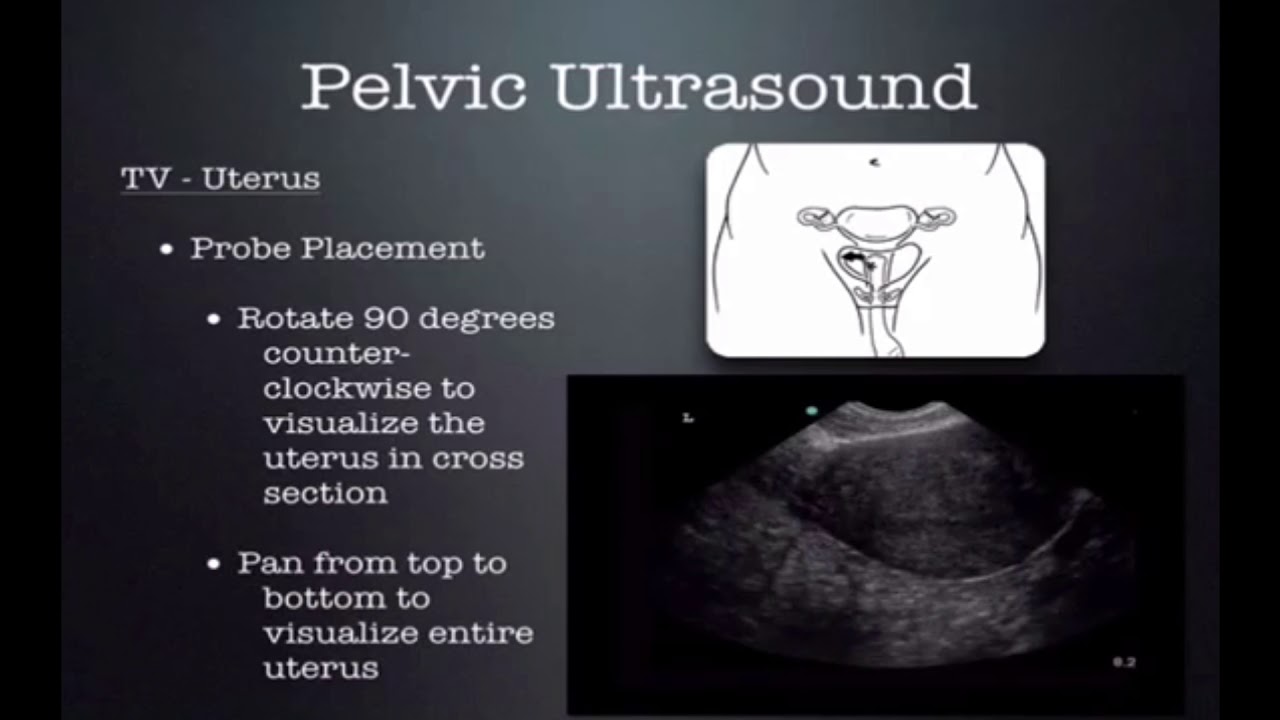
Point-of-care pelvic ultrasound is the most difficult POCUS examination to learn due to its invasive nature, unfamiliar probe, and intimate patient circumstances. However, it is one of the most commonly used examinations. Approximately 1-2% of US pregnancies are ectopic, accounting for 4% of pregnancy-related deaths. Mortality has declined from 1.15 to less than 0.5 deaths per 100,000 live births. Research by Metier & Company (2006) demonstrated that bedside transvaginal ultrasound improved early detection and decreased morbidity, with only 2.5% of ectopic patients sent home compared to 20% without ultrasound. Patient wait times decreased from 3 hours to 1 hour. Two approaches exist: transabdominal (non-invasive, less resolution) and transvaginal (detects findings up to a week earlier, detailed adnexal evaluation). Systematic technique: start with uterus in long axis, identify endometrial stripe, follow to cul-de-sac or cervix, turn 90 degrees for short axis, evaluate adnexa in both planes.
Click to watch →
Dr. Ada Gu: Advanced POCUS Applications
265
Point-of-care ultrasound (POCUS) is a critical emergency medicine tool for evaluating reproductive-age women with acute abdominal pain, particularly for detecting ectopic pregnancy, intrauterine pregnancy, and hemoperitoneum; the FAST scan is highly sensitive for detecting >100cc of intra-abdominal fluid, and serial POCUS should be performed every 30 minutes for unstable patients with changing clinical courses, as demonstrated in a case of ruptured ectopic pregnancy where bedside ultrasound findings guided urgent surgical intervention.
Click to watch →

Chương trình Hỏi đáp cùng chuyên gia cùng BS.CKI. Nguyễn Quang Trọng với chủ đề Siêu âm đau bụng cấp
3.7K
Three major gynecological emergencies affecting only women are pelvic inflammatory disease (PID), ectopic pregnancy, and ovarian torsion. PID shows characteristic 'cogwheel' ultrasound appearance of the fallopian tube. Ectopic pregnancy requires systematic evaluation including transvaginal ultrasound, beta HCG measurement, and clinical assessment. The beta HCG doubling time is crucial: ratio below 0.87 suggests abnormal pregnancy, 0.87-1.606 indicates ectopic pregnancy, and above 1.606 suggests normal intrauterine pregnancy. Ovarian torsion is a surgical emergency characterized by twisting of the ovary, with color Doppler showing absent or reduced blood flow. Proper probe selection is essential for effective ultrasound examination. For abdominal scanning, a convex probe (curved array) is typically used, with settings optimized for the specific organ system being evaluated. Morison's pouch (hepatorenal recess) is a critical area for detecting free fluid.
Click to watch →
FAST Pelvic Setup
Sagittal Bladder Scan
Uterus and Cul-de-sac
Sweep for Free Fluid
Transverse Probe Rotation
Inferior to Superior Sweep
Uterus and Ovary Check
FAST Pelvic Setup
0:15- 1
Begin FAST exam focused on female pelvic view.
- 2
Use phased array transducer with proper probe marking.
Diagnostic Limitations and False Positives in Female Pelvic FAST Exams
While the FAST (Focused Assessment with Sonography for Trauma) exam is a standard emergency protocol, critics and clinical studies highlight significant limitations in its application to the female pelvis. A primary concern is the high rate of false positives in reproductive-age females, who often have physiological free fluid (due to ovulation or menstruation) in the pouch of Douglas, which can be mistaken for traumatic hemoperitoneum. Conversely, the pelvic FAST exam has low sensitivity for detecting retroperitoneal bleeding, pelvic fractures, or specific uterine and ovarian injuries. Consequently, many trauma specialists argue against over-relying on pelvic ultrasound in stable patients, advocating instead for Computed Tomography (CT) as the diagnostic gold standard. They caution that a negative FAST exam can provide a false sense of security, potentially delaying critical interventions for internal injuries that ultrasound fails to detect.
Basic principles of medical ultrasound physics, including transducer selection, echogenicity (anechoic vs. hyperechoic), and image optimization controls like gain and depth.
Gross anatomy of the female pelvis, specifically identifying the urinary bladder, uterus, rectouterine pouch (Pouch of Douglas), and vesicouterine pouch.
The clinical indications and primary objectives of the Focused Assessment with Sonography for Trauma (FAST) protocol in emergency medicine.
Standard probe orientation and scanning planes (sagittal and transverse) used in transabdominal pelvic imaging.
Clinical differentiation between physiological pelvic fluid (e.g., normal menstrual cycle/ovulation) and pathological fluid (e.g., hemoperitoneum, ruptured ectopic pregnancy).
Integration of FAST pelvic ultrasound findings into clinical decision-making pathways for unstable trauma patients (e.g., immediate laparotomy vs. CT imaging).
Mastery of the remaining views of the Extended FAST (eFAST) protocol, including hepatic, splenic, cardiac, and thoracic windows.
Introduction to transvaginal point-of-care ultrasound (POCUS) for advanced assessment of gynecological emergencies such as ovarian torsion or pelvic inflammatory disease.
564.1K views966likes2:13@SonositeOriginal Release: 2011-06-01
- We're gonna do the FAST exam, the pelvic view in a female now.
Again we have the phase array transducer, here's our probe marker right here, I'm gonna start off with a sagittal view.
And ideally, the (slurred) bladder is full.
So you go just above the symphysis pubis, so we're scanning down here sagittaly, and some of the anatomic landmarks that we're looking at, at the very top of the screen is the bladder, which is an anechoic structure, fluid filled, obviously with urine, it's got nice walls around it.
Just below that is the uterus, which is a pear-shaped organ.
You can see it well visualized because the bladder is full.
And just below that is a potential space, the Cul de Sac, where we can look for, very carefully, small amounts of free fluid.
When we scan this patient, in this view, in the sagittal view, from the patient's left to the patient's right.
And we're slowly slowly scanning to see if there's any evidence of free fluid.
Once we finish the sagittal view, we want to go ahead and do a transverse view.
What we're gonna do now is point the transducer marker toward the patient's right.
So we're gonna rotate that transducer, toward the patient's right.
Again, we see the bladder in the center of the screen.
We're now gonna scan from inferior, just below the symphysis, to more superior, and in between we're also gonna identify the uterus.
So we're going right to the very top of the bladder, to about mid-bladder, we can see the uterus here in the center.
A very small amount of free fluid here, which can be physiological as well.
So we're scanning all the way inferiorly, to more superiorly, we see the uterus, we actually see part of the ovary there, and then we're gonna go all the way more superiorly.
Up Next
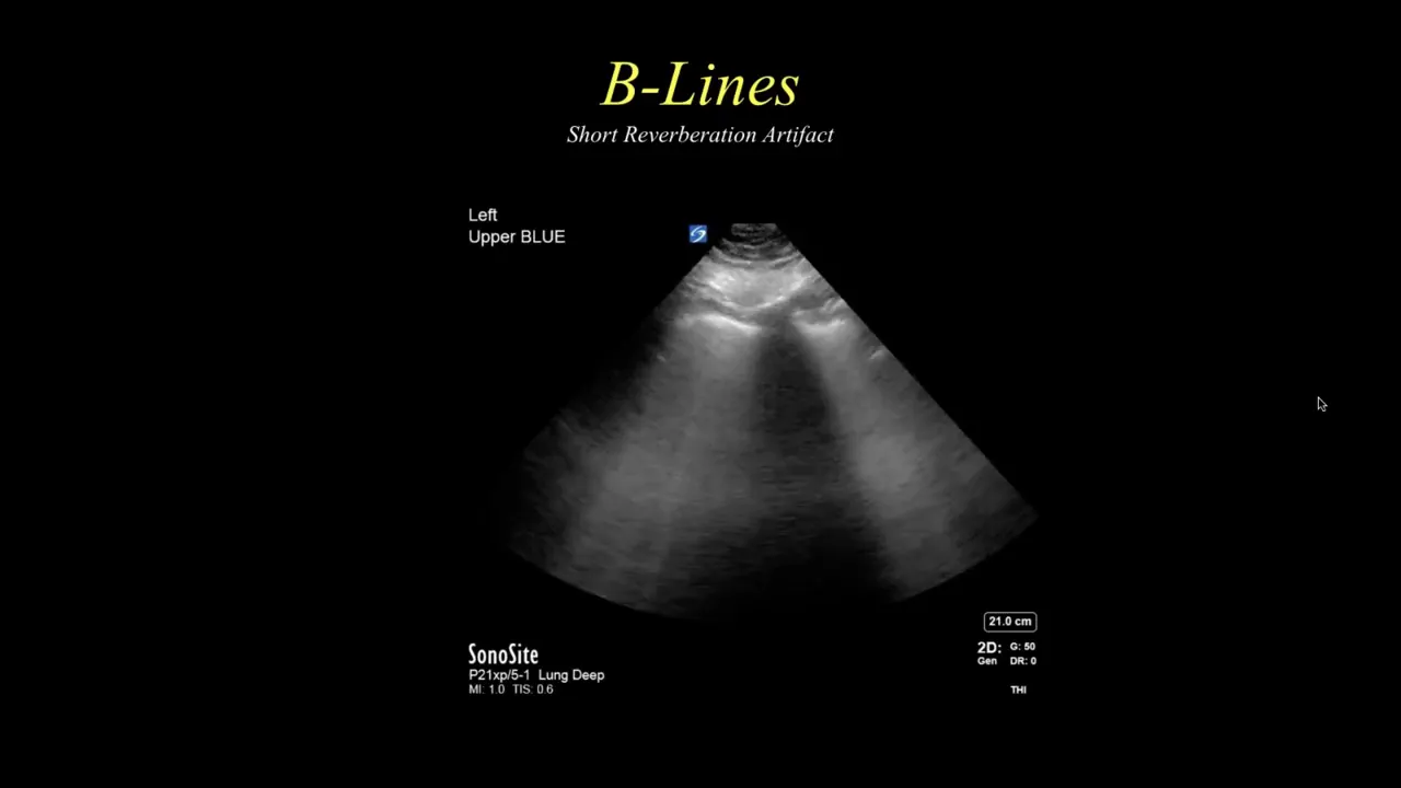
34:35
BLUE Protocol Bedside Lung Ultrasound in Emergency: A Practical Guide
@pocuscertificationacademy5053
26K views•2020-07-20

48:47
Graphic Medicine: Comics for Collaborative Healthcare Communication
@nationalpatientadvocate
189 views•2023-12-04

13:44
DVT Ultrasound Part 1: Focused Leg Vein Exam Anatomy & Technique
@Sonosite
258.4K views•2011-07-08

3:05
Stages of Labor and Vaginal Birth | Childbirth Animation
@nucleusmedicalmedia
52.1M views•2017-08-18
Related Study Plans & Knowledge Roadmaps
Structured learning paths in Medicine
medicine
Abnormal LFTs: Diagnostic Workup & Pathology
01Liver Anatomy and Basic Physiology
02Introduction to Liver Function Tests (LFTs)
03Hepatocellular vs. Cholestatic Patterns
+ 3 more learning modules
12h6 modules
Explore Roadmap
medicine
Analyzing Blood Smears: RBC, WBC & Platelets
01Introduction to Blood Components & Smear Preparation
02Normal Morphology of Blood Cells
03Erythrocyte Pathologies & Morphological Variants
+ 2 more learning modules
12h5 modules
Explore Roadmap
medicine
Mechanical Ventilation: Waveforms & Alarms
01Anatomy and Physiology of Breathing
02Introduction to Mechanical Ventilation & Core Modes
03Ventilator Settings and Waveform Interpretation
+ 2 more learning modules
12h5 modules
Explore Roadmap
medicine
Neuro Exam: Localization & Stroke Syndromes
01Foundations of Neuroanatomy and Vascular Supply
02The Neurological Examination: Step-by-Step
03Principles of Neurological Localization
+ 3 more learning modules
18h6 modules
Explore Roadmap