

















DROSHA is a key enzyme in microRNA biogenesis that processes primary microRNA precursors (pri-miRNAs) into functional microRNAs through a two-step cleavage mechanism; its crystal structure reveals remarkable similarity to DICER, another RNase III enzyme, but with a distinctive structural feature—a large bump near the branch junction—that limits its RNA binding capacity to shorter substrates compared to DICER's flat RNA-binding surface, providing mechanistic insights into how the microprocessor complex recognizes and processes pri-miRNAs.
Human DROSHA Structure & MicroRNA Biogenesis | RNA Scissors
Added:Understanding of the central dogma of molecular biology, specifically the distinction between coding (mRNA) and non-coding RNA (ncRNA).
Basic knowledge of RNA secondary structures, particularly stem-loop (hairpin) configurations and double-stranded RNA (dsRNA).
Familiarity with the concept of gene regulation and the general mechanism of RNA interference (RNAi).
Fundamental concepts of enzymatic cleavage, specifically the role of endonucleases and the RNase III family of enzymes.
The downstream processing of pre-miRNA in the cytoplasm by the DICER enzyme and its partner proteins.
The assembly and mechanism of the RNA-induced silencing complex (RISC) in targeting and silencing complementary mRNA transcripts.
The biomedical consequences of dysregulated microRNA biogenesis, including its link to oncogenesis, cardiovascular diseases, and genetic disorders.
Therapeutic strategies utilizing artificial microRNAs, miRNA mimics, and antagomirs (anti-miRs) for targeted gene silencing in medicine.
2.6K views47likes8:22Original Release: 2016-01-14
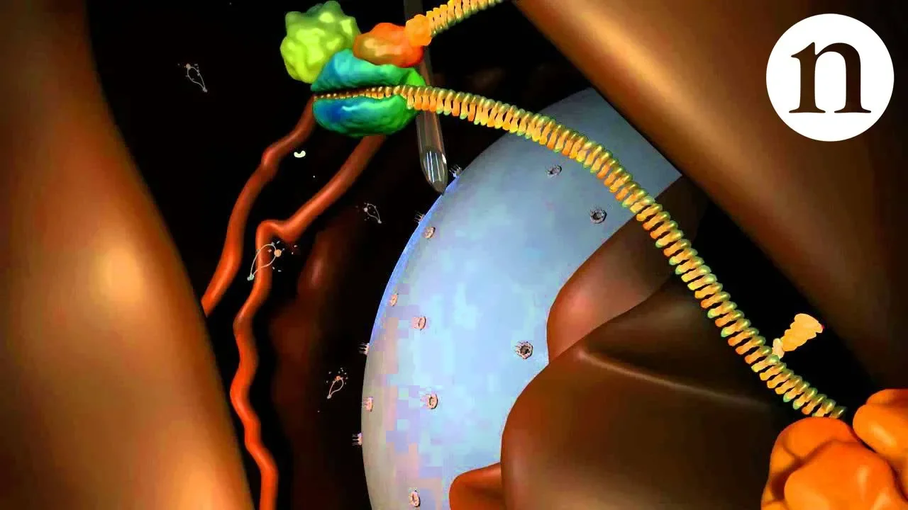
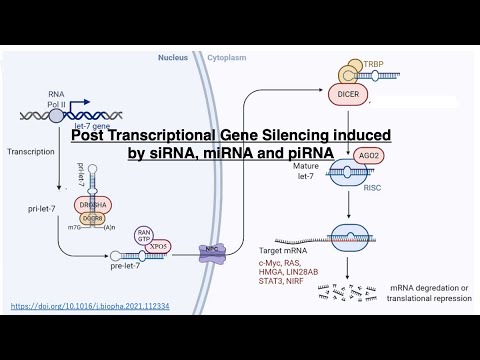
[Music] micro are small RNA of only 22 nuclear ties but they play big roles in gene regulation in animals and plants our group at IBS RNA Research Center and soul national university has been investigating how Micron is generated and regulated in the current study we serve the structure of droia a key biogenesis factor in the Micron pathway the structure together with the biochemical data explain how micro is are recognized and processed here's a primary train gri of micro also known as Prim micro which is transcribed from the micro gen by an enzymes called an polymer to a typical Prim takes a shape of stem Loof with three major parts the epical loops the stem and the single Tred tails on both sides later two parts together Define the Brier Junction because the micr sequence embedded in the stem region the prim Micon need to be processed by consecutive ACs of two other three types enzyme DOA and daa the initial processings Medi by DOA TOA makes a comp Lex with two molecules of ddci this complex called microprocessor microprocessors process a primary corate by cleaving the stem of primary corate at one helot turn away from the bjor junction the product microprocessor called pricon it then processed by daer another AR three enzyme ders measure two Hons from the end of the pricon and CLE UPS the Loof subsequently the R2 plus release form dier and loaded on acon 14 once loaded the an to presses and what train discarded what the other is maintained as M Micron this is how Micron is made in animal cells our group identified tra more than a decade ago however its detailed mechanism is still poorly understood due to the limited structural information here we serve the crystal structure of duia in complex with a small fragment of dcla this is the threedimensional model of DOA from the anus of the protein this part is the central domain and then two R3 domains are following and here is a double stranded RNA binding domain at the C terminal surprisingly the overall structure of Dua is very similar to that of dacer in dacer these are two R 3 domains and platform and P domains are at the bottom the connector Helix in red color is inside of platform similarly to daer the central domain of Dua it's also composed of three domains platform the parik domain and the connector the rebone diagrams show how throa and daer are folded the connector Helix inside of the platform is now shown very well platform and connector domains of two proteins are very well superposed next R diers are also superposed very well this structural similarity suggests that daa has diverged from an ancestor of daiser interestingly daa has more stoed posture compared with daer because of the conformational differences daer has a flat AR binding trap which can bind the long double TR RNA but josia has a big bump here to block the RNA binding so we can only accommodate a shorter R to understand the detailed interaction of duia with Prim mic RNA we built a model with RNA from previous studies we know that the first RNA is three domain of duia cause three prime strand of prime mic here and the second one cost five Prime strand here therefore this RNA should fit into the protein in this orientation not in the other way around this is the RNA bound model of Dua an interesting feature in Dua distinctive from other R3 enzymes is this long extended region from the first RNA 3 domain the reion is located near the basar junction and buried in the platform domain to constitute a big bump the distance between the bump and the cataly site of the firstage 3 domain is about 28om which is also the length of 11 base pair in contrast in the the da Model the double stand RNA can be extended up to the bottom of daer in our structure of DOA in complex with the c terminal tail of dd8 we found that doosa has two DDC binding site one on each RNA three domain one will be here and the other is here the interaction of the two ds8 with thosa are rather asymmetric now we will assemble the full microprocessor complex using the previous knowledge and the available partial structure of dc8 since the rscd domain ddcr 8 is known to interact with the epical element near the epical junction this should be located here then double Str RBD must be located between the rhd and C terminal tail and they are expected to inter with the upper stand of the prim micr RNA so here is the current model of the primary micr RNA binding by microprocessor microprocessor takes an elongated shape and covers the whole length of the primary micro RNA from the basil Junction to the epical junction this model is highly consistent with the previous result from the biochemical studies [Music] I've been investigating um [Music]
Up Next
7:58
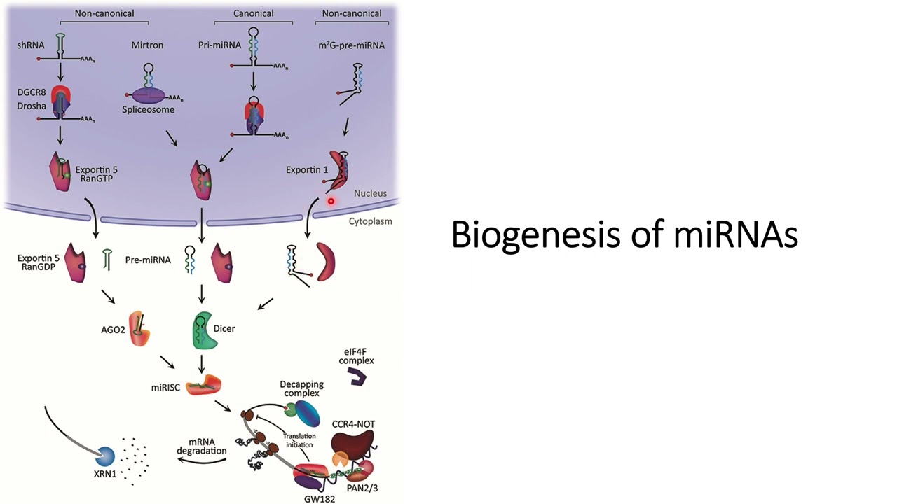
Biogenesis of miRNA: Canonical and Non-Canonical Pathways
@Informative_videos
246 views•2024-04-06

50:12
Bacterial Communication: Quorum Sensing and Biofilm Formation Explained
@JHUAAP
12.6K views•2011-06-28

5:01
Neurexin-Neuroligin Roles in Dendritogenesis
@cellvideoabstracts
3.4K views•2010-09-17

7:08
Bacteriophages: Earth's Deadliest Killers and Future Antibiotics
@kurzgesagt
34.6M views•2018-05-13
Related Study Plans & Knowledge Roadmaps
Structured learning paths in Biology
biology
RNAi: Biogenesis, RISC & Gene Silencing
01Molecular Biology Foundations: DNA to Protein
02Introduction to RNA Interference (RNAi)
03Molecular Biogenesis of miRNA and siRNA
+ 2 more learning modules
10h5 modules
Explore Roadmap
biology
Mycorrhizae: Signaling, Biophysics & Ecology
01Foundations of Mycorrhizal Symbiosis
02Molecular Signaling and Root Colonization
03Biophysics and Bioenergetics of Nutrient Exchange
+ 1 more learning modules
11h4 modules
Explore Roadmap
biology
Circadian Clocks: TTFL, SCN & Periphery
01Foundations of Molecular Biology & Gene Expression
02Introduction to Chronobiology and Actograms
03The Core TTFL: Molecular Architecture of the Clock
+ 3 more learning modules
14h6 modules
Explore Roadmap
biology
Population Genetics: Math, Drift & Selection
01Genetic & Probability Foundations
02The Hardy-Weinberg Principle and Baseline Models
03Mathematical Modeling of Natural Selection
+ 3 more learning modules
18h6 modules
Explore Roadmap