The Sliding Filament Theory explains that muscle contraction occurs when actin filaments slide inward toward the center of the sarcomere, shortening the muscle fiber without the filaments themselves changing length.
Sliding Filament Theory: Muscle Contraction Explained
Added:Deep Dive
The basic anatomical structure of skeletal muscle, including muscle fibers, myofibrils, and the functional unit known as the sarcomere.

Plus One Biology Public Exam | Chapters 11,13,19,7,17,12 | Exam Winner +1
79.7K
The sarcomere is the functional unit of skeletal muscle, bounded by Z-lines. It contains thin filaments (actin) and thick filaments (myosin). The I-band contains only thin filaments, the A-band contains only thick filaments, and the H-zone contains only thick filaments. The M-line is at the center of the sarcomere.
Click to watch →

Phoenix 2.0: Biology Most Important Video for NEET 2025 | Udaan
315.7K
Myofibrils are divided into repeating units called sarcomeres, which are the functional units of contraction. Each sarcomere contains: (1) Thin filaments (actin) - attached to Z-lines, (2) Thick filaments (myosin) - located in the center of the sarcomere, (3) H-zone - the central region containing only thick filaments, (4) I-band - the region containing only thin filaments. The sarcomere is bounded by Z-lines.
Click to watch →

LOCOMOTION & MOVEMENT in 1 Shot | NCERT Line by Line | Zoology Chapter 5 | NEET
109.6K
The sarcomere is the functional unit of contraction in skeletal muscle: (1) Sarcomere is the segment between two Z-lines. (2) It contains actin (thin) filaments and myosin (thick) filaments. (3) The arrangement of these filaments creates the characteristic bands: I-band (light, contains only actin), A-band (dark, contains myosin), and H-zone (central part of A-band, contains only myosin). (4) During contraction, the sarcomere shortens as actin filaments slide inward toward the center. (5) The Z-lines are pulled closer together, reducing the length of the sarcomere.
Click to watch →

Locomotion and Movement | Muscular System One Shot | L1 | NEET 2022/23 | Seep Pahuja
313.7K
The sarcomere is the basic contractile unit of skeletal muscle. It is bounded by Z-lines and contains thick (myosin) and thin (actin) filaments. The sarcomere shortens during muscle contraction as the thin filaments slide toward the center of the sarcomere.
Click to watch →

Plus One Zoology | Excretory Products And Their Elimination / Locomotion And Movement
72.4K
The sarcomere is the basic contractile unit of skeletal muscle. It is located between two Z lines (Z discs). The sarcomere contains thick filaments (myosin) and thin filaments (actin) that slide past each other during muscle contraction.
Click to watch →
Deep Dive
The identities and structural roles of the contractile proteins actin (thin filaments) and myosin (thick filaments).

LOCOMOTION AND MOVEMENT in 1 Shot - All Theories & PYQs Covered | PUC 1 | NEET
32.2K
The two contractile proteins are actin (thin filament) and myosin (thick filament). Actin exists as G-actin (monomeric) and F-actin (polymeric). Thin filaments contain F-actin, tropomyosin, and troponin. Myosin consists of six polypeptide chains (two heavy, four light) forming a tail (light meromyosin) and head region (heavy meromyosin). The head contains ATP binding and actin binding sites.
Click to watch →

Structure of Contractile Proteins | Structure Of Myosin And Actin | Locomotion And Movement | NEET
1.1K
Contractile proteins in skeletal muscle include myosin (thick filament) and actin (thin filament). Myosin is a polymerized protein formed by approximately 300 meromyosin monomers, each consisting of a globular head (heavy meromyosin) with ATPase activity and actin-binding sites, and a tail (light meromyosin). The globular heads project outward from the filament surface at regular intervals as cross-arms. Thin filaments consist of two helical actin filaments (F-actin) formed by polymerization of globular actin (G-actin) monomers, along with regulatory proteins tropomyosin and troponin. Tropomyosin wraps around actin filaments, while troponin (a complex of three subunits: TnT, TnI, and TnC) attaches to tropomyosin and regulates muscle contraction by controlling myosin binding sites.
Click to watch →
Human Physiology 4| NEET 2024 Preparation Biology By Dr Rishabh Choubey Sir | Bio Guru
65.2K
A sarcomere is the basic contractile unit of skeletal muscle, located between two Z-lines. It contains thick filaments (myosin) and thin filaments (actin). The arrangement of these filaments creates the characteristic striated appearance. Myosin filaments are bipolar with two heads containing ATPase activity and actin-binding sites. Actin filaments are composed of globular actin monomers that polymerize into fibrous actin. Each G-actin monomer has an active site for myosin binding. The sarcomere shortens during contraction as thin filaments slide past thick filaments, generating force.
Click to watch →

Cell & Tissue MCQs | PR Yadav Book | Anatomy & Physiology | Voogly Classes | NORCET | NCLEX
62.7K
Skeletal muscle contains contractile proteins: actin (thin filaments) and myosin (thick filaments). Tropomyosin is a regulatory protein that controls muscle contraction. Striated muscle shows dark bands (A bands, myosin) and light bands (I bands, actin). The sarcomere is the functional unit between two Z lines. The H zone contains only thick filaments.
Click to watch →
Locomotion & Movement | NEET 2026 | NCERT 360° | Zoology | LIVE @Aakash_NEET
2.8K
Muscle contraction depends on two contractile proteins: actin (thin filaments) and myosin (thick filaments). Actin filaments consist of two F-actins, each a polymer of globular G-actin monomers. F-actin formation requires magnesium ions. Actin filaments are regulated by tropomyosin (fibrous protein) and troponin (globular trimeric protein with three subunits: Troponin I inhibits actin-myosin interaction, Troponin C binds calcium, Troponin T binds tropomyosin). Myosin filaments are composed of meromyosin monomers containing heavy meromyosin (head and short arm) and light meromyosin (tail). The myosin head contains ATP-binding sites and an active ATPase enzyme.
Click to watch →
Deep Dive
The role of Adenosine Triphosphate (ATP) as the primary chemical energy source required for cellular work and conformational changes in proteins.
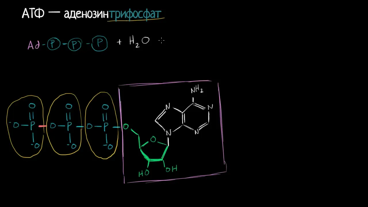
АТФ.Аденозинтрифосфат (видео 1)| Фотосинтез | Биология
33.5K
ATP (adenosine triphosphate) is the primary energy currency of cells, consisting of adenosine (adenine + ribose) and three phosphate groups connected by high-energy bonds; when these bonds break during hydrolysis, electrons transition to lower energy states, releasing energy that powers cellular processes like heating, protein conformation changes, and other biochemical reactions.
Click to watch →

DNA 🧬 Structure & Function - Nucleosides & Nucleotides - Biochemistry & Biology Series
174.8K
ATP (adenosine triphosphate) serves as the primary energy currency of the cell, storing high energy in its three phosphate groups. The energy derives from electrostatic repulsion between negatively charged phosphates—the more phosphates, the greater the repulsion and stored potential energy. ATP hydrolysis releases this energy through exothermic reactions, an exception to typical chemistry rules. When muscles contract, ATP breakdown powers movement via myosin heads. Approximately 25% of ATP energy powers actual work, while 75% generates heat essential for maintaining optimal enzyme temperatures. Creatine kinase rapidly regenerates ATP from ADP during brief exercise, providing immediate energy for anaerobic metabolism when oxygen delivery decreases.
Click to watch →
Dr. Benjamin Bikman - 'Ketones: The Metabolic Advantage'
374.1K
ATP (adenosine triphosphate) represents the cellular or chemical currency that cells use to perform work. Cells utilize ATP for various functions including muscle relaxation and contraction (crossbridge cycling at the microscopic level), and neurons use ATP to maintain proper electrolyte flux for conducting electrical impulses along axons. Essentially, ATP enables all cellular work and activity.
Click to watch →

احياء الخامس علمي 2024/ الفصل الثاني / المحاضرة 2 / التنفس الخلوي
85.7K
ATP (adenosine triphosphate) is the primary energy currency of cells, essential for all cellular activities including muscle contraction, nerve impulse transmission, and protein synthesis. ATP stores energy in its high-energy phosphate bonds. When ATP loses a phosphate group, it becomes ADP (adenosine diphosphate) and releases energy. Conversely, ADP can be converted back to ATP when it gains energy. This reversible conversion allows cells to store and release energy as needed. The breakdown of glucose molecules releases energy that is captured in ATP, making glucose the most important energy source for cellular metabolism.
Click to watch →

ATP the cell's energy currency | Smart syllabus | Chapter 7 | 9th class Biology |ALP |Lec 2
91K
ATP (Adenosine Triphosphate) is the primary energy currency of cells, functioning like money in human society. Cells require ATP to perform essential activities including movement, nerve impulse transmission, active transport, metabolic processes, cell growth and division, and macromolecule synthesis. ATP consists of three major components: a five-carbon sugar called ribose, a nitrogenous base called adenine, and three phosphate groups. When adenine combines with ribose, it forms adenosine (a nucleoside). Adding one phosphate group creates adenosine monophosphate (AMP), two phosphate groups create adenosine diphosphate (ADP), and three phosphate groups create adenosine triphosphate (ATP). The nitrogenous base adenine contains nitrogen atoms in its structure, making it a nitrogen-containing base. This molecular structure is fundamental to understanding how ATP stores and releases energy.
Click to watch →
Deep Dive
The general concept of the neuromuscular junction and how action potentials trigger the release of calcium ions from the sarcoplasmic reticulum.
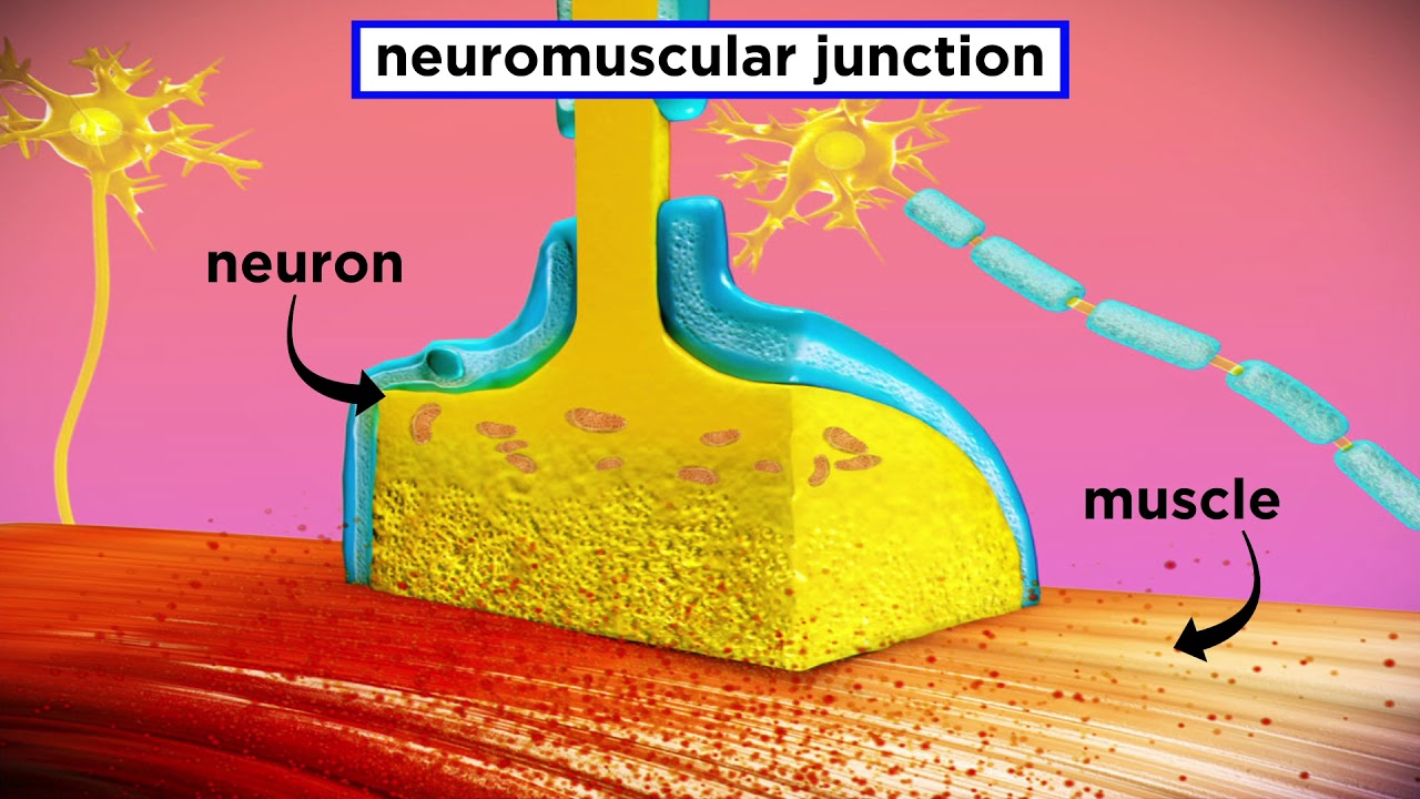
Muscle Contraction Mechanism: Sarcomeres & Neuromuscular Junction
2M
Muscle contraction occurs through the sliding filament model, where action potentials generated at the neuromuscular junction trigger calcium release from the sarcoplasmic reticulum; calcium binds to troponin, shifting tropomyosin to expose actin binding sites, allowing myosin heads to form cross bridges and pull thin filaments toward the sarcomere center, shortening the I bands and H zone while maintaining constant A band length.
Click to watch →
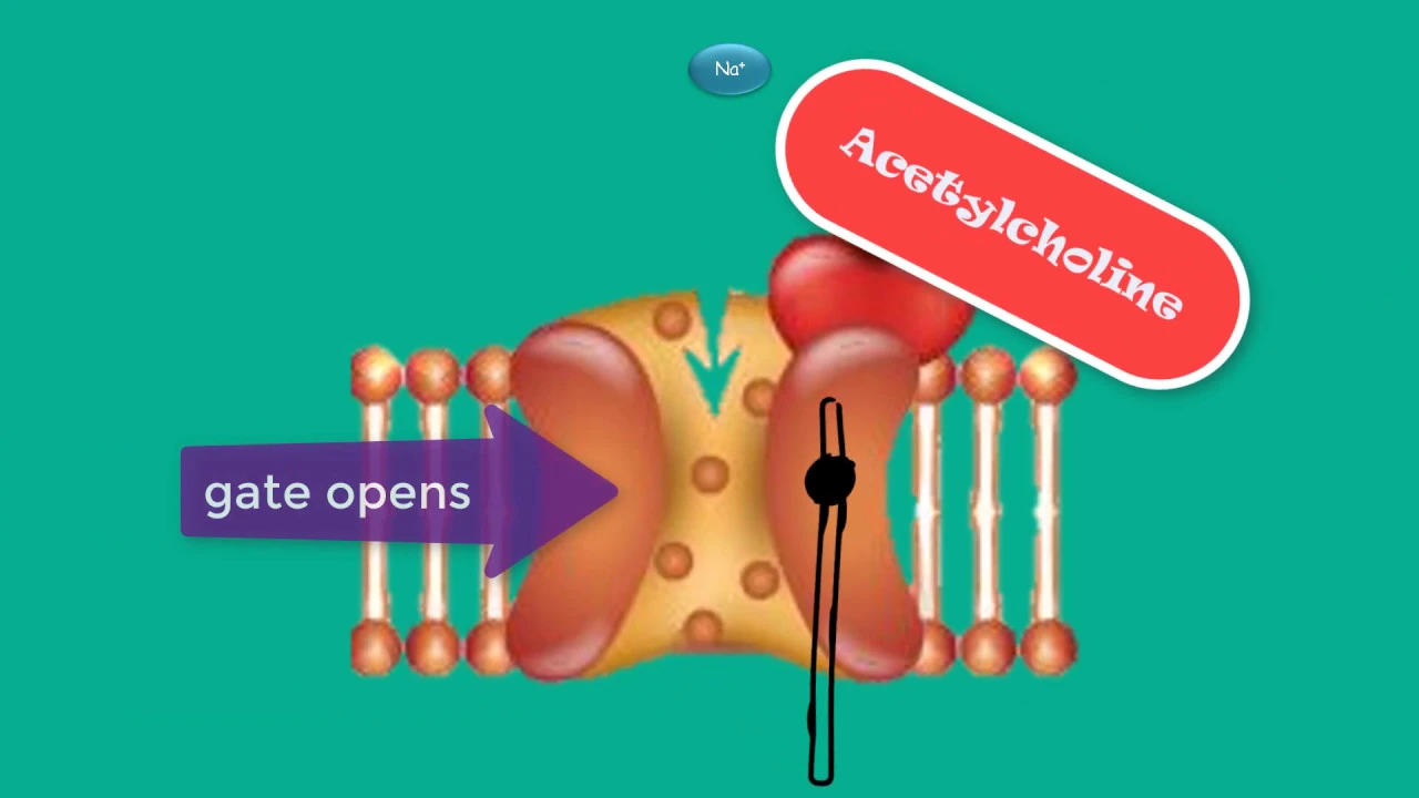
the neuromuscular junction Initiation of muscle contraction
51.5K
Muscle contraction begins when a brain signal travels through the spinal cord to a motor neuron at the neuromuscular junction, where action potentials carried by sodium ions trigger voltage-gated calcium channels; calcium release causes synaptic vesicles to fuse with the presynaptic membrane and release acetylcholine, which binds to nicotinic acetylcholine receptors on the muscle fiber, opening ligand-gated sodium channels that initiate depolarization and propagate an action potential across the sarcolemma and T-tubules, ultimately triggering voltage-gated calcium channels on the sarcoplasmic reticulum to release calcium ions that bind to troponin, causing tropomyosin to shift and expose myosin binding sites for the crossbridge cycle to begin.
Click to watch →

Locomotion and Movement I Biology Free lectures l Biomentors online
5.4K
The neuromuscular junction connects motor neurons to muscle fibers. Acetylcholine is released from motor neurons and binds to receptors on the sarcolemma, generating action potentials. These action potentials travel along the sarcolemma and into T-tubules, triggering calcium release from the sarcoplasmic reticulum.
Click to watch →

Locomotion and Movement Part 1| NCERT Lines + PYQs Solving | NEET 2025 & 2026 Biology | Gopika Ma'am
20.1K
The neuromuscular junction is where motor neurons meet muscle fibers. Motor neurons release acetylcholine, which binds to receptors on muscle membrane, triggering action potential. Action potential causes sodium influx through T-tubules, which leads to calcium release from sarcoplasmic reticulum. Calcium binds to troponin (specifically TPC subunit), causing conformational change that moves tropomyosin and exposes the myosin binding site on actin.
Click to watch →

Skeletal Muscle | Physiology of Muscle Contraction | Neuromuscular Junction | Structure | Ch 2 U 2
136.6K
The action potential causes the sarcoplasmic reticulum (a specialized endoplasmic reticulum in muscle cells) to release calcium ions into the sarcoplasm (cytoplasm of muscle cells). This calcium release is essential for initiating muscle contraction, as calcium ions bind to troponin to expose binding sites on actin.
Click to watch →
Prerequisite Knowledge
- Concept 01The basic anatomical structure of skeletal muscle, including muscle fibers, myofibrils, and the functional unit known as the sarcomere.
- Concept 02The identities and structural roles of the contractile proteins actin (thin filaments) and myosin (thick filaments).
- Concept 03The role of Adenosine Triphosphate (ATP) as the primary chemical energy source required for cellular work and conformational changes in proteins.
- Concept 04The general concept of the neuromuscular junction and how action potentials trigger the release of calcium ions from the sarcoplasmic reticulum.
Subsequent Learning
- Step 01Excitation-Contraction Coupling: The complete sequence of events from neural stimulation to the physical contraction of the muscle fiber.
- Step 02The biochemical explanation behind Rigor Mortis, specifically how the depletion of ATP prevents the detachment of myosin heads from actin.
- Step 03The mechanical classification of muscle contractions, including isotonic (concentric and eccentric) versus isometric contractions.
- Step 04The physiological causes of muscle fatigue and the metabolic pathways (such as creatine phosphate and anaerobic glycolysis) that replenish ATP during sustained activity.
- Step 05Pathophysiology of muscle disorders, such as Myasthenia Gravis or Duchenne Muscular Dystrophy, which impair the contraction process.
Opening
Transition
Wrap-up
Opening
0:30Playing Section
- 1
Initial segment begins with vague sounds and unclear context.
- 2
No substantive content or topic is established yet.
The Winding Filament Hypothesis
While the classic Sliding Filament Theory explains muscle contraction through the relative sliding of actin and myosin filaments powered by cross-bridge cycling, it struggles to account for certain phenomena, particularly the increased force generated during eccentric (lengthening) contractions, known as residual force enhancement. To address these limitations, the Winding Filament Hypothesis was proposed. This model introduces the giant structural protein titin as a dynamic, active participant rather than a passive spring. According to this theory, calcium influx during muscle activation binds to titin, increasing its stiffness, while the rotation of cross-bridges winds titin around the thin (actin) filaments. This winding action stores elastic energy and explains the high forces and efficiency observed during active muscle lengthening, offering a vital refinement and challenge to the traditional two-filament sliding model.
Deep Dive
Excitation-Contraction Coupling: The complete sequence of events from neural stimulation to the physical contraction of the muscle fiber.

Excitation Contraction Coupling | Skeletal Muscle Contraction | Cross Bridge Cycling | Myology
196.8K
Excitation-contraction coupling is the process by which an action potential triggers muscle contraction through a series of coordinated events: the action potential propagates through T-tubules, activating the dihydropyridine receptor which mechanically opens the ryanodine receptor on the sarcoplasmic reticulum, releasing calcium ions that bind to troponin C and shift tropomyosin aside to expose actin binding sites; myosin heads then undergo cross-bridge cycling using ATP hydrolysis to pull thin filaments inward (sliding filament model), shortening the sarcomere and causing muscle contraction; muscle relaxation occurs when calcium is pumped back into the sarcoplasmic reticulum by SERCA pumps, lowering intracellular calcium and stopping cross-bridge formation.
Click to watch →
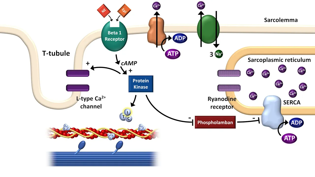
Cardiac Excitation-Contraction Coupling
167.1K
Cardiac excitation-contraction coupling is the process by which electrical activation of the cardiomyocyte cell membrane via an action potential triggers mechanical contraction through actin-myosin cross-bridge cycling; this occurs when calcium influx through L-type calcium channels in T-tubules during phase 2 of the action potential triggers calcium release from the sarcoplasmic reticulum via ryanodine receptors, which then binds to troponin C to shift tropomyosin and enable myosin heads to bind to actin, with relaxation occurring as calcium is pumped back into the sarcoplasmic reticulum by SERCA and regulated by the autonomic nervous system.
Click to watch →

Phoenix 2.0: Biology Most Important Video for NEET 2025 | Udaan
220.6K
A motor unit consists of a single motor neuron and all the muscle fibers it innervates. The neuromuscular junction is the site where the motor neuron communicates with the muscle fiber. Acetylcholine is released from the motor neuron and binds to receptors on the muscle fiber, triggering muscle contraction. The motor end plate is the specialized region of the muscle fiber membrane where the motor neuron synapses. Excitation-contraction coupling links the electrical signal to the mechanical event of muscle contraction. At rest, the muscle fiber membrane has a resting membrane potential. When acetylcholine binds to receptors at the motor end plate, it causes depolarization. If depolarization reaches threshold, an action potential is generated and propagates along the muscle membrane and into the T-tubule system. The T-tubule system conducts action potentials into the interior of the muscle fiber. Calcium released from the sarcoplasmic reticulum binds to troponin C, causing a conformational change that moves tropomyosin and exposes the myosin binding sites on actin. This allows myosin to bind and initiate the cross-bridge cycle.
Click to watch →

Fisiologia: Aula 11 - Músculo esquelético - Acoplamento Excitação Contração
88.2K
This lesson covers the fundamental mechanism by which skeletal muscle converts electrical signals into mechanical contraction. The instructor introduces the t-tubule system and sarcoplasmic reticulum as the two key structures that facilitate communication between excitation and contraction. The t-tubules are invaginations of the sarcolemma that penetrate deep into the muscle fiber, while the sarcoplasmic reticulum stores calcium. The dihydropyridine receptor (DHP receptor) in the t-tubule membrane is mechanically coupled to the ryanodine receptor on the sarcoplasmic reticulum, enabling calcium release when the muscle depolarizes.
Click to watch →
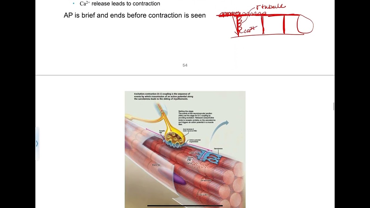
9.4 - Excitation-Contraction Coupling
161
Excitation-contraction coupling is the process that connects electrical signals to mechanical contraction in muscle cells. After an action potential propagates along the sarcolemma and down T-tubules (extensions of the plasma membrane), it triggers voltage-gated calcium channels in the sarcoplasmic reticulum to open. This releases calcium ions into the cytoplasm near myofibrils. The calcium then binds to troponin, causing tropomyosin to rotate and expose myosin binding sites on actin filaments, allowing crossbridge formation and initiating contraction.
Click to watch →
Deep Dive
The biochemical explanation behind Rigor Mortis, specifically how the depletion of ATP prevents the detachment of myosin heads from actin.

Biology Ch#16-Lecture#19 Sliding filament model (F.Sc 2nd Year)
37.8K
ATP hydrolysis powers the cross-bridge cycle by providing energy for myosin head repositioning and enabling cross-bridge detachment. After the power stroke, ATP binding causes myosin to detach from actin, allowing the cycle to repeat. Without ATP, cross-bridges remain locked to actin, preventing muscle relaxation. This explains rigor mortis, where post-mortem muscle stiffening occurs because ATP depletion prevents cross-bridge detachment. The phenomenon demonstrates that continuous ATP supply is essential for muscle relaxation and that muscle contraction requires ongoing energy.
Click to watch →

Sliding filament model | cross bridges control | controlling actin and myosin interaction by Ca++
96.1K
Muscle contraction proceeds through a cyclic molecular mechanism involving ATP hydrolysis, cross-bridge formation, power stroke, and detachment. Myosin heads attach to exposed actin binding sites, then hydrolyze ATP to ADP and phosphate, storing energy. Release of phosphate triggers the power stroke, where myosin pulls actin filaments toward the sarcomere center, shortening the muscle. New ATP binding causes cross-bridge detachment, returning myosin to its cocked position. This cycle repeats as long as calcium and ATP are available. Rigor mortis demonstrates ATP's essential role: after death, ATP depletion prevents cross-bridge detachment, permanently locking muscles in contraction. The all-or-none principle states individual fibers fully contract or not at all, with overall contraction strength depending on fiber activation numbers.
Click to watch →
Fisiologia da Contração Muscular - Aula 15 - Módulo VII: Histologia e Fisiologia Humana | Prof. Gui
133K
Rigor mortis occurs because after death, blood circulation stops, halting oxygen delivery to muscles. Without oxygen, cells cannot produce ATP through cellular respiration. Without ATP, myosin heads cannot detach from actin filaments, causing permanent cross-bridge binding that locks muscles in a contracted state. This rigidity persists for 24-36 hours until muscle proteins begin degrading through lysosomal release, causing the cadaver to become flaccid again.
Click to watch →

Locomotion and Movement | NEET | Muscle Fatigue, Oxygen Debt, Rigor Mortis | Neela Bakore Tutorials
47.6K
Rigor mortis results from complete ATP exhaustion after death. ATP is essential for both forming and breaking actin-myosin cross-bridges. Normally, ATP binding causes myosin heads to detach and reset. After death, ATP is rapidly depleted, preventing detachment. Myosin heads remain permanently attached to actin filaments in the contracted state, causing muscles to become rigid and maintain whatever posture existed at the moment of death.
Click to watch →
Rigor mortis | Postmortem changes in the muscle | mechanism of Actin & Myosin protein filament.
21.1K
Rigor mortis is a postmortem phenomenon where muscles become stiff after death due to the depletion of ATP, which normally allows myosin and actin filaments to separate during muscle contraction; immediately after death, muscles relax (primary relaxation), then rigor mortis develops approximately 1-2 hours later as ATP stores are exhausted, causing myosin heads to remain permanently bound to actin filaments, creating stiffness that progresses from eyelids and face to neck, trunk, and finally extremities before resolving into secondary relaxation.
Click to watch →
Deep Dive
The mechanical classification of muscle contractions, including isotonic (concentric and eccentric) versus isometric contractions.

The Hill model - isotonic and isometric contractions
3.2K
Muscle contractions are classified by tension-load relationships and length changes. Isometric contraction occurs when tension equals or is less than the load, with no muscle length change—sarcomeres shorten while stretching the series elastic element. Isotonic concentric contraction occurs when tension exceeds the load, causing muscle shortening and movement. Isotonic eccentric contraction occurs when tension is slightly less than the load, causing muscle lengthening while maintaining near-maximal tension. In all cases, cross-bridge cycling continues. Eccentric contractions cause greater muscle damage and soreness. Understanding these types is essential for comprehending how muscles perform different movement tasks—from holding positions (isometric) to lifting (concentric) and lowering (eccentric) loads.
Click to watch →

SOMA 16002 EC2
1.1K
There are two main types of muscle contractions: (1) Isotonic contractions - where the muscle changes length, including concentric (origin and insertion get closer together, accelerating movement) and eccentric (origin and insertion get further apart, decelerating movement); (2) Isometric contractions - where there is no movement occurring, the muscle contracts but does not change length. The sliding filament mechanism explains how muscle fibers contract and bind to shorten the muscle during concentric contractions, and unblock to lengthen during eccentric contractions.
Click to watch →

Physiology - Chapter 12 - Muscle
1K
Isotonic contractions involve muscle shortening against a load, where the sarcomere shortens during concentric contraction (bones come together) or lengthens during eccentric contraction (bones move apart). Isometric contractions involve tension development without length change, where elastic components (titin and nebulin) stretch while the sarcomere remains at constant length. Both are true contractions, but isotonic involves sarcomere shortening while isometric involves elastic component stretching. Antagonistic muscle pairs work together through these contraction types to produce coordinated movements.
Click to watch →

Muscle Physiology Revision for Medical Students | Last-Minute Test Prep!
213
Muscle contractions are classified by length change: isometric (tension develops but length remains constant, e.g., holding a heavy object) and isotonic (tension remains constant but length changes). Isotonic contractions include concentric (muscle shortens, e.g., lifting a weight) and eccentric (muscle lengthens, e.g., lowering a weight).
Click to watch →

CH9 - Muscles & Muscle Tissues - Part 3
905
Isotonic contractions involve muscle length change: concentric contractions shorten the muscle while generating force (e.g., biceps picking up a book), while eccentric contractions lengthen the muscle while generating force (e.g., lowering a book). Isometric contractions generate tension without changing length, occurring when the load exceeds maximum muscle tension (e.g., holding a plank). The mechanical events are the same in both types, but the results differ based on whether the muscle shortens, lengthens, or maintains length.
Click to watch →
Deep Dive
The physiological causes of muscle fatigue and the metabolic pathways (such as creatine phosphate and anaerobic glycolysis) that replenish ATP during sustained activity.

2BAC - BIOF : UNITE 1 CONSOMMATION DE LA MATIERE ORGANIQUE ET FLUX D'ENERGIE
71.8K
Muscle contraction requires continuous ATP supply from three pathways: (1) Creatine phosphate system - rapid but limited (depleted in <1 minute); (2) Anaerobic glycolysis - moderate speed, produces lactate; (3) Aerobic respiration - slow but sustainable. During exercise, systems activate in sequence: CP first, then anaerobic glycolysis, then aerobic respiration. Lactate accumulation from anaerobic glycolysis lowers muscle pH, causing fatigue. Prolonged intense exercise leads to insufficient ATP regeneration, resulting in muscle exhaustion.
Click to watch →

COURS DE TERMINALE SPÉCIALITÉ SVT CHAP.17: ATP et CONTRACTION MUSCULAIRE
641.5K
The creatine phosphate system provides rapid ATP regeneration for short bursts (10 seconds) through phosphate transfer between creatine phosphate and ADP. Creatine is synthesized in the pancreas and kidneys or obtained from dietary sources like meat and fish. For longer efforts (1-2 minutes), lactic acid fermentation takes over: glucose breaks down anaerobically through glycolysis (producing 2 ATP per glucose), and pyruvate converts to lactate while regenerating NAD+ for continued glycolysis. This pathway doesn't require oxygen or increased blood flow, making it suitable for moderate-duration efforts but producing limited ATP.
Click to watch →

HAP101 F2020 10_3 Muscular Tissue NARRATED
324
Muscle contraction requires ATP for myosin energization, detachment, and calcium sequestration. Pre-formed ATP powers contraction for only a few seconds. Creatine phosphate stores energy in phosphate bonds and regenerates ATP for about 15 seconds. Anaerobic glycolysis produces ATP for approximately two minutes. Aerobic cellular respiration in mitochondria generates ATP for minutes to hours through pyruvate metabolism, Krebs cycle, and electron transport chain. Muscle fatigue results from calcium depletion, energy reserve exhaustion, substrate depletion, lactic acid buildup, and motor neuron failure. Oxygen debt is the extra oxygen consumed after exercise to replenish creatine phosphate, convert lactic acid to pyruvate, and reload myoglobin. Muscle tone maintains firmness through alternating motor unit contractions. Isotonic contractions maintain constant tension while changing length (concentric shortening, eccentric lengthening), while isometric contractions increase tension without length change.
Click to watch →

Live Tutorial - Muscular System
18.4K
Muscle cells store limited ATP (~3-6 seconds). Creatine phosphate serves as an immediate energy buffer: excess ATP transfers phosphate to creatine during rest, storing energy. During activity, creatine phosphate donates phosphate back to ADP, rapidly regenerating ATP. For sustained activity, glycolysis breaks glucose anaerobically, producing pyruvate. When oxygen is insufficient, pyruvate converts to lactic acid. Both glycolysis and oxidative phosphorylation together produce ~36 ATP per glucose molecule, providing long-term energy for prolonged muscle activity.
Click to watch →

Lecture15 Muscle Physiology
252K
Aerobic metabolism (oxidative phosphorylation) in mitochondria produces the most ATP per glucose molecule and is the primary energy source during prolonged moderate activity. Muscles use glycogen stored within muscle cells as the initial fuel source, supplemented by glucose and fatty acids delivered by the bloodstream. Fatigue occurs through multiple mechanisms: depletion of ATP and creatine phosphate stores, accumulation of lactic acid lowering pH, and failure of the sodium-potassium pump due to ATP depletion. These factors reduce ion balance and impair muscle function.
Click to watch →
Deep Dive
Pathophysiology of muscle disorders, such as Myasthenia Gravis or Duchenne Muscular Dystrophy, which impair the contraction process.

Nervous System - Mysthenia Gravis (MG)| For NORCET | ESIC | RRB | DSSSB | RPSC | CHO Exams |Raju Sir
47.2K
In Myasthenia Gravis, muscle weakness occurs because the muscle cells do not receive sufficient acetylcholine. The deficiency results from genetic defects disrupting production, inefficient use, decreased receptor numbers, and receptor damage. Without adequate acetylcholine binding to receptors, the muscle cannot contract effectively, leading to progressive muscle weakness that worsens with activity and improves with rest.
Click to watch →

360 Degree Series | Most Imp. MCQ’s #865 | NORCET & All Nursing Exam Special | Siddharth Sir
16.1K
Myasthenia Gravis is an autoimmune disease characterized by: (1) Receptor changes in the neuromuscular junction; (2) Disruption of acetylcholine function; (3) Muscle weakness due to inadequate acetylcholine transmission. The disease typically occurs when the thymus gland fails to disappear after puberty (thymic hyperplasia), leading to autoimmune dysfunction that attacks acetylcholine receptors, preventing proper muscle contraction.
Click to watch →

Myasthenia Gravis || Nerve Muscle Physiology || hindi || Ashish Agrawal
65.9K
Myasthenia gravis is an autoimmune disorder characterized by muscle weakness that worsens with activity and improves with rest. The condition involves antibodies attacking acetylcholine receptors at the neuromuscular junction, causing a decrease in receptor number while increasing signal transmission speed. This leads to impaired neuromuscular transmission. The disease commonly affects extraocular muscles, facial muscles, and masticatory muscles, explaining symptoms like drooping eyelids and facial weakness. Respiratory muscles can also be involved, potentially causing respiratory failure.
Click to watch →

MSN, PEDIA, PHARMA | NORCET Series #761 | All Nursing Exams Special Class By Raju Sir
21.7K
In myasthenia gravis, the damaged acetylcholine receptor sites cannot properly bind acetylcholine. When 10 acetylcholine molecules are released, only 2-4 may bind to the damaged receptors instead of the normal 6. This insufficient binding results in inadequate muscle contraction, leading to muscle weakness. The disease causes progressive muscle fatigue and weakness that worsens with repeated use.
Click to watch →

Pharma-Once Daily | ANS Drugs | L2 | Pharmacology | 2021| Let's crack NEET PG | Dr. Priyanka Sachdev
317.9K
Myasthenia gravis is an autoimmune disorder characterized by the production of autoantibodies against nicotinic acetylcholine receptors at the neuromuscular junction. These antibodies destroy approximately 90% of the available receptors, leaving insufficient receptors for normal muscle contraction despite adequate acetylcholine release. Cholinergic drugs (specifically anticholinesterase inhibitors) treat this condition by inhibiting acetylcholinesterase, thereby increasing acetylcholine concentration at the neuromuscular junction and allowing remaining receptors to be utilized more effectively. This provides symptomatic relief rather than curing the underlying autoimmune process.
Click to watch →
Opening
Transition
Wrap-up
Opening
0:30- 1
Initial segment begins with vague sounds and unclear context.
- 2
No substantive content or topic is established yet.
The Winding Filament Hypothesis
While the classic Sliding Filament Theory explains muscle contraction through the relative sliding of actin and myosin filaments powered by cross-bridge cycling, it struggles to account for certain phenomena, particularly the increased force generated during eccentric (lengthening) contractions, known as residual force enhancement. To address these limitations, the Winding Filament Hypothesis was proposed. This model introduces the giant structural protein titin as a dynamic, active participant rather than a passive spring. According to this theory, calcium influx during muscle activation binds to titin, increasing its stiffness, while the rotation of cross-bridges winds titin around the thin (actin) filaments. This winding action stores elastic energy and explains the high forces and efficiency observed during active muscle lengthening, offering a vital refinement and challenge to the traditional two-filament sliding model.
The basic anatomical structure of skeletal muscle, including muscle fibers, myofibrils, and the functional unit known as the sarcomere.
The identities and structural roles of the contractile proteins actin (thin filaments) and myosin (thick filaments).
The role of Adenosine Triphosphate (ATP) as the primary chemical energy source required for cellular work and conformational changes in proteins.
The general concept of the neuromuscular junction and how action potentials trigger the release of calcium ions from the sarcoplasmic reticulum.
Excitation-Contraction Coupling: The complete sequence of events from neural stimulation to the physical contraction of the muscle fiber.
The biochemical explanation behind Rigor Mortis, specifically how the depletion of ATP prevents the detachment of myosin heads from actin.
The mechanical classification of muscle contractions, including isotonic (concentric and eccentric) versus isometric contractions.
The physiological causes of muscle fatigue and the metabolic pathways (such as creatine phosphate and anaerobic glycolysis) that replenish ATP during sustained activity.
Pathophysiology of muscle disorders, such as Myasthenia Gravis or Duchenne Muscular Dystrophy, which impair the contraction process.
27.1K views125likes1:46@adamcurry3514Original Release: 2015-01-21
a oh created using punon
Up Next

11:37
Cocoliztli Mystery: Salmonella Behind Aztec Epidemic?
@WeirdHistory
368.8K views•2022-03-09

50:12
Bacterial Communication: Quorum Sensing and Biofilm Formation Explained
@JHUAAP
12.6K views•2011-06-28

5:35
Enteric Nervous System Explained: The Gut's Brain | Neurobiology Lecture
@alumniu6029
438 views•2018-09-12

7:08
Bacteriophages: Earth's Deadliest Killers and Future Antibiotics
@kurzgesagt
34.6M views•2018-05-13
Related Study Plans & Knowledge Roadmaps
Structured learning paths in Biology
biology
RNAi: Biogenesis, RISC & Gene Silencing
01Molecular Biology Foundations: DNA to Protein
02Introduction to RNA Interference (RNAi)
03Molecular Biogenesis of miRNA and siRNA
+ 2 more learning modules
10h5 modules
Explore Roadmap
biology
Mycorrhizae: Signaling, Biophysics & Ecology
01Foundations of Mycorrhizal Symbiosis
02Molecular Signaling and Root Colonization
03Biophysics and Bioenergetics of Nutrient Exchange
+ 1 more learning modules
11h4 modules
Explore Roadmap
biology
Circadian Clocks: TTFL, SCN & Periphery
01Foundations of Molecular Biology & Gene Expression
02Introduction to Chronobiology and Actograms
03The Core TTFL: Molecular Architecture of the Clock
+ 3 more learning modules
14h6 modules
Explore Roadmap
biology
Population Genetics: Math, Drift & Selection
01Genetic & Probability Foundations
02The Hardy-Weinberg Principle and Baseline Models
03Mathematical Modeling of Natural Selection
+ 3 more learning modules
18h6 modules
Explore Roadmap