













![[SEMINAIRE] Imaging life at high spatiotemporal resolution - Eric Betzig](https://i.ytimg.com/vi_webp/FDqXarfZulM/maxresdefault.webp)









This lecture presents methods for imaging brain circuit development in vivo using transparent model organisms like Xenopus and zebrafish, which allow researchers to visualize and track individual neurons and their connections in real-time without invasive procedures, enabling studies of synaptic formation, neural plasticity, and circuit refinement processes that were previously impossible to observe.
Imaging Brain Circuit Development in Vivo | Ed Ruthazer | Frontiers in Neurophotonics
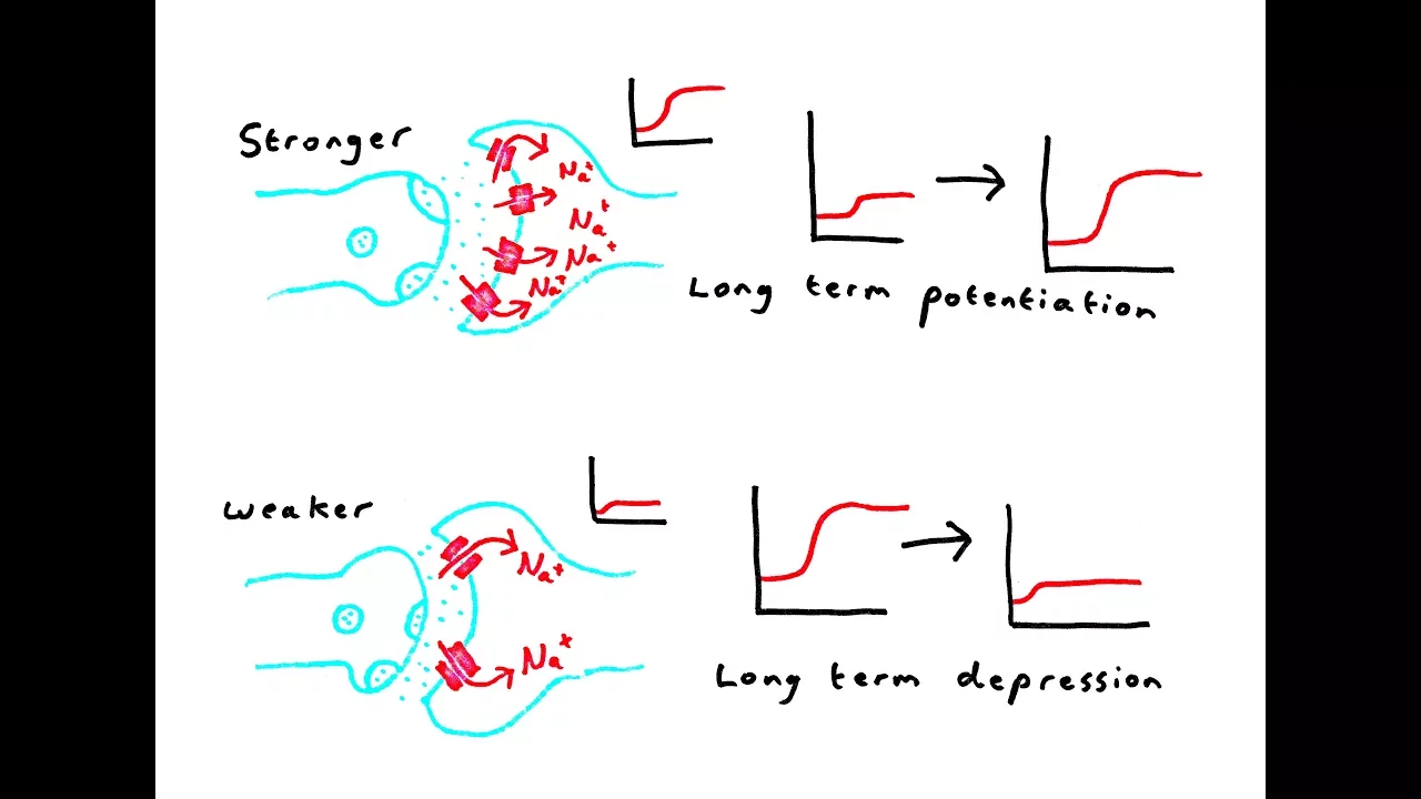
Added:Fundamental principles of synaptic plasticity, including Long-Term Potentiation (LTP) and Long-Term Depression (LTD).
Basic optical physics and fluorescence microscopy techniques, such as confocal and two-photon excitation microscopy.
The core concepts of optogenetics, specifically how light-activated proteins like channelrhodopsin are used to manipulate neuronal activity.
The biology and developmental advantages of using transparent model organisms, specifically Xenopus laevis tadpoles and zebrafish larvae.
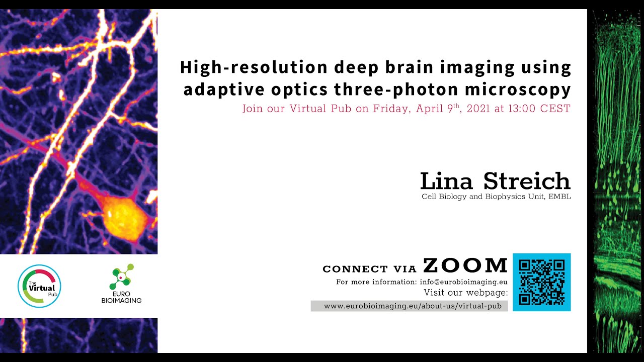
Advanced deep-tissue imaging modalities, such as three-photon microscopy and adaptive optics for in vivo mammalian brain imaging.
Translational research applying circuit development insights to understanding neurodevelopmental disorders like autism spectrum disorders and schizophrenia.
Computational neurobiology methods for reconstructing 3D connectomes and analyzing high-dimensional calcium imaging data.
The application of live-imaging assays in high-throughput pharmacological screening for neuroprotective and neuroregenerative drugs.
422 views2likes1:40:30Original Release: 2021-05-21
and has no um secret motives or any uh or uh um and he's he's always he's just the greatest so uh thanks ed for being here and um go do it okay yeah thanks paul um it's true i'm an open book but uh um what i'm gonna talk to you guys about today um is this is actually my kind of pitch for the kind of work that we're doing in the lab and it's increasingly um less necessary given the kind of work that i saw student presentations and from paul's lab are focusing on that you know this this move to uh doing a lot of in viva work in in zebrafish is really exciting i'm happy to see it taking off at uh at servo um and so i'm gonna what i'm going to tell you about is some of the methods and techniques that we've developed for doing um imaging in the brain in in mostly in xenopus but a little bit in zebrafish and i think the goal here is that it's a system which is really accessible anybody can set it up in their in their labs it's not actually very expensive um whether it's zipperfish or or xenopus there's different advantages to to the two systems and so you know i'm going to do a pitch for xenopus here you know catch you guys while you're still early on in your careers for people and a lot of you guys are engineers right and developing tools for optogenetics and and imaging and this is where i think our model is really uh beneficial because first of all it's inexpensive you know we don't have the kind of costs of maintenance that the mouse labs have but also it's really easy to take essentially you know to start with a plasmid and go all the way from plasmid to in vivo 3d light sheet image in a matter of days and so if you're somebody who's developing tools for imaging were you know either either on the optic side or on the genetic side um it's a really powerful uh test bed so i'm gonna bring you through a little bit of the history and what we've been doing and hopefully uh get you guys excited about it so can you all see just my my screen can you see my mouse yes good okay thanks guys and also please uh feel free to like chime in with questions at any point just interrupt speak up or i'll put the chat window up make sure that i can see the chat sorry it's covered over at the moment there we go okay all right so this is my home base it's mcgill university and i work up here at the montreal neurological institute so you just have to take a stroll down through the reddit gates extend all the way down to the micro neurological institute and you reach my lab what i'm going to start this talk with is just a little bit of history about imaging synapses in vivo and um and cells in vivo so this i think i always show this every year is what i believe to be the first example of in vivo imaging of a synapse and it was done in the 1980s by uh dale purvis and jeff lichtman this is this is from jeff lichman's study and it's you know considering uh that technologies like two foot on imaging didn't exist at the time or was was not in extensive use it's kind of impressive what what he was able to do he he screened for a large number of dyes fluorescent dyes that would actually stain synaptic elements so uh they ended up using a styro dye here that was apparently taken up by mitochondria and because mitochondria are so dense um at synaptic terminals when you um stain this is a peripheral muscle the sternomastoid muscle so right by the neck and if you uh stain this with the the styro dye uh what happens is it takes up the dye you can still image while it's alive and they're able to then capture images of a motor neuron end plate on on the muscle so it's not the central nervous system but it is a synapse and it's an important one for understanding synaptic development and you know the images that they were able to collect are they're not stunning but you can really make out um the sort of pretzel shape of the of the end plate and um they were one of the things i love in this paper is that they they give the dates in which they were able to image repeatedly at the same site and you know you can see that many months apart they are able to go back and find the same structure at that that synapse so you know this was sort of pioneering work allowed them to make a lot of conclusions about what happens at synapses during development but really this kind of work took off with the advent of genetic tools and in particular the use of gfp and gfp derivatives to label motor neurons and neurons in general so this is the same system now but in this case imaging yfp and cfp labeled you know stochastic uh expression of motor neurons and in this case you have two this is development and so what you have are two motor neurons terminating at the same end plate in the adult animal there's only a single motor neuron that takes over the entire end plate and so you can see this process of takeover in which one of the inputs actually gradually displaces the other one causing it to retract over time this is another example of that where the retraction bulb is very clear so this idea of synaptic um uh um you know this takeover notion and and competition at a synapse and the ability to actually image this in vivo and so you know one of the nice things also at the neuromuscular junction is that you can stain the postsynaptic side with low concentrations of fluorescent bungerotoxin which binds acetylcholine receptors and at low concentrations it doesn't block function very much but it still allows you to visualize and read the post-synaptic sides you've got the pre and post in the same structure um and lickman made an incredible uh career out of this kind of work now you'll notice that the scale of this structure is enormous compared to a typical central nervous system synapse right which is on the order of a fraction of a micron so when we want to study more complex motor units it's possible to do um imaging at the light level and make out you know a lot of the this these there's just a few uh motor neurons labeled and their projection to different terminals on the muscles themselves you can look at single axons or you can look at this group of axons terminating in a region and you know follow them over time during during development or in adulthood the um the challenge though is that if you want to study this process developmentally and what changes over time you really need to be able to reconstruct single axons so you know in this case you can sometimes pull out a single expressing motor unit but it's more useful if you can get multiple motor units in the same prep and so you know at this level of resolution it's really easy to do um segmentation of the images and digitally separate out the individual axons that project to the different motor units and generate these kinds of beautiful reconstructions of complex inputs to the to the periphery so you know this is the kind of work that allows us to come up with a wiring diagram but obviously in the peripheral nervous system it's much much easier because of the scale and the fact that the axons aren't so densely packed together now when we move to the central nervous system the problem becomes much more challenging right so you know this is a typical image of hippocampal cells um these are gfp expressing lines from gupping thung which are essentially the you know the standard um the community for imaging there are many different lines that have expression um that end up in different cell types but as you can see if you want to really understand the complex structure of something with the hippocampus it becomes this forest of of cells and you can you know find specific lines that have very sparse low density expression and you can use those to pull out morphological information um but really if we want to understand how this circuit functions we need to be able to get you know both high resolution and also single cell resolution images from from the prep and people have argued you know this this is an example of an image i think you guys in your lobby you have this beautiful spinal cord section from ramone um and you know all of his his drawings are really impressive in part because he was able to extract or or infer function and the direction of information flow uh just by looking at the anatomy and part of the way he was able to do that is to be able to visualize multiple cells in a structure so to get the circuit information but his understanding of the the fact that these were individual neurons functioning as individual units came from being able to see one cell at a time using the golgi stain and it's hard to sort of overstate the importance of the golgi stain for this advance so this is an image of you know what a golgi stained neuron looks like uh these are drawings from kahal and there's this really nice um text that kahal wrote in his in his histology where he kind of describes the the first impressions he had upon looking at a golgi stain material bear in mind that before that people were using stains that would reveal kind of gross structure of the of nervous system tissue but this ability to see single cells was a really important breakthrough so i'll read it out loud because i think it's really nice he said against a clear background stood stood black threadlets some slender and smooth some thick and thorny in a pattern punctuated by small dense dots all was sharp as a sketch with chinese ink on transparent japanese paper and to think that this was the same tissue which when stained with carmen or longwood left the eye in a tangled picket where sight may stare and grope ever fruitlessly baffled in his efforts to unravel confusion and lost forever in twilight doubt here on the contrary all was clear and could plain as a diagram a look was enough dumbfounded i could not take my eyes from the microscope and i think any of you who have done imaging know that you've had this experience of just seeing this extraordinarily beautiful and complex cell under the microscope and so for kahal seeing these you know in many cases for the first time um the passion and ideas started to flow so one of the reasons that kahal was able to to do this kind of work in addition to the existence of the of the golgi stain was that he had access to really high quality german optics in his microscope and so the sharpness of the images that he was able to collect was actually superior to what golgi was able to collect on his own microscopes and it may be one of the main reasons why kahal actually was able to come up with this neuron doctrine the idea that cell that the nervous system consists not of a mesh of of interconnected wires but rather of individual cells separated by synapses just that higher level of resolution so the things that you really want for biological imaging then is improve signal-to-noise ratio right that's the that's the name of the game for imaging and vivo no matter whether it's functional or structural and there's basically two sources of noise the biological noise that's the complexity of having millions of cells within this tiny chunk of brain tissue and you want to be able to study the structure of just one of those and then of course there's micros microscope noise which um essentially makes it uh difficult to you know it's the issue of having improved improved optics improved ability to to resolve um the structures that you're looking at and we've had a lot of of um important innovations in in the last couple of decades for biological noise this ability to get genetic expression in single cells and for microscope noise obviously two photon microscopy has been in light sheet microscope micro oh my gosh would be techniques that allow you to essentially extract the complexity of three dimensions and focus on single dimensions is a really important part but every improvement that we can make will allow us to understand more and more about the system so what i'm going to advocate for today is to kind of try to get the best of both worlds what i say here is that a true reductionist approach to studying the brain um brain development and function requires both cellular and circuit level analyses so we want to be able to see single cells but to really know what's going on i think we also want to look at them in the intact circuit so this image which i just randomly pulled off the web consists of just these beautiful dissociated neurons in culture but i'm wondering what are these neurons thinking about this is a great model for studying yeah exactly there's a great model for studying neurons and and the processes that occur at the single cell level but if you want to study and of course even contacts between cells but if you want to study the complex circuit you really need to go to a more complex structure the the in vivo system and so you know it's kind of like asking um here here's the spark plug um and this is a dissociated high performance automobile how how fast can this go so we really want to get back into the sports car and so approaches to to get us there have obviously involved the ability to pull out within the intact brain or within um original tissue uh the individual cells that and and pull them out um optically so the approach that many people um have have shown and have have you often see this in talks about the nervous system is was developed by jean-luc and again working with sainz and lickman this is the brainbow approach and the idea here is that you can get stochastic expression of a number of different fluorescent proteins in different ratios in cells so that each individual cell has its own kind of color profile right its spectral profile and then you can use spectral analysis to separate the individual cells now it turns out this is much harder to do well in practice than than in theory but it does generate these beautiful images of the the complexity of the central nervous system so here's the hippocampus that we were looking at before now shown in its full glory all of these cells in the dentate gyrus and what a forest this is so i like the term that jeff lickman uses which is if we want to study neurons in their natural setting we need to all become neuronal naturalists and this actually is a quote that i stole from stephen smith who was one of the first people to do in vivo photon microscopy in vivo veritas i think if we really want to know what's the um the function and the role of cells in the nervous system we need to go back to the individual prep but it's hard to do in vivo imaging right so um if you put a mice a mouse under a microscope you've got to control body temperature make sure that it's respiration is good anesthesia monitoring right all of these these issues if you're working with an animal that's that's anesthetized to get stable images you've got to make a cranial window um and there's a number of different approaches for that but it's all quite um open to um to complexity like infection inflammation things that are often overlooked the the consequences of of heating there's a limited ability to image really deep so you know it becomes increasingly invasive and expensive to image deep into the brain you may have to insert you know grin lenses or perform tissue aspiration or have you know very very um high-tech inexpensive approaches three-photon excitation and of course there's respiratory artifact and other and other movements and i appreciate that um at the servo and in this course you guys are probably learning all sorts of approaches to get around these problems so you know this is really fantastic and allows us to move these things forward and of course the ideal scenario is to do all this in a weak behaving animal and that makes things all the more complicated right but what i'm going to suggest is that there's at least for the early steps of this process there there is a better way and you know keep to yourself but um we can actually image in vivo uh without having to do any of this stuff above by taking advantage of the the xenopus and superfish models so you know i i'm kind of glad that i'm speaking today because um the person who follows me i guess is still tim murphy is that right so tim is the example of the opposite of this right he's the one who's figured out how to do all of this stuff in a really elegant way and so you know you'll hear the other side and he makes it look easy but um you know i'm going to sort of show you the the the dummies version so these are the aquatic species that we're using in the lab um they're very easy to rear they're inexpensive and as you can see their their bodies and brains are highly transparent so you know this is a xenopus tadpole during the stage when the retinotectal system which is the visual system that we study is really undergoing its its sort of greatest um uh development and segregation and so it's a really you know easy prep you can take the animal stick it right under the microscope um it's big enough that you know somebody with um you can you can you can see it quite easily yes it'll be recorded so um uh obviously zebrafish as well even more transparent than xenopus there are some subtle differences you can see that the size difference is pretty substantial the brain of the xenopus tadpole at this stage is about the size of the entire zipperfish head and so for things like electrophysiology um it's much easier to record from cells in the xenopus tectum obviously the genetics of zebrafish makes it more powerful for for genetic-based approaches so what i'm going to show you is a little bit of the work that we've been doing to try and you know identify single cell structure in the retinotectal system and then look at functional imaging so this this is basically the retinotectal projection of xenopus the system that we're that we're largely focusing on um we use as our main method for expressing green fluorescent protein we use electroporation and i'll show you why um it's so convenient shortly this is a stage 48 tadpole which as i said is that stage when there's an enormous amount of remodeling going on in the optic tectum and we can just you know inject plasmid into the eye electroporate and this is what you get so this is the optic nerve projecting into the tectum this is the structure here and these are the terminals of the axons from the eye okay this is a histological section from the optic tectum itself and you can see that it's laid out in this nice layered structure so you've got the neuropill region here where the synapses are formed and then a dense cell body region here and i think if you look here these are the the main components of the retinal axons which come in from the contralateral eye terminate on the dendrites of the tectal neurons and then there are these radioglial cells that are actually functioning as astrocytes in this population and they have these fine processes that extend and it's pretty straightforward to use electroporation to label these cells and so you get images like this so this is a two photon image of three retinal ganglion cell axons that happen to be labeled growing into the optic tectum and what's nice here is that we caught them at three different development stages within the same prep so this axon has a big growth cone at the tip and is growing towards its target this one has already reached its target and has extensively ramified but they're still highly dynamic at this point and so if you look i don't know how well you can see but you can see the axon the growth cone extending through the neural pill and then the fine processes of the axons extending and retracting branches oops this is a tactile postsynaptic neuron and you can see that they too are extraordinarily dynamic this is an image that i got from kurt haas's lab and what happens in the developing nervous system is that the axons grow into their target the dendrites actually extend and retract making occasional contact with the axons and when the dendrite contacts the axon uh often a synapse is initiated and that will result in at least transient stabilization of that contact and the entire structure of the retinotectal projection is built up through this kind of intermittent contact leading to either stable or dynamic interaction and then the glial cells these radial glial cells which have this nice brushy um appearance in the neuropill when we imaged those the first time we were quite surprised to see how dynamic they are so this is just a 30 minute loop and i apologize for the blue i don't know why that happens um a 30 minute loop showing that they're extending and retracting fine processes which we've shown have actually actually made contact with the synapses themselves so you've got this nice model where you've got the pre post and the glial component all easily accessible and imageable and then of course there's this fourth element which we've only started studying recently and that is the microglia so this is an example everybody's favorite kind of uh imaging project which is to induce a small lesion in the optic tectum or in the brain and then watch how the microglia actually pile on so this is a an mpeg gfp transgenic line but it's also been labeled using um a dye ib4 isolectin the red is the isolectin you can see that the isolectin doesn't give as good labeling of the microglia but it's perfectly useful when you have an you know you want to study microglial interactions with um with neurons and you don't have the transgenic line um available again in the video weird um okay um is there anything i wanted to say here no just to make the point that in might in zebrafish and in xenopus unlike in mammals the microglia are this wandering population and so when the lesion occurs like this ah i'm really sorry about that it does seem to produce this pylon where cells come in from quite distant locations and are attracted to the site so it doesn't take a large number of microglia to surveil the entire optic tectum structure in these animals it's probably on the order of two three dozen can i ask you a question yeah important temples i know it's just a single image but like a few slides ago that brain slice looked extremely similar to uh to zebra fish so i was just wondering like how similar are both brains this one right yes i think they're they're extremely similar the scale is a little different so one one neuronal cell soma here is on the order of 10 microns diameter in a zebrafish it's more like four or five microns and so that's one of the reasons that i still kind of rely on on xenopus is that it's quite easy to do whole cell uh voltage clamp recordings in these cells it's a bit harder to do that sort of thing in the small neurons of the zipperfish um but in fact in terms of the layout of the structure it's really similar the zebrafish tectum develops a laminar structure much earlier but by the time these guys are adults they'll have a nine layered optic tectum just like a mammal okay thanks for the question yeah okay so what are the techniques techniques that we can use to do single cell labeling obviously you know there's the standard genetic approaches where you might inject dna into the developing um embryo and then have stochastic expression in the animal but if you want to target your expression a really powerful technique and this works in zebrafish as well as in in xenopus but it also works extremely well in mammalian tissue is single cell electroporation this is a technique that was developed by kurt haas um back when he was working with with holly klein we were actually postdocs together and the basic approach you know it's it i my postdoctoral supervisor who similarly developed a a very similar approach said that this is the kind of thing that requires uh knowledge of molecular biology and electrophysiology not to not to execute it but to have come up with the idea and that is basically that you you set up as if you were going to record from the cell but then instead of recording you you ion to freeze and so the prep involves then bringing a micro pipet essentially a um a wholesale voltage clamp type glass micropipette filled with plasmid dna solution right up against a cell so in this case we can visualize the cells very easily as in as you would in a zipper fish as well um you go right up next to the cell that you're interested in you pass some pulses of current and there are different approaches that are used normally in the demo we show that you can use either high frequency 200 hertz pulses to get electroporation or you can use kind of one and a half millisecond pulse with a capacitor stuck in the path so in this case uh the capacitor is not not shown but we would stick a capacitor in parallel for the stimulator output and that produces this exponential um curve which is quite nice because you disrupt the membrane of the cell with the with the peak of the curve and then you continue to force dna into the cell in with the decline and what you end up with is you know roughly 12 to 24 hours later are nice single labeled cells if you do this improperly you end up with clusters of cells but you know just by changing your positioning or the shape of your pipette it's very easy to get this optimized and you know you have beautiful single cell resolution of golgi type quality and this is just an example of the approach that my postdoctoral supervisor developed in the lab when i was there and it's the same thing it's basically um single cell electroporation but in this case it's in a organotypic slice of cortex so the approach works perfectly well in cortical sections as well and you can do it in vivo but it's tough right because you you can't necessarily see the tip of your pipet and you need to have your pipet right up against the cell that you're electroporating for this to work basically the way this works is that when the cell is is away from when the tip of your pipet is not touching the cell the plasmid will just flow into the extracellular space and as soon as you get right up against the membrane it disrupts the membrane enough by passing current through it that you generate micropores within the plasma membrane that allows the plasmid to enter the cell and in fact that we think that the plasmid actually doesn't just enter the cell it has to enter the nucleus to get expression and so this is one of the reasons the sort of low efficiency of the whole procedure is why it's relatively straightforward to get single cell labeling so if you wanted to do this in you know in vivo you need to be able to position your pipette really carefully and so one approach that can be used is this shadow patching approach where you inject a fluorescent dye into the extracellular space and then using the contrast image you can bring your um electroporation pipette right up against a single cell shown in contrast here and electroporate either dye or or dna into that cell but that's you know it starts to become a little bit more complex right so there's another way to get um labeling using electroporation that doesn't give you single cells but it's really really efficient and easy and that's the bulk electroporation approach it's you know it's the thing that that probably most mouse labs would be using you can do it in the spinal cord you can do it in early development in utero and label cortex very easily or hippocampus here in the tadpole we'll use bulk electroporation just across the the ventricle of the plasmid of the tectum and you can see that you get unidirectional unidirectional delivery of plasmid into the cells on one side and you can see the axons projecting across to the contralateral side there's the occasional cell that gets labeled but for the most part um this is a nicely unidirectional approach so it's easy you inject plasmid you you know you've got lots and lots of cells labeled the next day so for electrophysiological approaches this is great you know you have lots of cells to record from if you wanted um and i think the same is true with uh in utero electroporation of course the time is this is 24 hours or 48 hours later um in mouse it will be a lot longer the negative though is that you've got you know a little bit lower co-transfection efficiency it's probably around 85 to 90 percent it's still pretty good but you can't assume that just because a cell has one one construct it has the other if you wanted to inject two constructs and of course this issue getting back to the you know getting single cells is really where it's at um that's problematic here so this is just an example of hippocampus that had been in utero electroporated and so these are just you know this contrast of the two images the whole brain electroporation and young animals gives really really dense labeling of not every cell but a large number of cells in the optic tectum this is a two photon step through the optic tectum from top to bottom and then this is the single cell electroporation technique on roughly the same scale so the question is can we take the benefits the advantages of this bulk electroporation approach and be able to get the strengths of single cell electroporation and so one idea we had was that well what if we just reduce the concentration of plasmid that we electroporate would that kind of reduce the frequency of electroporated cells to the point where we would have stochastic and and very sparse labeling and to some extent that works but it's problematic because as you lower the concentration of plasmid you also lower the brightness of expression of whatever it is that you're you're trying to express and so this is just an example where we go from you know 2 micrograms per microliter of dna down to 0.1 micrograms per microliter you can still get you know single cells and some labeling but it's much less expression and it's not great for that that high resolution you know the high signal to noise that we're really going for so in in the lab we developed this approach called we call crimsicle which is cremediated single cell labeling by electroporation and it's you know it's not brilliant uh it's basically just this idea that if you electroporate two plasmids there's some probability that um only one plasmid will get into the cell and so if you have a really high concentration of one plasmid and a really low concentration of the other the number of cells that have both plasmids is going to be very low and you'll get very sparse co-expression and in this case the two plasmids that we use are a gfp which has a stop codon introduced between two loxp sites so that essentially it doesn't express in the absence of creator companies and then the other plasmid is qui recombinase itself and when we express prerecombination we use very very low dilution so we basically have somewhere between uh 5 000 to 10 000 times more gfp plasmid than cream plasmid that we electroporate and the consequence of this is that you end up so as you reduce the concentration of cree that you're co-electroporating you end up getting this situation where you've got pretty much single cells another advantage to this is that this approach tends to label very immature neurons neurons that are often quite difficult to target with single cell electroporation because the cells that tend to get labeled by bulk electroporation are the ones closest to the ventricle and so those are usually the most immature so you know this approach works not only in you know in the optic tectum of of tadpoles it would work in fish i i'm pretty sure although we haven't tried it um but it works in mouse so many years ago mike crair at yale wanted to do some labeling of single cells in the retinocollicular projection and he was having this problem that he was getting you know large numbers of axons labeled every time he did electroporation um in the in the retina and so we suggested that he try this this crimsical approach and the consequence was that he was able to get really nice single cell labeling in the um in the retina and then be certain when he would reconstruct an arbor in the colliculus or in the lateral geniculate nucleus that it was really coming from a single cell in the eye so it does work in mammalian tissue the other approach that has been kind of a clever way to get single cells out of bulk electroporation or bulk genetic expression is to take advantage of optical highlighter proteins right and so you you've probably all seen this kind of approach there um there are many different optical highlighters the ones that we've looked at are photoactivatable gfp this is still really a very very useful and very very efficient highlighter protein and then their photo switchable proteins like kaede or kikjar which i guess maybe have the advantage that you can see the cell before you photo activate it but you just switch switch colors right and many times these kinds of proteins are used in um super resolution imaging so i think you guys probably have access to all kinds of really great optical switchable proteins so the approach that you know this is a paper um not from us but in zebrafish in which they used a kite expression expressing line and were able to show that you could basically at will identify cells for highlighting shine a 405 nanometer laser uh uv light onto the the cell of interest and basically convert it to red and so you know single cell morphology can be extracted in this approach um the problem with using 405 obviously is that you don't get the non-linear optic advantages of single single plane targeting which may be important for this kind of approach but you get the idea we've used photoactivatable gfp and we just co-expressed it so this is basically a retinal retinotectal projection we've electroporated m cherry and photoactival gfp into the retina and so this is the optic tectum you can see the axons from many many uh tactile neurons projecting into the optic tectum here this is a skin cell it's autofluorescence so just ignore it but basically this is what the image looks like before activation we then pick one site right here at the tip of one axonal terminal and we concentrated our photoactivation of photoactivatable gfp through the two-photon at that location and we were surprised we thought that we would probably get just local expression but the turns out that fluorescent proteins in neurons are they they diffuse really really rapidly throughout the cell i was quite shocked by how how rapid the diffusion is so this just a few minutes later results in an image like this where you can see the entire axonal arbor filled from this one location in the optic tectum which is pretty amazing and so from this approach you can see that it's possible to pull out a single cell and all its morphology from bulk electroporation so those two approaches are really quite powerful the the cremsicle method doesn't really allow you to pick which cell will be expressing this one obviously has the advantage that you can target individual cells for lighting them up and a technique developed in heroic buyer's lab which i think is just really very clever is the fujima approach which kind of combines functional imaging with this photoactivatable fluorescence labeling the idea is to express so this fujima construct basically is a gcamp 6 plasmid that is co-expressing photoactivatable gfp and so what happens is you can image evoked activity or whatever kind of of activity you're interested in studying in the um the cell bodies so it's a nuclear targeted gcamp so you pick the cell body of interest based on its activity profile so we want to study direction selective neurons so you find a cell that exhibits direction selectivity in the gcam signal and then you just photoactivate right over that that soma and you end up labeling filling the entire cell with photoactivatable gfp and you can do this over and over again and end up with a nice complex wiring diagram for functionally identified cells i really like this idea and then of course you know the ability to make transgenic animals makes um everything much easier and and um i think in xenopus there's this p transgenesis kit so you've got a wide spectrum of different uh constructs you can use cree um sorry um uh i think with crispr it's gotten even easier these days to make you know transgenic animals um and it doesn't matter the species so much right we're trying to make knock-ins now in the lab we've we've had some luck we think with um uh the nmda receptor so that's kind of cool um but i think you guys have seen i saw some of the students showing images from this paper from from heroic buyers group where it's possible to use basically um enhancer lines that express um basically uh cree or in this case it's um it's gal4 in a subset of the cells in the you know in the brain and then by taking advantage of variegated expressions so a bit of stochasticity in the in the expression of gfp it's possible to get animals that just have one or two fluorescent cells you can then take a kind of universal atlas a statistical atlas of the zebrafish brain and impose all of the different reconstructions into that atlas and i think what what buyer showed which i was kind of blown away by this is that you can you know by doing this approach almost uh painstakingly one by one you can reconstruct a very complex wiring diagram it's not it's not a complete diagram but it's the level of complexity and um uh sophistication that's possible to attain using methods like this is really quite amazing and i think this makes zipperfish quite a powerful model for for circuit bashing the other approach that's kind of a cool thing and i haven't seen it used very much and maybe that it's technically too challenging but i i really love the idea is if you just express um calcium indicators or activity reporters in cells in the brain in in theory there should be a much much higher degree of correlation in the activity of um or the calcium within a single cell than even in neighboring cells and so in principle by generating correlation maps of of images you should be able to pull out the structure that underlies that that um that particular cell and so um this is work in which this was done in in xenopus in the in the um olfactory system and you know they they expressed uh in this case it was a a different um calcium indicator than than we're using now but uh by looking at the calcium signal across an entire image of the olfactory bulb and then looking at correlations within the image they're able to basically determine that you know these are all inputs to this particular glomerulus and you can do this over and over again looking at the correlations of different locations and extract just by by the by by imaging over time the sites with the highest degree of correlation in the image and that gives you pretty good structural reconstruction so this is a video this is again the olfactory bulb in xenopus so we pick one site look at the calcium activity and then do a correlation map throughout the entire structure and you can get images like this and then you pick different glomeruli different sites and altogether you're able to identify different inputs to to identified structures you know these are these are still pretty small structures like 40 50 micron diameter structures in the brain so this i mean this is super cool it doesn't we've tried a lot to get this to work and it's not so easy to get high signal to noise but i think this this is worth investigating further um and of course the classic image that everybody it's like the mandatory image for a presentation on on this kind of uh method is the work from misha irons uh basically doing whole brain light sheet imaging of gcap and that's i think one of the real um the greatest advantages of a transparent model like this is that you really can do whole brain imaging at high temporal resolution and i really like some of the other approaches that i heard about the hi-lo approach for example that you guys are doing look sounds really cool so right so rapidly scanning through the brain and generating the nice three-dimensional reconstructions of activity so in our lab we're not doing light sheet but in part because the animals are you know we're interested in the visual system and so the light sheet we feel would introduce maybe some some uh artifact but we do do rapid focus you know high-speed 2-photon through the image of through the optic tectum in order to collect this kind of information it's not as fast but it's still pretty good and so you know the goal i think for people like me is to be able to watch the animal actually watching the stimulus right and so this kind of thing which just five years ago was was kind of a fantasy was a this was to me my dream experiment this is from uh uh muto at all from japan this is the group that developed g camp seven and uh but an earlier version of gcam7 and it's it's just quite amazing that you can actually make out in the optic tectum if i go back and play this again so this is a paramecium which is what superfish like to eat as you guys probably know and if you take a the g camp expressing animal this is the optic tectum here you can actually see the representation of that paramecium in the brain of the the animal in in real time it's super cool you're watching this animal watching the prey so that's kind of the goal for for my group to be able to um do that kind of thing um and use that methodology then not just to get pretty pictures but to understand um what are the mechanisms of map formation in the brain so there's one advantage in xenopus that is actually i don't think it's possible in zebrafish although we haven't tried i think there are reasons why it won't work in zipperfish but xenopus are you know they're externally fertilized just like zebrafish and just like nice video to show the process on the left it's the initial post fertilization image and you can see the the cleavage and then the division into into two cells and four cells and eats those um and then on the right are later stages of development of the embryo and all this occurs over a matter of days right so you can get to a pretty mature embryo that the embryos that i show you in um in the earlier slides were about a week a week and a half after this initial fertilization um so you know the subsequent process of going from a tadpole to a to a small froglet is kind of cool and there's there's a interesting xenopus unique issues here as the eyes actually move on the head you'll notice as the animal develops the visual system is actually has to keep up it has to basically remap itself in order to accommodate the fact that it's looking at a different part of the world and we can see remarkable changes in the anatomy that occur in parallel it's kind of cool to understand what's going on but what i wanted to show you actually was this cleavage issue so when the you fertilize we do a lot of in vitro fertilizations to generate tadpoles and at that first step of of cleavage you get two cells right two blastomeres and this is something that in xenopus the blastomeres are big they're physically separated and it's pretty easy to go in and micro inject dna or morpholinos or or mrna into one of the two blastomeres and they're not connected so you end up basically expressing or whatever you want in that one blastomere and all of its progeny all of its descendants and if you're lucky this doesn't happen 100 of the time but about 25 to 30 of the time you inject one side of the animal and you end up with a half animal expressing your gene of interest in just one half this is a whole tadpole i don't know if you can see it but you know the right half is completely not visible under fluorescence because it's not expressing um the the fluorescent protein the left half is where we injected the fluorescent protein and you can make out the even the brain is is like harlequin divided right in half which is pretty cool which is especially cool for us studying the visual system because the retinotectal projection is a crossed projection so the axons that originate on this side almost exclusively terminate on the contralateral side there's very rare exceptions and so that means that in one animal we can look at the presynaptic cells on one side and we can look at just the postsynaptic cells on the other side and ask questions not only about structure but also about function so gcamp injected like this gives you this is the retinal ganglion cell axons imaged on the contralateral side so that's the neuropill region and we're just flashing lights of different intensity different luminance into the the eye but it's kind of beautiful to be able to see all the retinal ganglion cells and pretty much no postsynaptic cells in this image and so we can extract information about topographic maps functional properties and this is where i would really like to be able to use that correlation based structural imaging if if there's any way to get that to work because you know there's the possibility of pulling out hundreds of individual rental ganglion cells from from an image like this this is the contralateral side and the ipsilateral side to the injected side so here you've got your tadpole this in this case this would be the side uh sorry this would be the side that was injected so we zoom into the optic tectum there's the cell body layer and one of the nice things about this mrna injection approach so we inject mrna against gcamp 6 and that every single cell on that half of the animal is expressing g-cap which means the glial cells are expressing it right the retinal axons from the other side are not but the tectal neurons are and so we can then use you know cell segregation methods to pull out both the individual neurons this is just spontaneous activity but also the end feet of the glial cells are really quite clearly visible in this case and so we've been taking advantage of that to look at neuron glia interactions to you know image calcium evoked by visual stimulation and see how it correlates with neuronal activity um and again you know this is where i wanted to kind of highlight if you're somebody who's making new fluorescent proteins so you know robert campbell's group is is making really cool uh new fluorescent proteins calcium probes and other kinds of probes what is possible is for us to get the plasmid converted into mrna that's you know a week of subcloning at the most to generate mrna inject into one of the two cells or even into both of the cells and you can then get basically an animal in which we're able to see all of the neurons in this case the olfactory bulb expressing this this was a near-infrared gecko 2 that robert's lab had generated and so we then took these animals to the light sheet microscope let's see if this will play we have to hit play and so this is this is light sheet imaging of this near infrared fluorescent protein it has an interesting property of being bright in the non-calcium bound state and then getting dark when it binds calcium which has some advantages it has disadvantages with respect to uh fluorescence uh bleaching over time but you know we were able to do all of this in under two weeks you know from from receiving a plasmid in the mail to capturing a 3d light sheet image which i think you know i would say is almost impossible to imagine doing in a mouse okay so um for my lab we're interested in studying the emergence and development of topographic maps so the approach that we've used is to take advantage of our ability then to express gcamp in these different compartments present visual stimuli to the animals on a video monitor while the animals sitting under the two-photon microscope embedded in agarose and you know we can either do kind of reverse correlation type stimuli where we flash a bar at different locations in the visual field or more often what we do is we sweep bars i don't know how well the videos come through on zoom but it's just a bar sweeping across the visual field and this is an approach that's actually used for mapping um you know it's been used for a long time in fmri and in intrinsic signal imaging in that if we present the stimulus at a regular duty cycle you know if the bar sweeps across the field once every 10 seconds then if we pull out the 0.1 hertz response within the within the tectum so i guess i have some images this is what it looks like as it sweeps so this is sped up quite a bit but we've got in one case the stimulus sweeping from front to back and in the other case the stimulus sweeping from back to front and i think you can make out the kind of movement of response across the optic tectum and then we just do the for each voxel we do a fourier power spectrum we pull out just the signal at the frequency of the visual stimulus itself right of the sweeping bar and that essentially is a great way of controlling the you know raising the signal to noise so we're getting rid of any sort of irrelevant responses and then by looking at the phase at that frequency it tells us you know where in the in the cycle was the stimulus this and that corresponds to then the position in the visual field so right as the bar sweeps across the field there's gonna i don't know why it does that there's gonna be early elements um that have an early phase and then later phase corresponds to things that are at the other end of the sweeping bar and we can generate then maps like this these are topographic maps at the subcellular level of the visual system in you know in the optic texture so this is a map for the postsynaptic cells this is a map for the presynaptic inputs using these approaches and i think what's nice is that not only do we have the ability now to see maps at stages where when people were using you know extracellular electrophysiological recordings in the old days they would have claimed that there was no map simply because this the structure is so small right i mean this is a probably 150 microns in this axis and maybe 400 microns along this axis and so the seeing distance of a typical extracellular recording electrode doesn't give you the resolution you need but with optical approaches we can see right quite nicely and so we can do elevation maps and azimuth maps within the brain look at how different manipulations might alter the formation of the map one of the things that was a bit surprising to us was that if you look at these two maps so this is an as an azimuth map an elevation map it turns out that the animals don't see very much over the top of their heads but they do tend to see a lot more below the orientation of these maps is not orthogonal i sort of was expecting that they would be perfectly orthogonal to one another but it we found that if we image the same animal over time i don't have this this data to show you but what happens is that the maps actually become increasingly orthogonal over time as the map develops so that may be one of the properties that emerges with development so what i showed you before was a single optical section but if we do optical sections through the entire tectum you know rapidly while presenting these stimuli we can generate not just a single optical section map but the map in 3d throughout the entire neuropill of the tectum and that gives a lot more information i think this is one of the first times that a three-dimensional um topographic map has been studied from the initial development of that projection in fact we're able to follow these maps in these animals from almost the day the axons arrive in the optic tectum until much much later when a complex map is formed so there's a lot of of power now for understanding the formation of maps this is the elevation map in that same animal so i don't know i mean probably this is for me this this is hard hard one data i've been wanting to to get this information for years and years and it was really only gcamp uh actually gcamp6 made this possible i think that um as these indicators improve it just becomes almost trivial to get this kind of data that we struggled to get in the old days so they're the two sort of almost orthogonal axes of azimuth and elevation represented in 3d so i'm really happy that we were able to get this information and now we can go in and do manipulations right genetic manipulations or even activity manipulations so the first thing that we would do is to ask um what happens if you block in mda receptors right and we know that nmda receptor blockade has a pretty dramatic effect on the retinotectal projection in terms of single cells so even the dynamics of axons when you wash on nmda receptor blockers you see an actual increase in the rates of branch addition and branch retractions over a very short period of time so these are images from holly klein collected many years ago um and what she showed is that when you you know apply apv you get an increase in the rates of additions and retractions um in the arbors suggesting that nmda receptors are important for stabilizing the projection and the blocking nmda receptors lead to this increased dynamism um and we thought that if we blocked nmda receptors the map would be totally messed up and we were quite shocked that what we found was that the maps were almost as good in nmda receptor blocked animals as in as in wild type so this is just single optical sections from a control animal and an mk to one reared animal this animal was reared in mk21 from the you know earliest innervation of the retinal axons this is the postsynaptic projection this is the presynaptic projection and you can see that there are actually pretty good maps in there already this is the azimuth map this is the elevation map really they're not that different um we can quantify this the way we quantified it was by looking at a measure which we call local discontinuity and that is essentially how grainy is the is the map that's generated in the neural pill so for any one voxel if you look at the surrounding immediate vicinity it should if in a really smooth map there should be very little difference between the um preferred position in in space that drives the you know the receptive field center that drives that voxel and all of its neighbors whereas in a more granular less refined projection you might expect to find that there's a high higher difference between that central voxel and its neighbors and what we found was that it is true mk21 rearing actually significantly increases the amount of granularity the discontinuity both in the postsynaptic side and in the presynaptic side and it's true for azimuth and it's also true for elevation so nmda receptors are important for refining the map but i think what's more striking here is that the difference between pre and post-synaptic maps is much much greater than the difference between you know nmda treated and not and so one of the reasons that the presynaptic map is more granular than the postsynaptic map almost certainly is that the postsynaptic cell is integrating across many inputs right and selecting um appropriate inputs and so it's more of a it's a higher level right it's a it's the refined um processed version of the raw inputs coming from the axon so that makes sense that it should be less discontinuous but the fact that there isn't when we do statistics on this there's no interaction between pre-post versus mk21 treated and control which which indicates that this difference between pre and post is not due to nmda receptor function it's probably due to some you know other processing going on in the dendrites and in the cells so that was a bit of a surprise after so many years of single cell imaging and being convinced that nmda receptors were the thing that refined the map it turns out that you know they have a role but it's much smaller than we initially thought um so what are the nmda receptors doing given that they have this this role in in sharpening the map how are they doing it so you know one approach is to just look at single cells and and image like i showed you uh dynamics or structure and ask what blocking nmda receptors does to that i think the problem with that approach is that we know that these mechanisms are highly activity dependent and nmda receptors of course are glutamate receptors so if you block nmda receptors you're kind of screwing up patterned activity within the network maybe not you know not as badly as as would happen if you block the ampa receptors but it's not a clean experiment because you're not really um just getting rid of nmda you're getting rid of activity at some level so in order to understand this a little bit better what nmda what is the contribution of nmda receptors we took advantage of a gain of function rather than a loss of function approach so in these experiments so basically nmda receptors consist you know of four subunits typically n1 and glue n2 or here nr1 and r2 and our two subunits are the subunits that bind glutamate it's the gluon1 subunit the nr1 subunit that actually binds the coagonist glycine or d-serine and so we reasoned that if we provided an excess amount of d-serine in the system that we would be able to enhance nmda receptor activation so rather than blocking it we would be able to really enhance it but the dependence on released glutamate would still be there and so we wouldn't be creating abnormal patterns of activity per se we would just be enhancing whatever activity is present and the amount of calcium that enters into cells during um glutamate binding so we tried rearing animals in um in glycine in sorry in d-serine we could have done glycine but the problem with glycine obviously is that it's a it's an inhibitory neurotransmitter as well and so it would have given a bit of a confound confounding result d-serine doesn't really have a lot of other targets it may have a few but much much fewer and so what you can do then is you record from a cell in the optic tectum this is an evoked nmda receptor response so you'll notice that this is in the absence of magnesium so that's why we can get an inward current when we stimulate so we stimulate the optic nerve we record from a technical neuron and just ask you know what is the nmda component of that response this is with ampa receptors blocked and if we wash on d-serine we find a pretty substantial enhancement of that of that response so this tells us two things it tells us that desiring is effective at enhancing the currents through nmda receptors but also it tells us that in a normal animal the nmda receptors don't have saturating levels of dieserine present or of glycine present and so by adding more we can actually truly enhance an mda receptor function this is just to show that the d serine wash on which causes about a 200 percent increase in the size of the currents can be washed off pretty pretty much and so we reared animals for two days in uh indeed syrian just putting it in the bath and the consequence of that is that we get an accelerated level of synaptic maturation so the way we measure synapse maturation in this system is by looking at the ampa nmda ratio and mini frequencies so if a synapse starts off having just nmda receptors right then what we believe happens is that in response to patterned activation that drives repeatedly drives nmda receptor activation it causes the trafficking of ampa receptors to the surface and the two consequences of that are that you'll have a higher mini frequency as you unsilence many synapses and you may also have an increased amplitude although not necessarily if the main effect is is unsilencing silent synapses that were initially nmda only and then the other effect is of course the nmda ampa ratios will change so this is just the mini data so spontaneous activity recording from tectal cells and we see a small but non-significant increase in d-serine reared animals in the amplitude but a very large increase in the frequency indicating that silent synapses had become matured and unsilenced and these are the ampa nmda ratios so you see that for any given set of inputs the relative contribution of ampa increases relative to nmda in d syrian reared animals indicating that the d-serine treatment resulted in ampa receptor trafficking to synapses so we're driving maturation of synapses by allowing nmda receptors to be enhanced what are the morphological consequences so for postsynaptic cells if we just look at the structure of those cells over over four days what we find is that d-serine rearing causes the cells to grow much more slowly so they tend to be more you know more stable and less exploratory and that's you know these are just two examples of cells over four days with the d serine added after day zero and you can see that on average the the length of the arbor and the number of branches increases over time and the shoal analysis really clearly shows that the most distal branching processes um is is not taking place in the in the d syrian rear case suggesting that the inputs onto that cell are essentially you know as the cell is growing it's sort of telling itself okay i've got enough input i don't have to grow any more i don't have to search for more inputs perhaps and that's just one interpretation presynaptically we see the predicted change in dynamics so the opposite of what is seen with epv treatment when we look at d serine treated animals we see a substantial decrease in the number of branches added and the number of branches lost over a one hour time lapse imaging period so again that suggests that there's a higher degree of stabilization we think that this is likely due to a retrograde signal that's generated from the postsynaptic cell and feeding back to the presynaptic cell i don't have the data to show you that but we've done knockdowns using this half animal approach of nmda receptors just in the postsynaptic cells and when you do that um these effects actually are lost so you the d-serine treatment only affects morphology of axons if the postsynaptic cells express nmda receptors so what is the effect on morphology over time over days um normally a cell goes from having a very simple arbor to you know this denser more complex arbor over over this three-day four-day period d-serine rearing has this remarkably powerful effect on limiting the arborization of the axon so just as it made the um the dendrites remain simpler the axons are almost frozen in their position they're still actually dynamic you can go in and watch them um you know with some level of dynamism but it's much lower than a control so they're presumably healthy but it's almost as if they're hyper stabilized and we can show that this is not making the cell sick it's really an nmda dependent effect by just going in and adding mk801 uh to block nmda receptors together with the d serine application which should you know prevent that enhancement of nmda activation and when you do that you basically rescue the effect so you get normal or relatively normal arbors so this is the the morphometric analysis branch number is pretty much flatlined for de-syrian animals but continues to increase if you block nmda receptors or look at controls and that's true for arbor size as well okay so um that's kind of the the way that we think that nmda receptors may be contributing to stabilization of the developing system and we you know it makes sense that correlated activity will activate nmda receptors and drive the stabilization what about synaptic pruning there's been you know the elimination of synapses so for years people have been talking about the immune system and microglia and the role that they may play in you know in synapse pruning or in refinement of projections and the data have been pretty um i would say indirect right you you do something that knocks out a particular microglial gene or or alters microglial function and you see a disrupted projection and again we thought okay here's an opportunity to use live imaging to look at the interactions between microglia and axons and see whether there really is something happening there so this is an example of an eight-hour time-lapse sequence of you know bulk labeled retinal ganglion cell axons and you can kind of see them extending and retracting over time although again this is because it's bulk label it's hard to make out individual things um and then in red it's the microglia labeled with that ib4 lectin right so you can see that the microglia are not just wandering around outside of the neuropill but every once in a while they'll dive in and and wander into the neural bill right so there's opportunities for interaction and we can actually you know kind of go in and find individual cases where the the microglia are interacting directly with the axons and this is this is pretty cool because if you were to just image this uh image of the axons without seeing the microglia you would see these occasional movements of the tissue and you might think that's just artifact you know the respiratory not respiratory but blood flow or an unstable image but when you have the microglia there what you can see is that in many cases the microglia are actually sort of forcing their way through the neural pill as they wander through and explore you can see them sort of pushing axons out of the way as they grow in which is really amazing i wouldn't have thought that that degree of you know that that that amount of space was available for them to move through so we see both you know microglia wandering into the neural pill and microglia extending processes into the neuropill region where they contact the axons is there a possibility then that they're actually you know phagocytosing axons during this process we use the term trogocytosis which refers to not eating entire cells but just nibbling like taking little bits and pieces off of the cell while it's still intact and um tony lim a postdoc in the lab was actually able to do these like long heroic imaging sessions where he captured the microglia in the act of trogocitosis so here you can you can see the white represents co-localization of gfp and the red dye alexa 594 and i don't know if you saw it but in this case the microglia starts off with a low level of green fluorescence associated with it some of that is autofluorescence but after interacting with the axon it actually has a higher level it's maybe even clearer in this case i don't know if the videos play i hope that they play smoothly so here's an axon that comes in spends some time interacting sorry a microglial cell spend some time interacting it wasn't red at all when it green at all when it came in and now it's wandered off with a bit of green associated with it so you know this this could be um an increase of autofluorescence it could be the actual transfer of gfp from the pre-synaptic cell to the microglia how can we assess this more more quantitatively um you know what's what's actually happening so if you imagine that microglia are interacting um with some kind of you know come hither signal eat me signal that the axons are are expressing then we ought to be able to to alter that signaling and alter then the amount of transfer of material into the microglia and that would be a pretty strong indication then that it really is transfer so one mechanism that's been widely cited is the complement pathway so there's these complement proteins that are in the extracellular space and they can occasionally bind to the surface of cells sometimes they're active mechanisms by which they're drawn to the cell but sometimes there's just redox reactions that will spontaneously cause them to become associated with the surface of cells and they probably have to be cleared away or inhibited in some way to avoid the cells becoming completely decorated with them over time but if a cell does accumulate enough complement c3 on its surface then it will recruit microglia through the the complement the cr3 receptor c18 and bring microglia in presumably to phagocytosed that bit of axon so if we imagine that the cells might be able to fight this off by expressing a complement inhibitory molecule that might be a nice way that activity could control the interactions of microglia with the um with the axons and prevent this this pruning from taking place right so the question is does such a complement inhibitor really exist so one thing we can do is to just try to over express complement c3 on the surface of cells artificially and see whether that increases the the nibbling is that nibbling idea really complement mediated and a simple way to do that is just to you know clone complement c3 from xenopus and make a construct which will put complement on the surface of cells specifically at synapses so what tony did was to make a synapto-brevin vamp 2 construct which has complement c3 on its you know fused on its um extracellular um or luminal surface and so these cells that are expressing this construct will be extensively decorated with complemented synapses and so what happens to those cells what tony saw was that over the course of a few days the um complement c3 expressing cells actually grew considerably less than the control cells so suggesting that there may be some interaction then with the microglia so this is the quantification i don't know if it's large enough to see but you can see that the size of the arbors and these are pretty high ends there was was much smaller for the complement expressing cells than for the controls whoops and um arbor complexity as well was greatly reduced in those cells so this at least is a kind of proof of the concept that complement may mediate axonal pruning or prevent the arborization of the axon when it interacts with microglia in some way what about that inhibitor so tony did a bioinformatics screen looking for xenopus proteins that might have complement inhibitory domains and he identified a number of candidates including the homologue of human cd46 this is called amphibian regulator of complement activity i think arca3 and arca3 was immediately stood out as a good candidate because it's expressed at high levels in neurons this is a cd46 from the alan um data set in and i think in this is mouse or human um and so you can see it's highly expressed in neuronal populations these are i think excitatory and inhibitory neurons represented in two colors so excitatory here and inhibitory here and the other candidates that he found that had these complement inhibitory domains were expressed at very low levels in neurons and so he really wanted to focus on on this particular one um and so comp you know this amphibian regulator of complement activation that's what is called arca3 um really does have the closest homology to human cd46 there's some slight differences i mean structurally they both have repeated complement inhibitory domains on the extracellular surface but the cd46 only has four of them whereas arca3 actually has eight such domains but it's actually quite common among um uh cd46 homolog homologues for the number to differ between species so that's not too surprising so okay we think we're looking at the amphibian homolog of cd46 so tony expresses cd46 then in the axon and he asks what is that going to do to arborization and to microglial uptake of gfp so how do you measure the uptake of gfp without sitting there at the microscope for eight hours hoping to catch one of those lucky interactions um one thing is to just measure the amount of green fluorescence associated with microglia so what he does is on the first day he electroporates retinal ganglion cell axons with a ph stable gfp he labels the microglia with ib4 lectin and then he images in the optic tectum so this is what you see you see the microglia in red two axons here labeled in green and then on day four he counts the amount of green fluorescence associated with the microglia so the way he does that is he creates a mask from the microglia and just asks how much green fluorescence is associated with that masked microglial territory and then he does the same thing the next day on day five okay and so that question then is between day four and day five on average how much did microglial green fluorescence increase the other nice thing about this approach is that he can count the number of axons of green axons in the um in the tectum these are rental ganglion cell axons and if an axon dies then all bets are off right you don't want microglia to pick up the green fluorescent protein from a dead apoptotic neuron and that's you know half of the neurons will die from apoptosis during this time period so that's a real confound but what's nice is that we can follow those axons pretty much from the moment they arrive in the tectum until the end of the experiment and know that none of them died none of the gfp positive axons have died and then we can just do this quantification so here on the left are just control cases expressing gfp and if we compare day four to day five fluorescence you can see that there's a wide range of different fluorescence levels in the microglia but there's no increase over time in contrast in animals that had one to four axons expressing gfp there's a significant increase from day four to day five in the microglia associated fluorescence that's pretty good indication that it's really coming from the axons itself and then you know there's an even greater increase if you start with with more axons that have been labeled now if we express arca 3 together with gfp in those axons pretty much all bets are off we lose the uptake of gfp from the microglia suggesting that in fact this complement inhibitor is preventing the microglial phagocytosis or troglocytosis of gfp so what the purpose of that troglocytosis is is not entirely clear we can't say for sure that it's involved in true synaptic pruning but one thing we can do is go in and just look at the axons and ask you know do they grow more or less when they express arc iii and it's already quite apparent that arca-3 expressing axons are much larger over time so suggesting less pruning of the arbor although it could be contact mediated signaling it's hard to say for sure so they're both both branchier and larger overall so it does really suggest that that complement mediated signaling is calling the microglia over and causing this interaction okay so that's what i wanted to show you um obviously we couldn't have done any of it without the really great team of people in the lab tony uh who did that microglia study is also an avid 3d printer so he made this um this image from this photograph and this is tony here marion did the um the d serine uh rearing experiments together with zara um and i just wanna you know acknowledge the the support over the years and thank everybody for uh for your attention thank you very much ed for an excellent talk again this year thanks any questions for ed i think it's pretty straightforward oh good so uh you've talked a lot about the optic tectum and and all but is it a future plan to try those experiments in other regions of the brain to compare the arborization or nmda or whatever yeah so i mean the architecture in fish and frogs obviously is the like it's the most salient structure there it's it's like a quarter of the entire grain at these developmental stages so it is kind of an obvious first target but i think the other area that's of quite a great interest is the olfactory bulb it doesn't seem to be as much nmda receptor interactions there but in terms of you know a nice model system where it's possible to observe developmental plasticity learning uh the olfactory system is actually quite cool and so and especially for activity imaging right so i think at some point it would be nice to get into that as well um i don't know i i think for now that's that's enough to keep us busy we barely understand the optic tectum yet but uh what were you thinking was there a brain area of uh particular interest i don't know i was just curious one thing that you know one of my colleagues does that i think is really quite nice is the optic tectum it's it's called optic tectum but it's actually a multimodal structure right it's an it's an integration center for tactile and auditory as well as visual inputs and so it's kind of nice to be able to study that multimodal integration at the level of the tectum and so again with calcium imaging perhaps using different some of some of robert's different color reporters it might be possible to look at different inputs and the activity there great thanks thanks i also have a question regarding the famous tectum yes uh could there be a way to correlate the the synaptic pruning and all the the molecules that you name to the the retinotopic maps of like the the moving bars and see how well they maturate and with the discontinuity and stuff yeah yeah so i mean we've tried it for nmda receptors we've also done it for d serine it turns out a little bit surprising to me was that d-serine actually makes the receptive fields slightly smaller um but it's an interesting result um but yeah the one of the cool questions would be what if we removed the effort and cues that are thought to guide the initial layout of the map in studies that were done decades ago it's been suggested that the activity dependent you know elements components are much more important for example in a regenerating map than they are in a that first initial map in part because molecular guidance cues are setting up the map so you take away the molecular guidance cues now you've created a situation where perhaps activity is is the only game in town and you know then is it conceivable that we could have you know we could exercise much greater control by presenting different patterns of visual stimuli like it would be very nice if we could shape the map by presenting you know correlated stimuli in different parts of the visual field right do you think that initial map would be completely random it's a good question i don't think so i i think um there's a straw i mean assuming the activity component is there there's strong reason for it to be continuous but it might be that it's got you know almost the same probability of being front to back as back to front um that's something that would be and what is the behavioral consequence of an inverted map for example i don't know those are those are kind of fun questions i'm not sure that that will work nature probably has a lot of built-in safeguards to prevent that kind of thing but there is evidence at least for regenerating projections in the adult so if you sever the optic nerve in an adult frog or fish and allow the axons to grow back in they initially grow everywhere they just cover the entire tectum and then over a course of weeks they've pruned back to re reconstruct a map and it's probably using activity rather than efference at that point although it's unclear okay great thank you all right everyone so enjoy tim's talk and thanks for your attention thanks ed that was great uh the microglia stuff was very cool um very impressive thanks thanks for uh uh the the d series so so i i missed uh a little bit of the for a few seconds uh the d-searing is just bathing it's just the information in the water or is it the special yeah no we just we just put it in the water um so is there any way that have you ever come up with a clever way to have some sort of more local b-serine uh injections so that you can control some regions it's hard right it's an amino acid but what we have what we have done is injected the enzyme that degrades it so there's a d amino acid oxidase and because that's a protein right it'll mostly stay where you put it so we inject that into the tectum and we can get no i don't know if we haven't found a way to get it in say you know one part of a cell but we could imagine decorating a cell with d amino acid oxidase that might be interesting the other thing is you know endogenously we believe that the dieserine is released it's probably released by both neurons and glia um but it's it's complex but it's probably synthesized by neurons and then transported to glia for vesicular release so we are trying to do some experiments where we block vesicular release with tetanus toxins and dominant negative vamp to you know make the microglia uncapable incapable of releasing the serine and then ask what happens in that in that case and there we can get you know very localized we could just you know electroporate 10 micro 10 radial glia in one part of the tectum and compare it to other parts nice so when you uh this is just sort of random but the rna experiment uh when you you know the gecko one uh what stage were you injecting the rna right after fertilization so you know we add sperm wait a half hour and then inject and and you were imaging how many hours or days later that's about a week later so you still have expression uh a week later after that it's quite amazing that this is a problem if we want you know we want to study a a functional gene right because you'll have it expressed for the entire time up to that point so ways to control the the turning on of my of mrnas would be kind of cool but um but for gcamp surprisingly it doesn't seem to do any harm and it its expression is really highly persistent it stays expressing for about 10 days or so we have some transgenic uh tadpoles that are expressing gcamp under a beta tubulin promoter and sadly they stop expressing at the same time so it doesn't get doesn't buy us anything except a slightly brighter signal so is that is that just because the protein's so stable or is the rna surviving i'm not sure um we haven't done enough different proteins to to know for sure what the relative stability is is this why the pfizer vaccine works well yeah i definitely was thinking about this right that we're more or less i mean if we were to put it in lipid um if we were to lip effect then we'd have the same the same result right one thing we haven't done i've been begging someone in the lab to do this is to electroporate mrna it's a trivial experiment but i think it would be way more efficient you know the dna has i think it has to get into the nucleus when you electroporate it the rna of course does not and so i i think if you wanted to target a region of the brain for high levels of expression in a spatially and temporally controlled manner that might be fantastic or inject it with a lipid yeah yeah that's true okay we should let the students go because they have to uh come back in five minutes all right everyone have a good day have a good weekend thanks a lot thank you so much thanks bye-bye i'm gonna say hello to cynthia yeah we'll do she's still she's doing okay yeah she actually her paper is almost out she's it's in review right now so what is it what is it about it's um it's this uh maternal immune activation model that's the same story that we've had for years and yeah it's it's gotten better um but one of the one of the things we had a big microglial part of that story early on and gradually we had to take it out because we realized that microglia were were not uh we were getting rid of microglia using um morpholino and we discovered that they were they actually come back after about four days so that's why she's made these transgenic lines that should have no microglia at all so like the next step is to really figure out what the role of microglia is in this whole maternal immune activation it doesn't look like it's that important it looks it actually looks like her she gets high levels of tnf of of um il1 and the il1 expression is probably highest in the skin right because okay the animals are being bathed in this lps and so i actually think it's it's skin il1 that's getting into the bloodstream and making its way to the brain and then having these very rapid effects on neuronal remodeling nice yeah and in some ways that makes it more like the maternal immune activation right where the maternal immune system is is generating the cytokines and causing you know autism susceptibility in the in the offspring so maybe it's a better model i was kind of hoping the microglia we're going to do everything but it's not so simple very cool yeah i mean i was listening to your students presentations and um i would have loved to try some of this stuff with you know the like the um the hi-lo imaging that sounds like it's really it's really a neat a neat uh know what imaging i mean of course valerie made a nice uh you know it's trying to make it work but um i'm not sure whether it'll be all that useful for zebra efficiency to be honest i guess it's a question of um temporal resolution is that the problem and and and but even the signal i mean it's still i i don't know she hasn't collected enough data she has spent uh this project has been sort of difficult uh on the technical side and it's it's extremely slow so i to be honest i i don't really know i see welter credit she made it she made it sound very exciting so yeah she's very good at it very very good yeah so i anyway it i mean it's fine it's fine if she does that but i'm not so sure no i mean i think i think then yeah danielle hasn't been paying much attention so that's the problem but anyway dania has too much on his plate yeah i could imagine and so this is a product this is sort of a project that he's not so interested it's a you know a letter letting her and she's she's sort of the biochemist who doesn't really know optics and but she wants she wants to do it um but she she's not being coached very closely and so and so it's a bit slow you know i should really go and to introduce tim so uh but i want to talk more uh about zebrafish and microglia and stuff because you know we have we've got some ideas and i've got some new people so yeah no that's definitely that the the whole microbiome stuff sounds really cool and then yeah the calcium imaging is still the analysis is something that you guys i think are way ahead of us on so well this guy antoine that was asking you uh this question he's really keen so he's uh he's pushing things right now he's done some cool analysis of of existing data from nisha aaron's and you know he's been playing with this other thing from uh what's his name the german guy that you showed this paper um okay with fire buyer yeah and uh he's really he's really uh keen so uh eventually we should we should we should we should have a meeting and discuss what he's done and have your input and your ideas yeah yeah i mean one of the things we've struggled with is in these um lps reared zebrafish yeah um you know we've done analysis calcium imaging analysis of receptive fields and visual response properties and stuff like that but it's been you know it's been an exercise in data mining trying to get effects out of that we we ultimately ended up like filtering the data to the point where we were only looking at cells with responsiveness to particular you know like with orientation preferences or certain spatial frequency preferences and then asking how do they change like it really ended up being a heavily filtered data set to get any anything meaningful out of it okay and i think it's part of that has to do with just the fact that we're not asking the right questions so right well that that ends up often being the case all right well let's let's have uh let's get together soon yeah yeah sounds great thanks so much and see you later
Up Next

2:20
Mindset and Brain Activity: How Growth Mindsets React to Mistakes
@PsychologicalScience
366 views•2011-05-29

21:55
Deciphering Neural Circuits Controlling Anorexia in Mice | Richard Palmiter
@AllenInstitute
2.3K views•2011-10-10

4:10
Vagus Nerve (CN X): Anatomy, Nuclei & Functions Explained
@Alilamedicalmedia
305.2K views•2022-10-31

13:03
How Exercise Benefits Your Brain: Science Explained
@TED
11.4M views•2018-03-21
Related Study Plans & Knowledge Roadmaps
Structured learning paths in Neuroscience
neuroscience
Appetite Regulation: Brain Circuits & Hormones
01Foundations of Endocrinology and Brain Anatomy
02Introduction to Energy Homeostasis
03Peripheral Signals: Leptin, Ghrelin, and Insulin
+ 2 more learning modules
14h5 modules
Explore Roadmap
neuroscience
Neuroeconomics: Value, Risk & Brain Mechanisms
01Foundations of Neuroscience and Brain Anatomy
02Introduction to Neuroeconomics and Subjective Value
03Dopaminergic Prediction Errors & Reinforcement Learning
+ 3 more learning modules
18h6 modules
Explore Roadmap
neuroscience
Spatial Navigation: Cells, Grids & Brain Maps
01Foundations of Brain Anatomy and Memory
02Place Cells and Head-Direction Cells
03Grid Cells: The Brain's Coordinate System
+ 2 more learning modules
12h5 modules
Explore Roadmap
neuroscience
BCIs: Neural Decoding & Prosthetic Control
01Neuroscience Foundations: How the Brain Signals Intent
02Signal Acquisition: Recording the Brain's Activity
03Neural Decoding: Translating Brain Waves into Data
+ 2 more learning modules
25h5 modules
Explore Roadmap