This video demonstrates how to perform a supramolecular chemistry NMR titration to measure binding constants between host and guest molecules. Key steps include: (1) planning the experiment by simulating binding isotherms and defining 10-20 titration points; (2) preparing samples using a micro balance, with host solution (~1 mole) and guest solution 40-100x more concentrated; (3) recording NMR spectra after each guest addition using a micro syringe; (4) analyzing data by charting multiple proton signals that shift upon binding and applying global analysis for improved model fitting; (5) evaluating results using residual plots. The process enables determination of binding constants through systematic NMR monitoring of molecular interactions.
Supramolecular NMR Titration: Measuring Binding Constants
Added:Fundamentals of NMR Spectroscopy, including chemical shifts, peak integration, and how molecular environment changes affect spectra.

NMR (Nuclear Magnetic Resonance) Spectroscopy is a branch of chemistry that uses radio frequency radiation to study samples and record their spectra. The technique involves placing samples in a magnetic field and exposing them to radio frequency energy, which causes nuclei to absorb energy and transition to higher energy states. NMR works only for nuclei with non-zero nuclear spin (I ≠ 0). The nuclear spin quantum number I depends on the number of protons and neutrons: if both are even, I = 0 (NMR inactive); if either is odd, I = 1/2, 3/2, 5/2, etc. (NMR active). Protons are classified into three types: (1) Homotopic protons - equivalent and give the same signal when any one is replaced by a different group, (2) Enantiotopic protons - mirror images that give the same signal in achiral environments, (3) Diastereotopic protons - not mirror images and give different signals. The number of NMR signals equals the number of chemically distinct proton types in the molecule. Chemical shift (δ) indicates signal position on the NMR spectrum, measured in ppm relative to TMS (tetramethylsilane, reference at 0 ppm). Shielding occurs when electrons reduce the effective magnetic field, causing signals at lower δ values (upfield). Deshielding occurs in electron-deficient environments, causing signals at higher δ values (downfield). Electronegative atoms (O, N, halogens) deshield nearby protons, while electron-donating groups shield nearby protons. Integration measures the area under each NMR signal peak, proportional to the number of equivalent protons giving rise to that signal.

Nuclear Magnetic Resonance (NMR) spectroscopy uses radio wave absorption to identify molecular components. Unlike infrared spectroscopy involving electron transitions, NMR focuses on nucleons (protons and neutrons) with odd nucleon counts that can absorb radio waves to flip spin states. Carbon-12 cannot be detected but carbon-13 (used in carbon dating) and hydrogen (one proton) work effectively. Atoms in identical chemical environments absorb the same frequency; different environments produce different absorption frequencies. This principle allows chemists to distinguish between structurally different molecules based on their unique nuclear environments.

NMR spectroscopy determines molecular structure by analyzing three key pieces of data: chemical shift (indicating the proton's chemical environment and distance from electronegative atoms, with deshielded protons appearing more downfield), integration (showing the number of chemically equivalent protons through the area under each peak), and splitting patterns (revealing neighboring protons via the n+1 rule, where n neighboring protons split a signal into n+1 peaks).

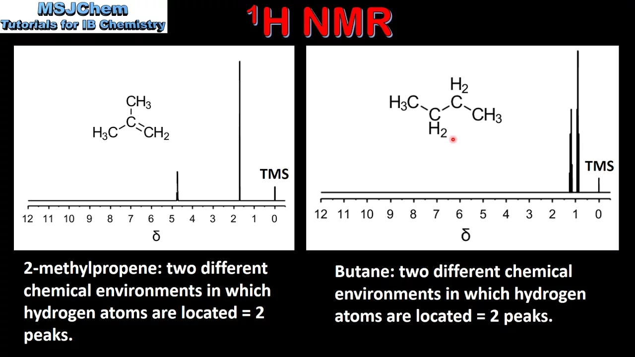
NMR (Nuclear Magnetic Resonance) spectroscopy analyzes organic molecules by detecting how atomic nuclei resonate in magnetic fields. Two main types exist: carbon-13 NMR reveals carbon arrangements, while proton/hydrogen-1 NMR shows hydrogen arrangements. Both work because nuclei with odd nucleons possess spin, enabling magnetic interaction. Four key rules govern interpretation: peak counting (non-equivalent environments), chemical shift values, integration traces, and splitting patterns. The number of peaks equals non-equivalent environments—ethane shows one peak (all equivalent hydrogens), propane shows two peaks. Symmetry dramatically reduces peak counts: butane shows two peaks instead of four because terminal methyls and central methylenes are equivalent. Benzene produces one peak due to perfect symmetry; substituted benzenes increase complexity based on substituent positions and symmetry lines.

NMR spectroscopy analyzes molecular structure through three key characteristics: chemical shift, multiplicity, and integral intensity. Chemical shift (δ) quantifies the difference between a nucleus's resonance frequency and a reference standard (TMS), calculated as δ = [(ν_sample - ν_reference)/ν_spectrometer] × 10^6, yielding values in ppm independent of concentration. Shielding occurs when electron clouds around nuclei oppose external magnetic fields, causing nuclei in different environments to resonate at different frequencies. Electron-withdrawing groups cause deshielding (larger δ, left side of spectrum), while electron-donating groups cause shielding (smaller δ, right side). Characteristic chemical shift ranges include: alkyl protons (<1 ppm), vinylic protons (4.5-6.5 ppm), aldehydic/protonic acids (9-13 ppm), and aromatic protons (6.5-8.5 ppm). Magnetic equivalence describes nuclei experiencing identical effective magnetic fields and producing identical signals. The n+1 rule states that a nucleus with n equivalent neighboring nuclei splits into n+1 peaks, with relative intensities following Pascal's triangle coefficients. Singlets occur when no neighbors exist or when coupling averages out (e.g., OH/NH groups).
Basic principles of Supramolecular Chemistry, specifically host-guest complexation and non-covalent interactions (such as hydrogen bonding, hydrophobic effects, and pi-stacking).

Supramolecular chemistry differs fundamentally from molecular chemistry: it involves pre-formed molecules (hosts and guests) associating through non-covalent interactions without bond-breaking. Hosts are larger molecules with converging binding sites, while guests are smaller with divergent binding sites. Molecular recognition occurs through geometric complementarity: guests fit into host cavities (capsular), sit on host faces (nesting), perch at edges, or get sandwiched between hosts. Flexible hosts can wrap around guests through conformational changes. Researchers study supramolecular complexes by examining recognition, catalysis, transport, ordering, interaction nature, symmetry, and packing. These principles enable applications in nanomaterials, nanomedicine, and molecular devices.

Molecular recognition involves specific binding of guest molecules to complementary host molecules to form host-guest complexes. Crown ethers bind cations through lone pairs on oxygen atoms. DNA is a supramolecular structure held together by hydrogen bonds between complementary bases. Pi-pi stacking interactions are not possible in cyclodextrins.
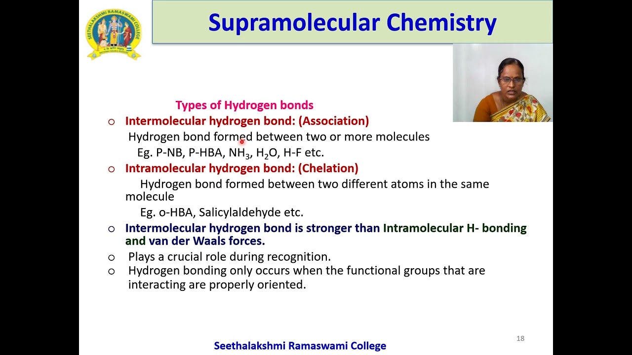
Supramolecular chemistry is an interdisciplinary field that studies molecular assemblies rather than individual molecules, focusing on non-covalent interactions such as electrostatic interactions, hydrogen bonding, pi-pi stacking, halogen bonding, and hydrophobic interactions to achieve molecular recognition and self-assembly; this chemistry enables the design of functional systems for applications in drug development, catalysis, nanoscience, and materials technology, with key examples including DNA base pairing through hydrogen bonds and host-guest complexes where complementary binding sites form stable associations through electronic and size complementarity.

Supramolecular chemistry involves the study of non-covalent interactions between molecules. Molecular recognition refers to the specific binding between substrates and receptors, governed by shape complementarity, charge complementarity, and large contact area for multiple interactions. Ionophores are molecules that facilitate ion transfer across membranes, such as valinomycin for potassium ions and crown ethers for metal ions. The hydrophobic effect describes how guest molecules displace water molecules from host structures, increasing entropy. Key non-covalent interactions include ion-ion (200-300 kJ/mol), ion-dipole (50-200 kJ/mol), hydrogen bonding (4-20 kJ/mol), dipole-dipole (5-50 kJ/mol), pi-pi interactions, and van der Waals forces.

Supramolecular chemistry is the study of molecular assemblies formed through non-covalent intermolecular interactions such as hydrogen bonding, hydrophobic effects, and electrostatic forces, enabling molecular recognition and self-assembly; it has diverse applications in medicine (targeted drug delivery, contrast agents for imaging), materials science (self-healing polymers, nanomaterials), and catalysis, with key principles including molecular complementarity, reversibility, and the lock-and-key mechanism of host-guest interactions.
Chemical equilibrium theory, including the definition of association constants (Ka), dissociation constants (Kd), and stoichiometric ratios (e.g., 1:1 binding).

For a bimolecular reaction A + B ⇌ AB, the equilibrium constant (Ka) is defined as [AB]/([A][B]) and is called the association constant because it describes the process of A and B associating to form the complex. The reverse reaction AB ⇌ A + B has an equilibrium constant (Kd) defined as ([A][B])/[AB], called the dissociation constant. These two constants are reciprocals of each other (Ka = 1/Kd), meaning knowing one automatically gives the value of the other.

Chemical equilibrium occurs when product formation reaches saturation and there is no further change in product formation or reactant concentration. In a closed system, reactions tend toward thermodynamic equilibrium where forward and reverse reaction rates are equal, resulting in zero net rate. The equilibrium constant (K) is the ratio of forward to reverse rate constants (K1/K2). For dissociation reactions (HA → H + A), the dissociation constant KD = [H][A]/[HA], while the association constant is the reverse. KD and KA are reciprocals of each other (KD = 1/KA).

In biochemistry, every ligand-protein receptor interaction has an association constant (Ka) and dissociation constant (Kd), which are numerical values representing the equilibrium state of the reaction; these constants are mathematical inverses of each other, with higher Ka values indicating stronger binding affinity (more molecules in the bound complex form at equilibrium) and lower Kd values also indicating stronger affinity, while lower Ka and higher Kd values indicate weaker binding; additionally, these equilibrium constants relate to rate constants through the relationship Ka = k_on/k_off and Kd = k_off/k_on, where k_on is the rate constant for complex formation and k_off is the rate constant for complex dissociation.
Dissociation constant (Kd) and association constant (Ka) are equilibrium constants that describe the extent of dissociation and association reactions. For dissociation: AB ⇌ A⁺ + B⁻, Kd = [A⁺][B⁻]/[AB]. For association: 2A ⇌ A₂, Ka = [A₂]/[A]². These constants help determine the degree of dissociation/association in solution.

Ka (association constant) and Kd (dissociation constant) describe the equilibrium between enzyme-substrate binding. Ka represents the association reaction where enzyme and substrate come together to form the enzyme-substrate complex. Kd represents the dissociation reaction where they separate. Conceptually, high Ka means strong affinity (favorable association), while high Kd means weak affinity (favorable dissociation). Since Kd is easier to measure experimentally, it is more commonly provided on tests. For enzyme comparison, low Kd indicates better affinity.
General concepts of titration, including how concentration changes affect the ratio of free and complexed species in solution.

Titration is the most important topic in analytical chemistry, involving the gradual addition of one solution to another using a burette and Erlenmeyer flask. Two types of solutions are involved: the standard solution with known concentration in the burette, and the unknown solution with unknown concentration in the Erlenmeyer flask. The endpoint of reaction is the experimental point where the reaction ends, while the equivalence point is a theoretical point where the amount of standard solution equals the unknown solution. Error in titration is the difference between these two points. Normal concentration (تركيز نورمالي) is the concentration containing one gram equivalent of solute per liter, measured in gram equivalent per liter. Molar concentration (تركيز مولاري) is the concentration containing one mole of solute per liter, measured in mole per liter. Indicators (الدلائل) are chemical substances added to the titration solution that do not participate in the reaction but change color at the endpoint.

For successful titration, conditions must be met: (1) Reaction must be simple and expressible by balanced chemical equation; (2) Reaction must be irreversible (one-directional) to ensure complete reaction; (3) Reaction must be rapid and instantaneous for immediate endpoint detection; (4) Appropriate indicator or method must be available for endpoint detection. Titration reactions include: acid-base titration (acid and base), salt-acid or salt-base titration, precipitation titration (forming solid precipitate), complexation titration (forming complex compounds), and redox titration (oxidation-reduction reactions). Two concentration types are used: molar concentration (mol/L or M) representing moles per liter, and normal concentration (eq/L or N) representing equivalents per liter. The equivalent weight is calculated as molecular weight divided by valence factor.
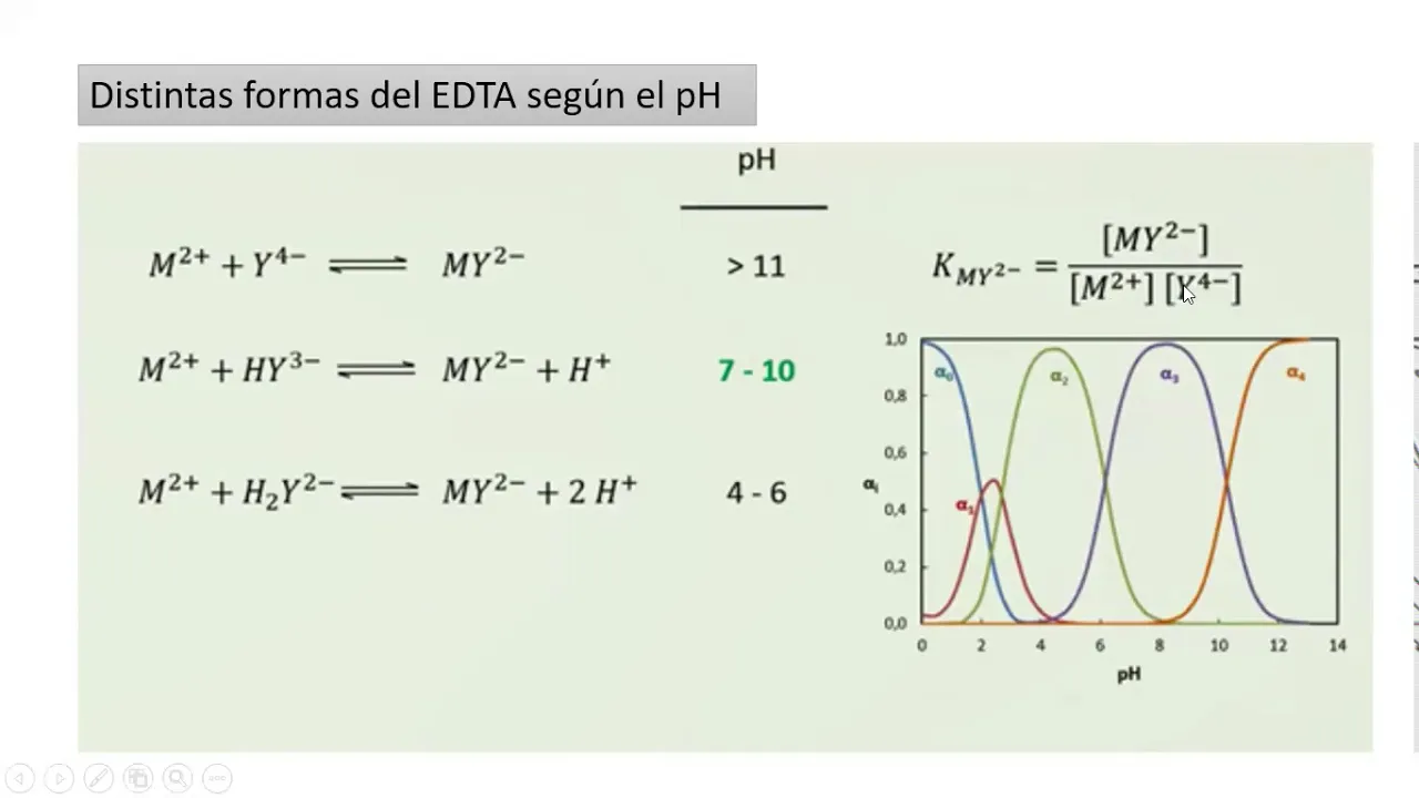
Titration curves plot pM (negative log of metal concentration) versus volume of titrant added. Before equivalence, free metal concentration comes from excess metal ions. At equivalence, [M] = [L]total / √K'f. After equivalence, free metal concentration depends on excess ligand: [M] = [L]excess / (K'f × [L]excess). The pH significantly affects the titration endpoint because K'f is a function of pH through the alpha functions. Different pH values produce different K'f values, leading to different pM values at equivalence. This is why pH must be carefully controlled and specified in complexometric titrations.

Complexation titration depends on: (1) pH - controls EDTA dissociation and metal ion complexation; (2) Concentration - higher concentrations improve complex formation; (3) Stability constant (Kf) - higher values mean more stable complexes; (4) Presence of interfering ions. The pKf (negative log of Kf) allows comparison of complex stabilities. Proper pH control ensures complete dissociation of EDTA for accurate results.

This segment covers calculating concentration ratios and analyzing titration data. The instructor derives the expression [NH3]/[NH4+] = 10^(pKa - pH) and demonstrates calculating the ratio at specific titration points. The instructor explains that this ratio changes throughout titration, being greater than 1 when pH < pKa and less than 1 when pH > pKa. The instructor demonstrates calculating the ratio at a specific point (pKa = 9.2, pH = 5.2), obtaining [NH3]/[NH4+] = 10,000, indicating the base form predominates. The instructor emphasizes that these calculations help understand the distribution of species throughout the titration process.
Prerequisite Knowledge
- Concept 01Fundamentals of NMR Spectroscopy, including chemical shifts, peak integration, and how molecular environment changes affect spectra.
- Concept 02Basic principles of Supramolecular Chemistry, specifically host-guest complexation and non-covalent interactions (such as hydrogen bonding, hydrophobic effects, and pi-stacking).
- Concept 03Chemical equilibrium theory, including the definition of association constants (Ka), dissociation constants (Kd), and stoichiometric ratios (e.g., 1:1 binding).
- Concept 04General concepts of titration, including how concentration changes affect the ratio of free and complexed species in solution.
Subsequent Learning
- Step 01Advanced data fitting techniques for complex stoichiometry, such as 1:2 or 2:1 host-guest systems, and cooperativity models.
- Step 02Complementary analytical techniques for measuring binding constants, such as Isothermal Titration Calorimetry (ITC), UV-Vis, and fluorescence titration.
- Step 03NMR exchange kinetics, studying how fast, slow, and intermediate chemical exchange rates on the NMR timescale affect peak shapes and signals.
- Step 04Real-world applications of host-guest binding studies, such as the design of molecular sensors, drug delivery vehicles, and self-assembling materials.
ITC Method
0:08- 1
Simulate isotherms online and define 10–20 titration points.
- 2
Measure host and guest solutions accurately with a micro balance and syringe.
- 3
Analyze shifted proton signals and fit data using online calculators.
Methodological Limitations of NMR Titrations and the Pitfalls of Overfitting
While NMR titration is a popular method for determining host-guest binding constants, it has significant limitations compared to techniques like Isothermal Titration Calorimetry (ITC) and is highly prone to mathematical fitting errors. NMR chemical shift changes are indirect measures of binding, susceptible to local environmental changes and conformational fluctuations that do not reflect true thermodynamic binding. Furthermore, direct NMR titration fails in the "slow exchange" regime (typical of high-affinity complexes), where peaks do not shift but instead appear and disappear, making simple curve-fitting impossible. Critics also highlight that web-based fitting tools (such as supramolecular.org) can easily suffer from "overfitting"—yielding excellent mathematical fits for incorrect binding stoichiometries (e.g., fitting a 1:2 complex to a 1:1 model) if not rigorously validated by independent thermodynamic methods like ITC, which directly measures heat changes and provides a complete thermodynamic profile (ΔH, ΔS, and K).
Advanced data fitting techniques for complex stoichiometry, such as 1:2 or 2:1 host-guest systems, and cooperativity models.

The method of continuous variation (Job's method) determines coordination complex stoichiometry. The procedure involves preparing solutions with varying mole fractions of reactants and measuring a property like electrical conductivity. If a complex forms, the plot of property versus mole fraction shows a maximum (break) at the stoichiometric ratio. If no complex forms, the plot is linear. This method uses additive properties to identify complex formation and determine the ratio of metal to ligand in the complex.

SEDFIT software enables multi-wavelength analysis through systematic parameter entry: load experiments, configure noise refinements, define extinction information with known values fixed and unknowns refined, set CK(S) distributions with sedimentation bounds and regularization, and specify stoichiometry matrices. Initial fitting fixes meniscus and frictional ratio to stabilize convergence. Stoichiometry determination integrates CK(S) distributions for each component in the mixture, with hydrodynamic information confirming true stoichiometry. Validation requires assessing chi-squared statistics, residual randomness, and agreement between input and calculated concentrations. The human lactoferrin/TP34 case demonstrated how multi-signal analysis revealed a 1:2 complex differing from ITC-suggested 2:1 ratio, illustrating the technique's power to reveal true molecular architecture.

Two methods determine metal-ligand complex stoichiometry: (1) Job's method of continuous variations keeps total concentration constant while varying mole fractions, plotting absorbance versus mole fraction to find the maximum point indicating stoichiometric ratio; (2) Mole ratio method keeps metal concentration constant while varying ligand concentration, plotting absorbance versus mole ratio to find the maximum. Both methods work best for 1:1 to 1:2 complexes; ratios above 1:2 introduce significant errors. The mole ratio at the maximum indicates the ligand-to-metal ratio in the complex.
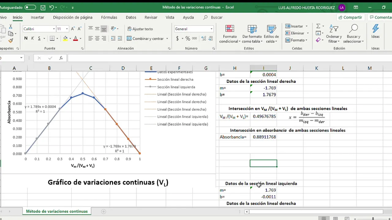
Job's method determines complex stoichiometry by mixing metal and ligand solutions at constant total volume (10 mL) with varying proportions. The volume ratio (metal volume / total volume) is calculated and plotted against absorbance at 390 nm. The resulting curve shows two linear sections: left (0 to 0.3) and right (0.7 to 1.0). Linear regression is performed on these sections to obtain slopes and y-intercepts. The intersection point of these lines reveals the stoichiometric ratio, where the x-value represents the mole fraction of metal at maximum complex formation.

Job's continuous variation method determines the stoichiometry of metal-ligand complexes by measuring absorbance at varying mole fractions of ligand while keeping total concentration constant; the complex composition is identified from the mole fraction at which maximum absorbance occurs, where a 1:1 complex shows maximum at 0.5 mole fraction, 1:2 at approximately 0.66, and 1:3 at 0.75.
Complementary analytical techniques for measuring binding constants, such as Isothermal Titration Calorimetry (ITC), UV-Vis, and fluorescence titration.

NMR spectroscopy identifies nuclei by concentration-weighted average chemical shifts under rapid exchange conditions, revealing binding-induced electronic changes and detecting hydrogen bonding/π-π interactions. UV-vis spectroscopy applies Beer-Lambert law: A_λ = L × Σ(ε_i(λ) × C_i), enabling linear least-squares determination of extinction coefficients. Fluorescence data follows similar principles: I_λ = Σ(Φ_i(λ) × C_i). Isothermal titration calorimetry measures heat evolution under isothermal conditions, providing complete thermodynamic parameters (K, ΔG, ΔH, ΔS) without requiring chromophores. Surface-enhanced and resonance Raman spectroscopy overcome weak scattering cross-sections in solution-phase studies. Recent advances correlate Raman band intensity with complex geometry in excited states to calculate binding constants quantitatively. Each technique offers unique advantages: NMR for structural insights, UV-vis for speed, ITC for universal detection of binding events regardless of molecular properties.

Isothermal Titration Calorimetry (ITC) is a label-free technique that measures heat changes during biomolecular binding reactions to determine binding affinity (KD), stoichiometry, enthalpy (ΔH), and entropy without requiring molecular labels; the method works by detecting temperature differences between a reference cell and a sample cell containing the biomolecule of interest, where small ligand injections cause measurable heat changes proportional to the amount of binding, allowing researchers to construct binding isotherms and extract thermodynamic parameters from the resulting data.

Isothermal Titration Calorimetry (ITC) is a powerful analytical technique that measures the heat changes occurring during macromolecule-ligand binding interactions to determine binding affinity, stoichiometry, enthalpy, and entropy from a single experiment; the method requires careful sample preparation including matching buffer composition and pH, degassing to eliminate air bubbles, and systematic titration with appropriate injection volumes and timing, followed by data analysis using fitting software to extract thermodynamic parameters that reveal the nature of molecular interactions.

Isothermal titration calorimetry (ITC) is a sensitive thermodynamic technique that measures the heat released or absorbed when two molecules interact by injecting a ligand into a solution containing a macromolecule; the differential power required to maintain constant temperature between the sample and reference cells reveals the enthalpy change, binding constant, stoichiometry, and equilibrium constant of the interaction, making it invaluable for studying weak biological interactions like protein-ligand, protein-nucleic acid, and antibody-ligand binding where heats of reaction are typically very small.

Isothermal Titration Calorimetry (ITC) is a technique used to measure the heat of interaction between molecules. In the experiment described, lysozyme (the analyte) is titrated with NAG3 (the titrant). The heat associated with each injection is measured, and modeling of the heat per injection curves gives an apparent association constant (Kapp). This technique is particularly useful when direct measurement of affinity is not possible, such as when the substrate interacts weakly with the enzyme.
NMR exchange kinetics, studying how fast, slow, and intermediate chemical exchange rates on the NMR timescale affect peak shapes and signals.
NMR signal intensity and line shape reflect molecular motion through relaxation properties. Since the 1940s, relaxation rates have been correlated with motion time scales. Chemical exchange between protein conformations produces distinct spectral signatures depending on exchange rate relative to NMR timescales: slow exchange shows separate states, intermediate exchange causes broadening/coalescence, and fast exchange yields averaged signals. The heteronuclear NOE measures picosecond motions, distinguishing structured (values ~0.7-0.8) from disordered regions (lower/negative values). T1 and T2 relaxation provide complementary information about molecular tumbling and size.

Dynamic NMR studies molecules that interconvert between conformations. When exchange is slow, separate peaks appear for each form. When exchange is fast, a single averaged peak appears. The coalescence temperature marks when peaks merge, indicating the exchange rate equals the NMR timescale. This technique reveals conformational dynamics and energy barriers between molecular forms.

This section covers exchange broadening and relaxation times in NMR spectroscopy. Exchange broadening occurs when nuclei exchange positions rapidly (such as OH protons in alcohols exchanging with solvent water). This rapid exchange causes the NMR signal to broaden. The rate of exchange affects the appearance of the signal: slow exchange gives separate signals, fast exchange gives a single broadened signal. The width of NMR peaks depends on the relaxation times (T1 and T2) of the nuclei. Shorter relaxation times result in broader peaks. The line width is inversely proportional to the relaxation time. Factors affecting relaxation include molecular tumbling rate, viscosity, and the presence of paramagnetic species.

Exchange processes in NMR spectroscopy cause signal broadening or disappearance when protons rapidly exchange between different chemical environments. Rapid exchange (e.g., -OH, -NH, -COOH protons) averages the chemical shifts, producing broad or invisible signals. Slow exchange preserves distinct signals. Understanding exchange rates is crucial for interpreting spectra of dynamic molecules and for choosing appropriate experimental conditions (temperature, solvent) to optimize signal visibility.

Chemical exchange (or proton exchange reaction) in NMR spectroscopy refers to the transfer of atomic nuclei from one molecular environment to another, causing changes in chemical shift and coupling constants (J values) as nuclei enter new environments; this phenomenon manifests differently in NMR spectra depending on exchange rate: slow exchange produces two distinct peaks, intermediate exchange produces broadened peaks, and fast exchange also produces broadened peaks but with different intensity patterns, as demonstrated by examples including amide-water proton exchange, hydrogen bonding in water, conformational changes in molecules like cyclohexane, and biological processes such as water molecule transport across cell membranes.
Real-world applications of host-guest binding studies, such as the design of molecular sensors, drug delivery vehicles, and self-assembling materials.

Host-guest chemistry is the study of non-covalent molecular assemblies where a host molecule (possessing a cavity or binding site) selectively binds to a guest molecule through various non-covalent interactions including electrostatic forces, hydrogen bonding, van der Waals forces, and pi-stacking; this principle explains biological phenomena like enzyme-substrate specificity and has diverse applications in pharmaceuticals (e.g., nitroglycerin-cyclodextrin complexes), environmental remediation, optoelectronics, and molecular sensing.

Supramolecular host-guest chemistry enables precise molecular recognition and functional applications through complementary binding interactions. Host molecules with appropriately sized cavities can selectively stabilize reactive or unstable species like white phosphorus, dramatically enhancing their stability from detectable to nearly undetectable concentrations. These systems enable controlled release and extraction of encapsulated species. Beyond stabilization, host-guest systems serve as molecular sensors, where binding events produce measurable signals like fluorescence changes for selective detection of target analytes. Extending these principles to catalysis, supramolecular hosts can enforce specific orientations and proximities between reactants, driving selectivity that would not occur in bulk solution. Coordination polymers extend these concepts to extended networks with long-range electronic communication, amplifying optical signals for sensing applications. Covalent organic frameworks (COFs) represent another class of porous materials with inherent hydrolytic stability and tunable pore geometries for gas separation and storage applications.

Host-guest chemistry, defined by Donald Cram, describes when a larger molecule (host) wraps around a smaller molecule (guest) through non-covalent interactions. Self-assembly is the spontaneous, reversible organization of molecules into ordered structures through non-covalent bonding. Binding sites require specific size, geometry, and functional groups for successful host-guest recognition. Binding sites must be complementary - fitting together like puzzle pieces. Cooperativity occurs when multiple binding sites work together to bind multiple guests, creating stronger combined interactions than independent sites.
Host-guest recognition can occur through two mechanisms: recognition-first (guest binds then host changes conformation) or conformational selection (host exists in multiple conformations and guest selects one). These mechanisms can be distinguished by analyzing concentration dependence of association rate constants. Guest identity affects which mechanism operates, as demonstrated with sodium versus potassium ions. This understanding allows rational design of supramolecular systems with specific recognition properties, enabling applications in sensing, catalysis, and drug delivery.

This comprehensive section covers the principles of molecular assembly and recognition. Self-assembly involves spontaneous, reversible association of molecules forming discrete or extended entities through non-covalent interactions. Molecular self-assembly creates covalent bonds under synthetic control, while supramolecular self-assembly relies on recognition-directed association. Self-organization extends to spontaneous emergence of order in space and time, involving collective behavior and dynamic processes. Homo-assembly involves identical components associating symmetrically, while hetero-assembly involves complementary components forming asymmetric complexes. Host-guest chemistry describes molecular recognition where hosts (with convergent binding sites) bind guests (with divergent binding sites) through non-covalent interactions. Nobel laureates Cram, Lehn, and Pedersen pioneered this field, developing systems ranging from clathrates (solid-state inclusions) to solution-stable complexes using crown ethers, cryptands, and cavitands. These principles enable selective binding of specific molecules, forming the foundation for applications in catalysis, sensing, and drug delivery.
ITC Method
0:08- 1
Simulate isotherms online and define 10–20 titration points.
- 2
Measure host and guest solutions accurately with a micro balance and syringe.
- 3
Analyze shifted proton signals and fit data using online calculators.
Methodological Limitations of NMR Titrations and the Pitfalls of Overfitting
While NMR titration is a popular method for determining host-guest binding constants, it has significant limitations compared to techniques like Isothermal Titration Calorimetry (ITC) and is highly prone to mathematical fitting errors. NMR chemical shift changes are indirect measures of binding, susceptible to local environmental changes and conformational fluctuations that do not reflect true thermodynamic binding. Furthermore, direct NMR titration fails in the "slow exchange" regime (typical of high-affinity complexes), where peaks do not shift but instead appear and disappear, making simple curve-fitting impossible. Critics also highlight that web-based fitting tools (such as supramolecular.org) can easily suffer from "overfitting"—yielding excellent mathematical fits for incorrect binding stoichiometries (e.g., fitting a 1:2 complex to a 1:1 model) if not rigorously validated by independent thermodynamic methods like ITC, which directly measures heat changes and provides a complete thermodynamic profile (ΔH, ΔS, and K).
hi i'm paula solution from the open data fit project and along with change web we will show you how to perform a nuclear magnetic resonance or an anima titration experiment start by using our website to simulate what your binding isotherms might look like define your target titration points aiming for between 10 and 20 points it's important to use a micro balance to measure all volumes about one mole of the host solution is enough as you'll use point six mils for the NMR tube then you've still got point four mils left to make up the guest solution that is typically between 40 to a hundred times more concentrated than the host after recording the first NMR spectra of the host eject the sample and then add the first portion of the guest solution using an accurate micro syringe when analyzing your data charting more than one proton signal that has moved allows you to use global analysis to improve your fit fit your data using our online calculator to the suspected binding model or models look at the various outputs including the residual plot to evaluate your results you can get more information from our website and the open data team thanks you for watching
Up Next

Molecular Machines: Nobel Prize in Chemistry 2016 Explained
@simpleshowfoundation
16.6K views•2016-12-08

Biocatalysis and Catalysis in Islatravir Synthesis | Merck Webinar
@SCIwheresciencemeetsbusiness
675 views•2020-10-08
1H NMR: Determining Number of Peaks from Structure
@MSJChem
59.2K views•2017-04-06

Edible Water Bottles: A DIY Guide to Sodium Alginate Spherification
@ryan
10.5M views•2019-06-21
Related Study Plans & Knowledge Roadmaps
Structured learning paths in Chemistry