Lateral flow assays are diagnostic test devices that detect the presence of specific substances (such as antigens, antibodies, or biomarkers) in a sample by using a combination of a sample pad, conjugate pad, nitrocellulose membrane with test and control lines, and absorbent pad, where the sample flows laterally through the membrane and produces visible lines indicating positive or negative results.
MiraVista Diagnostics Lateral Flow Assay | Lab Animation
Added:Deep Dive
Fundamental principles of antigen-antibody binding specificity and immunological affinity.

Sự kết hợp kháng nguyên-kháng thể ( buổi 4.1)
582
The binding between antigen and antibody is highly specific, analogous to a key fitting into a lock. This specificity is fundamental to immunological reactions and diagnostic applications. The binding occurs through chemical interactions between complementary chemical groups on the molecules. Four primary forces govern antigen-antibody binding: electrostatic forces (ionic bonds between oppositely charged groups), hydrogen bonds (sharing hydrogen atoms between electronegative atoms), Van der Waals forces (weak attractive forces from electron movement), and hydrophobic interactions. These forces work cooperatively to achieve both the strength and specificity required for effective immune recognition. The distance between binding sites critically affects binding strength, with closer proximity resulting in stronger attraction. Immune reactions can occur in vivo (within the body) or in vitro (outside the body). In vivo reactions are protective but can become pathological when they occur inappropriately, causing organ dysfunction or even death.
Click to watch →

Affinity|Avidity|Cross-reactivity|
680
Antigen-antibody binding occurs through specific molecular recognition where epitopes (antigenic determinants on antigens) interact with paratopes (antibody binding sites). This interaction is mediated by weak non-covalent forces including hydrogen bonds, electrostatic interactions, hydrophobic interactions, and Van Der Waals forces, which require close molecular proximity. Affinity measures the total strength of non-covalent interactions between a single epitope and a single paratope. This fundamental understanding forms the basis for comprehending immune recognition and antibody function.
Click to watch →
IMUNOLOGIA- Imunoensaios (parte 1)
11.1K
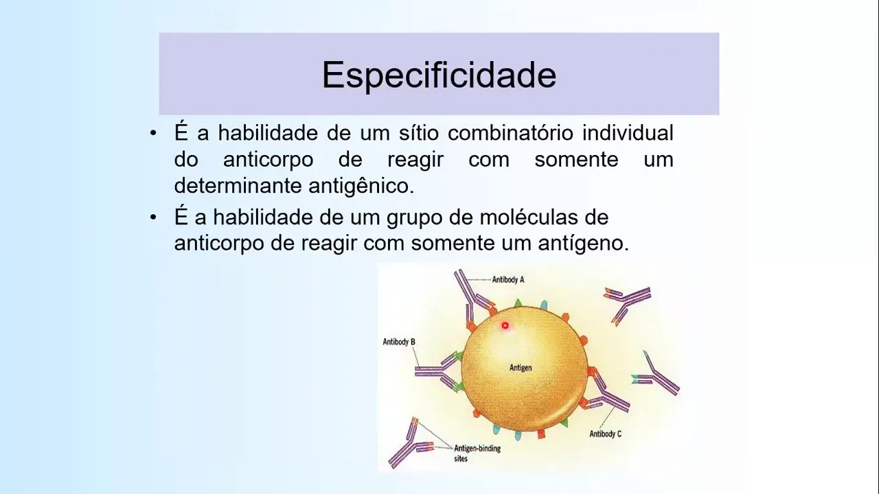
Antigen-antibody binding occurs through non-covalent interactions including hydrogen bonds, electrostatic bonds, Van der Waals forces, and hydrophobic bonds. Molecular complementarity between antibody combining sites and antigen epitopes determines binding efficiency. Greater complementarity increases attractive forces and decreases repulsive forces during molecular collisions. Affinity measures single-site binding strength, representing the net balance of attractive and repulsive forces. Avidity represents total multivalent binding strength across all antibody combining sites. Specificity refers to the ability of individual combining sites to react only with specific antigens. Cross-reactivity occurs when shared epitopes allow antibodies to bind to multiple antigens.
Click to watch →

Immunology Chapter 10: Precipitations & Agglutination Reactions
376
This section covers the fundamental principles of antigen-antibody binding that underlie all precipitation and agglutination reactions. Affinity refers to the initial attraction between a single antibody binding site and a single epitope, while avidity represents the overall strength of binding when multiple sites are involved. The law of mass action governs all binding as reversible reactions, with the equilibrium constant K indicating binding strength. Cross-reactivity occurs when antibodies react with similar antigens, and higher affinity results in more sensitive assays. These principles form the theoretical foundation for understanding how immunoassays detect and measure antigens and antibodies in clinical specimens.
Click to watch →

كل تمارين الإستجابة المناعية مع الحل المفصل | من دورة 2007 حتى 2019
53.1K
Antigens (foreign substances) and antibodies have specific binding sites that allow them to recognize and attach to each other. This binding occurs through structural compatibility (structural complementarity) between the antigen and the antibody's binding site. Each antibody is specific to a particular antigen, and this specificity ensures that the immune system can target specific threats without affecting the body's own cells.
Click to watch →
Deep Dive
The concept of capillary action and chromatography, specifically how fluids migrate through a porous membrane without external pump assistance.
CHROMATOGRAPHIE sur couche mince ✅ MÉTHODE 💪 Seconde
84.3K
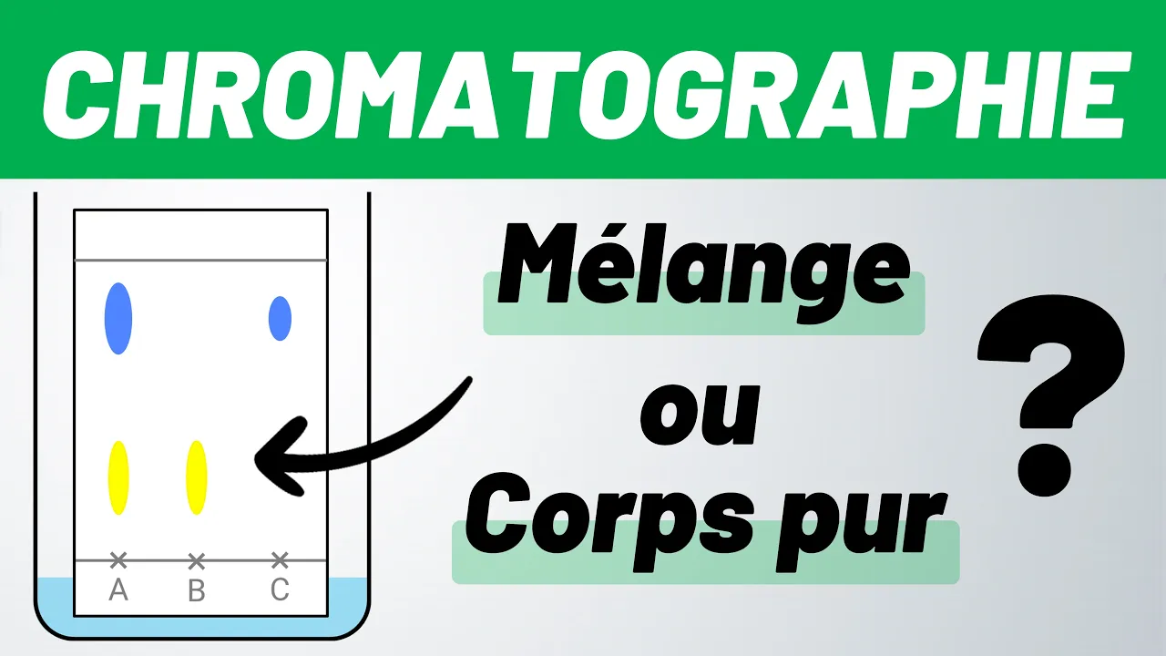
During TLC, the eluent migrates upward through the silica gel by capillary action. As the eluent moves, it carries the components of the mixture with it. Each chemical species migrates to a different height depending on its chemical properties. The mixture spot divides into several spots, indicating the presence of multiple components. When the eluent reaches approximately 1 cm from the top of the plate, the plate is removed and the eluent front is marked.
Click to watch →

Northern blotting technique
234.1K
After electrophoresis, RNA must be transferred from fragile agarose gels to durable membrane filters for hybridization. Aminooxy methyl filter paper is preferred over nitrocellulose due to its greater RNA binding affinity. The transfer uses capillary action: buffer flows upward through a sponge layer into the gel, creating pressure that pushes RNA fragments out of the gel matrix. These fragments migrate toward and attach to the membrane filter, which is impermeable to RNA. Paper towels above absorb excess buffer, maintaining flow direction. This blotting process creates an exact imprint of RNA fragments on the membrane for subsequent probe hybridization.
Click to watch →

Présentation de la Chromatographie sur Couche Mince (C.C.M)
187.5K
In TLC, migration occurs through capillary action where the eluent rises along the TLC plate, carrying dissolved chemical species with it. This phenomenon is similar to how water climbs up a paper towel when placed in a puddle. The eluent front moves upward, and different compounds travel at different rates depending on their affinity for the stationary phase versus the mobile phase.
Click to watch →

7 EXPERIMENTOS CIENTÍFICOS FÁCEIS DE FAZER EM CASA
4.3M
Water can move through porous materials like napkins and paper due to capillary action, which is the ability of water to flow upward against gravity through narrow spaces. This occurs because water molecules are attracted to the fibers in the material, creating a continuous chain of movement. Experiments demonstrate this principle through water ambulance demonstrations, colored water droplet dances, and rainbow creation using colored napkins. When water is added to a colored napkin placed on a plate, the water travels up through the napkin via capillary action, carrying color pigments outward in radial patterns that resemble blooming flowers.
Click to watch →
![[#하이라이트#] 살아있는 것처럼 우유를 마시는 석상 미스터리! #다빈치노트 EP.5](https://i.ytimg.com/vi/Yp25GpNuE-E/maxresdefault.jpg)
[#하이라이트#] 살아있는 것처럼 우유를 마시는 석상 미스터리! #다빈치노트 EP.5
291.8K
Capillary action is the ability of a liquid to flow in narrow spaces without external forces, caused by cohesion and adhesion between liquid molecules and the material surface. In porous materials like certain stones, capillary action allows liquids to be drawn upward through tiny channels and pores. The siphon effect is a related phenomenon where liquid flows from a higher point to a lower point through a tube due to pressure differences. When a tube connects two containers at different heights, the pressure difference causes liquid to flow from the higher container to the lower one. These principles explain phenomena like the milk absorption in the Ganesha statue.
Click to watch →
Deep Dive
Basic mechanics of conjugation, specifically how antibodies are conjugated to signal-producing markers like colloidal gold or latex beads.

Antibody-Nanoparticle Conjugation Webinar: Gold nanoparticles - optimization of conjugates
40.3K
Surface-coated gold nanoparticles enable one-step antibody conjugation kits that complete reactions in 20 minutes with minimal hands-on effort, generally requiring less antibody than passive methods. Oriented coupling positions antibody fragments (Fab', Fab) in favorable orientations for antigen binding: periodate-treated antibodies create aldehyde groups reacting with hydroxylamine derivatives, or thiol-containing fragments react with maleimide groups. This oriented approach often outperforms random lysine-mediated attachment. These strategies reduce antibody denaturation by preventing direct metal-protein contact and enable controlled molecule exchange through cleavable linkers.
Click to watch →

The science of rapid COVID-19 antigen tests
372
Antibodies are Y-shaped proteins with variable regions that can be engineered to bind specifically to particular antigens. During immune development, B cells randomly generate antibody combinations, and those successfully binding foreign antigens are selected for production. This specificity enables precise detection of viral components. In diagnostic tests, antibodies are conjugated to labels such as colored latex beads, fluorescent markers, or colloidal gold particles. Colloidal gold produces a red color through surface plasmon resonance. Control antibodies ensure test validity by verifying proper flow and binding throughout the test strip.
Click to watch →

Tutorial | BioReady™ Passive Conjugation Protocol
5.7K
This video tutorial demonstrates a three-step passive conjugation protocol for attaching antibodies to BioReady™ Bare (Citrate) Gold nanoparticles using pH titration: (1) mix antibody with gold nanoparticles at different pH levels (7, 8, 9), (2) test stability by adding sodium chloride to challenge weakly bound conjugates, and (3) select the most stable pH mixture for blocking and purification; stable conjugates appear red while unstable ones turn purple, gray, clear, or precipitate.
Click to watch →
Tutorial | Covalent Conjugation of Antibodies to Gold Nanoparticles
16.8K
This video demonstrates a step-by-step protocol for covalently conjugating antibodies to carboxyl gold nanoparticles using EDC and Sulfo-NHS crosslinkers. The process involves activating the nanoparticle surface with EDC (10 mg/mL) and Sulfo-NHS (16 μL/mL for 1 mL gold nanoshells), incubating with antibody (20 μg/mL), quenching remaining reactive groups with hydroxylamine, and purifying the conjugate by centrifugation. This covalent conjugation method enables rapid antibody screening for lateral flow assay development, reducing optimization time from days/weeks to hours compared to passive conjugation methods.
Click to watch →

Antibody Conjugation
647
Antibody conjugation is the process of attaching antigens to larger carrier molecules to stimulate a humoral immune response, producing high-performance monoclonal or polyclonal antibodies coupled to labels for use as reporter molecules in immunoassays like ELISA, Western blot, immunofluorescence, and flow cytometry. The protocol involves adding sodium bicarbonate buffer to primary antibody, dissolving succinimidyl ester dye in DMSO, slowly adding dye while vortexing, shaking for 1 hour at room temperature, adding hydroxylamine, and dialyzing in PBS. Conjugates are stored at 4°C with BSA and/or sodium azide. Four basic conjugation protocols exist for different fluorophores and proteins. Popular carrier proteins include KLH, BSA, and ovalbumin, which are highly immunogenic. Chemical cross-linkers like glutaraldehyde, carbodiimide, and succinimidyl esters control antigen orientation to carrier proteins. Since antigens attached to carrier proteins are digested along with carriers, antibodies produced may bind to carrier fragments, requiring purification for pure antibody solutions.
Click to watch →
Deep Dive
The functional difference between test lines (which capture target analytes) and control lines (which verify test validity) in rapid diagnostics.

المحاضرة الثانية - التقنيات المختبرية المتقدمة
799
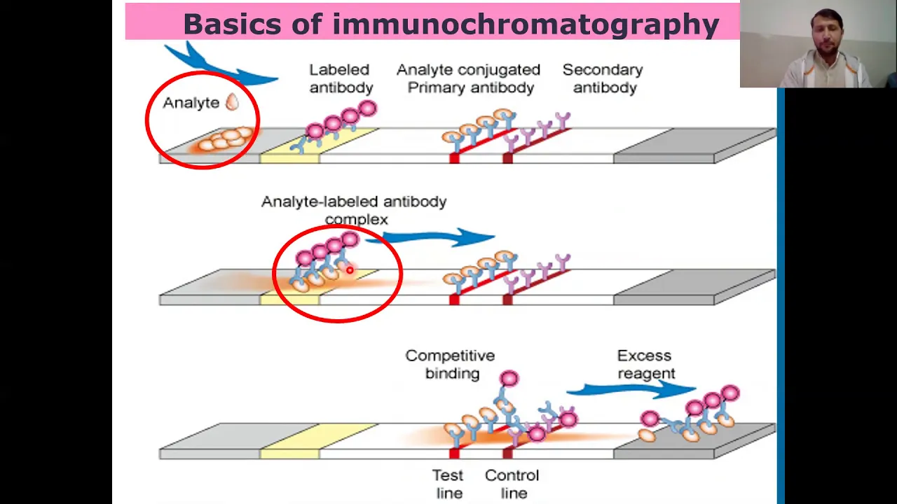
The nitrocellulose membrane contains two critical lines: (1) Test line - captures antigen-antibody complexes to indicate positive results, (2) Control line - confirms the test is functioning properly. The test line appears when the target antigen is present, while the control line always appears to validate the test. Both lines must appear for a valid result.
Click to watch →
Testes de Diagnósticos Rápidos ( TDR)
713
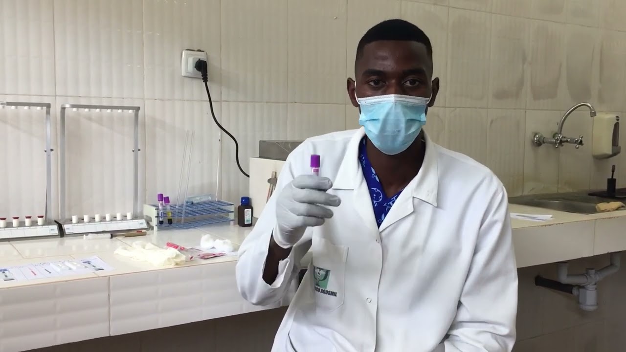
Rapid diagnostic tests have two identification areas: the control line (C) and the test line (T). The control line always reacts and must appear for the test to be valid. The test line indicates the presence of the target antigen or antibody. The control line is the last line that appears on the test, while the test line is the first line that appears.
Click to watch →
Immunochromatographic Assay or Technique I Guide to Immunochromatographic Assay
27.7K
The control line contains secondary antibodies that are not specific to the target antigen but serve to verify test validity. Regardless of whether the test is positive or negative, the control line should always appear if the test is functioning properly. If the control line does not appear, the test is considered invalid because it indicates a problem with the test procedure or reagents. This dual-line system ensures reliable interpretation of results.
Click to watch →

Testes Rápidos
204
Rapid test kits contain specific components including lancets, test cassettes, reagents, and pipettes. Each test type requires its own specific pipette to ensure accurate volume delivery. The control line (C) indicates the test is functioning properly, while the test line (T) indicates presence of the target antigen/antibody. All tests must show the control line to be valid. If the control line does not appear, the test is invalid and must be reported to the Ministry of Health. Positive rapid test results require confirmation testing with a different brand.
Click to watch →
ICT method in laboratory | working principal of lab kits
1.1K
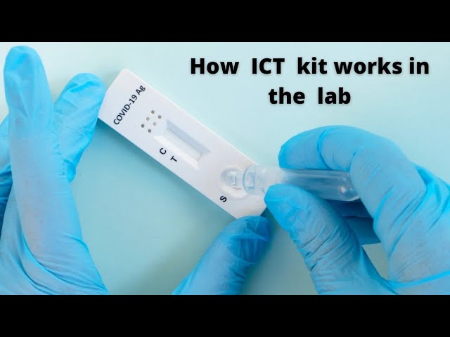
Lateral flow chromatography test strips have two lines: the control line and the test line. The control line contains antigens that react with the conjugate pad to produce a visible line, confirming the test is working correctly. The test line contains antibodies that react with target antigens in the sample. A positive result shows both lines, indicating the presence of the target substance. A negative result shows only the control line. The control line serves as a validation mechanism—if it does not appear, the test kit may be defective or the sample was not applied correctly. Each test kit must undergo quality assurance testing to ensure consistent and accurate results. Different diseases require different test kits because the antigens and antibodies are unique to each disease.
Click to watch →
Prerequisite Knowledge
- Concept 01Fundamental principles of antigen-antibody binding specificity and immunological affinity.
- Concept 02The concept of capillary action and chromatography, specifically how fluids migrate through a porous membrane without external pump assistance.
- Concept 03Basic mechanics of conjugation, specifically how antibodies are conjugated to signal-producing markers like colloidal gold or latex beads.
- Concept 04The functional difference between test lines (which capture target analytes) and control lines (which verify test validity) in rapid diagnostics.
Subsequent Learning
- Step 01Advanced assay design strategies, including multiplexing to detect multiple target analytes simultaneously on a single strip.
- Step 02Quantitative lateral flow assays, which utilize digital optical readers or smartphone-based detection to measure analyte concentration.
- Step 03Troubleshooting assay sensitivity and specificity, including how to mitigate issues like the 'hook effect' and non-specific background binding.
- Step 04Comparative analysis of lateral flow assays against laboratory standards like ELISA and PCR in clinical diagnostic settings.
Gratitude
Main Topic
Key Insights
Deep Dive
Audience Engagement
Gratitude
0:08Playing Section
- 1
Expresses thanks to the audience.
- 2
Sets a warm and appreciative tone.
- 3
Acknowledges the viewers' presence and support.
Molecular and Quantitative Diagnostics (PCR/ELISA) vs. Rapid Lateral Flow Assays
While lateral flow assays (LFAs) offer rapid, low-cost, point-of-care results, they are frequently criticized for their lower sensitivity and specificity compared to laboratory-based molecular and quantitative diagnostics. LFAs typically provide qualitative (yes/no) or semi-quantitative results and have a higher limit of detection, meaning they may miss early-stage infections or low concentrations of an analyte, leading to false negatives. In contrast, molecular techniques like Polymerase Chain Reaction (PCR) and enzyme-linked immunosorbent assays (ELISA) offer superior sensitivity, exact quantification, and lower detection limits. Proponents of laboratory-based methods argue that for critical diagnoses—such as systemic fungal infections—molecular assays and quantitative ELISAs remain the gold standards, as they significantly reduce diagnostic errors and allow clinicians to monitor disease progression and treatment efficacy with high precision.
Deep Dive
Advanced assay design strategies, including multiplexing to detect multiple target analytes simultaneously on a single strip.

Multiplexed Lateral Flow Assay Development: Benefits, Risks, and Strategies for Success...
141
A multiplex lateral flow assay measures one or more analytes in a single device through three approaches: single strip with multiple targets, multiple strips for single targets, or multiple strips with multiple targets. Key benefits include improved clinical sensitivity and specificity through combined biomarkers, enhanced analytical range via semi-quantitative test lines, reduced customer costs through consolidated testing, novel intellectual property creation through combined marker algorithms, and manufacturing efficiency gains from shared components. The Vigilant Biosciences saliva oral cancer test exemplifies successful multiplexing despite complexity, demonstrating how strategic application of these principles can achieve FDA breakthrough designation.
Click to watch →
Lateral Flow Assay (Sensors and Devices 02_2)
1K
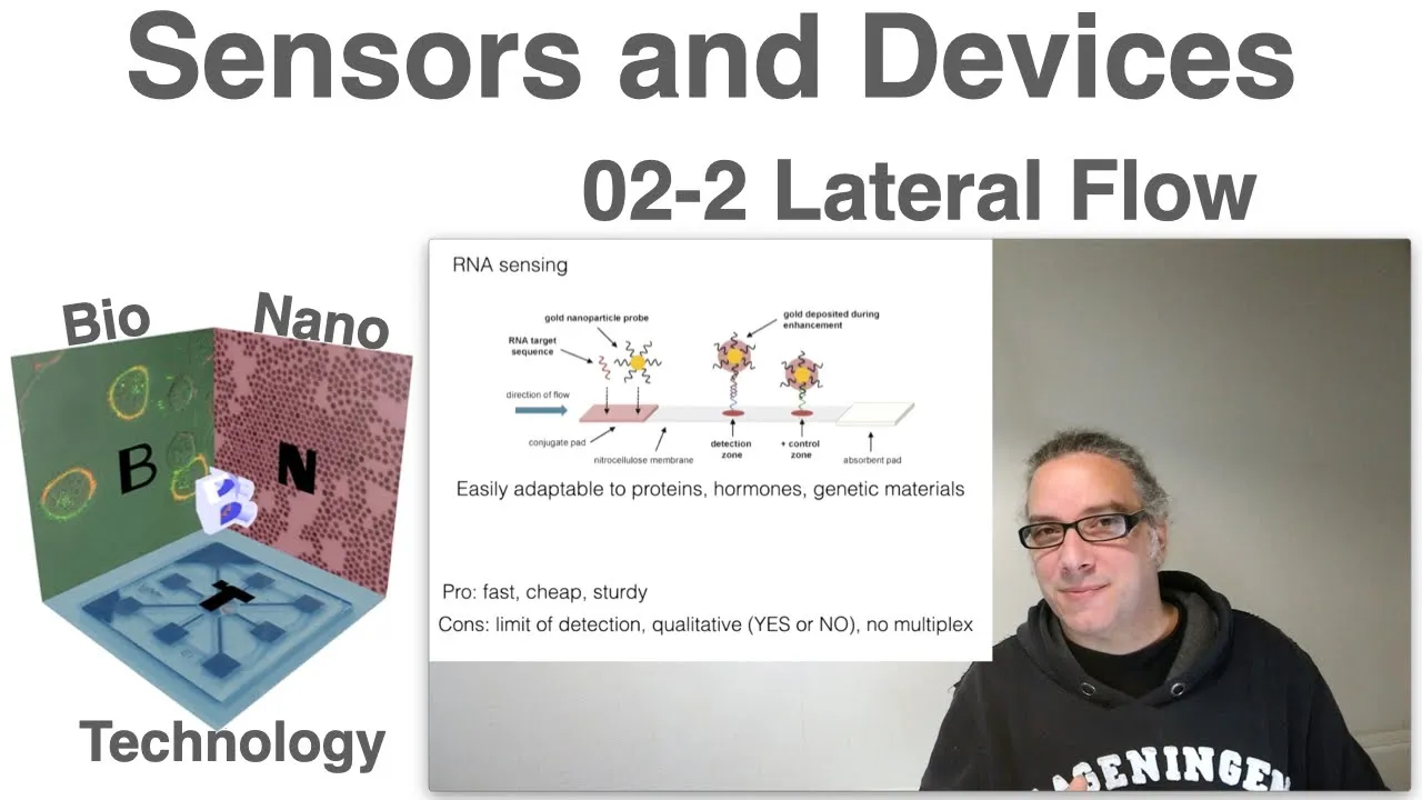
Lateral flow devices consist of four essential components working in sequence: the sample pad for initial treatment (pH adjustment, salt removal, viscosity control), the conjugate pad containing detection reagents that mix with the sample, the analytical membrane (typically nitrocellulose) where capture antibodies and control lines are located, and an absorbent wad to manage liquid flow. A breakthrough in multiplexing utilizes silver nanoparticles of different sizes, which produce distinct colors despite being identical in composition due to plasmonic effects. This enables simultaneous detection of multiple pathogens (e.g., yellow fever, Zika, Ebola) on a single strip for under 20 cents, demonstrating how paper-based sensors combine affordability, portability, and sophisticated analytical capabilities for global health applications.
Click to watch →
Webinar Crystal Digital PCR cdPCR
472
Optimal assay design requires amplicon size of 100-120 bp, GC content of 40-60%, primer length 17-25 nt, probe length 15-25 nt, primer Tm 60-62°C, probe Tm 66-70°C. Multiplex digital PCR offers significant advantages: reduced reagents, elimination of standard curves, decreased consumables, and faster setup. Multiple multiplexing strategies exist: independent gene targeting with different fluorophores, SNP region targeting, and universal reporter approaches. SYBR Green creates discrimination challenges because shorter amplicons fluoresce less intensely; TaqMan chemistry solves this with gene-specific probes producing distinguishable signals. Amplitude multiplexing uses same fluorophore at different concentrations but is more complex. Color combination strategies enable up to 21 targets on seven-color instruments.
Click to watch →

Moving xMAP Technology Forward: New Targets and High-Sensitivity Precision Pro Assays
904
Multiplex assays provide significant advantages over single-target assays: (1) Sample conservation - enables analysis of multiple targets from limited sample volumes, critical for mouse studies and precious human clinical samples; (2) Labor efficiency - reduces experimental workload by analyzing multiple targets in one run; (3) Cost-effectiveness - for three or more targets, multiplexing becomes more economical than running multiple ELISAs; (4) Temporal consistency - provides a complete snapshot of all targets at the same time point, preserving biological relationships between analytes that would be lost with sequential measurements.
Click to watch →
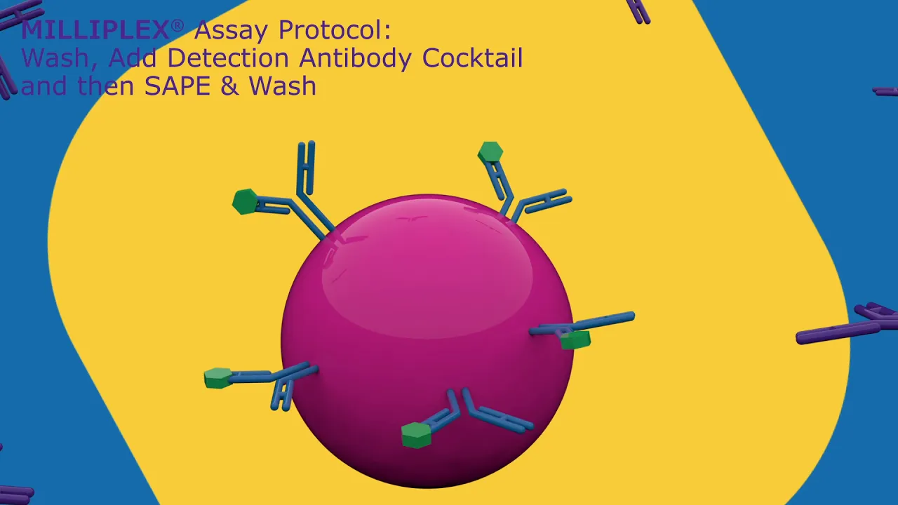
MILLIPLEX® Multiplex Assays
22.9K
Milliplex Multiplex Assays utilize Luminex's XMAP technology with color-coded microsphere beads to enable simultaneous detection of up to 48 different analytes in a single sample, offering significant time savings (4 hours vs. 10 hours for ELISA) and reduced sample consumption while maintaining comparable sensitivity; the technology employs sandwich immunoassay principles where beads coated with specific capture antibodies bind target analytes, followed by detection antibody and fluorescent reporter addition, with washing steps performed using magnetic plate washers before analysis on Luminex instruments.
Click to watch →
Deep Dive
Quantitative lateral flow assays, which utilize digital optical readers or smartphone-based detection to measure analyte concentration.

Smartphone Reader for Lateral Flow - DEMO
2.9K
Smartphone readers with patented image processing technology can quantify lateral flow test results by dynamically assessing image quality, focus, lighting, and orientation, eliminating the need for expensive dedicated readers while enabling data recording, location capture, barcode traceability, and automatic sharing through the phone's connectivity.
Click to watch →

Microfluidics 2.0: Point-of-Care Diagnostics for the Developed and Developing World
31.5K
Cell phone cameras equipped with simple optics can provide quantitative readout for paper-based assays. Image analysis software identifies fiducial marks, locates assay spots, and measures intensity over time. Initial rate measurements provide quantitative concentration information across multiple orders of magnitude, enabling sophisticated diagnostics with minimal instrumentation.
Click to watch →

Abingdon Health plc (OTCQB: ABDXF | LSE: ABDX) — Small Cap Growth Virtual Investor Conference 2026
429
Lateral flow testing technology has evolved beyond simple yes/no devices for single biomarkers to multiplexed capabilities that can test for multiple biomarkers simultaneously. The technology can now quantify results rather than just providing binary outcomes, using small readers or smartphone apps for measurement. The tests can work with multiple sample matrices including saliva, urine, and blood. Various chemistry formats and particle types can be used to generate signals, with common examples being pink (gold) and blue (latex) color indicators.
Click to watch →

TEDItalia - Un laboratorio grande quanto un francobollo: George Whitesides
106
Lateral flow immunoassays, exemplified by pregnancy tests, represent the most common paper-based diagnostic technology. These binary tests work well for conditions with clear yes/no outcomes but are limited for quantitative analysis. Wax printer technology enables mass production of paper-based diagnostic devices by printing wax patterns onto paper, which absorb when heated. These printers cost approximately $800 and can produce about 10 million tests per year when operating 24 hours daily. The printing process takes about two seconds per device. Smartphone camera phones can capture images of paper-based diagnostic devices, which are then sent to central laboratories for analysis. This approach eliminates the need for doctors to be present at the point of care, allowing healthcare workers to collect samples and transmit data while computers perform complex analysis.
Click to watch →

Sensors for Medical Diagnostics | Engineering Speaker Series
1.7K
Commercial rapid diagnostic tests (like pregnancy tests) use antibody-based assays that produce visible lines when positive. A smartphone-based optical reader replaces expensive laboratory equipment ($20,000-$50,000) by using the phone's camera and computational power. Optimized angled detection with custom amplification achieved one order of magnitude better limit of detection compared to standalone laboratory readers.
Click to watch →
Deep Dive
Troubleshooting assay sensitivity and specificity, including how to mitigate issues like the 'hook effect' and non-specific background binding.

Specific and Non-specific Binding in a ligand binding assay. Drug discovery methods
50.3K
In radioligand binding assays, specific binding refers to the tight, high-affinity interaction between a ligand and its target receptor, while non-specific binding occurs at low-affinity sites throughout the reaction mixture; these two types of binding can be separated by adding excess unlabeled (cold) ligand, which displaces the radioactive (hot) ligand from specific sites but not from non-specific sites, allowing researchers to calculate specific binding by subtracting non-specific binding from total binding and construct saturation curves to determine binding affinity through the dissociation constant (Kd), which equals the ligand concentration at which half of the receptors are bound.
Click to watch →
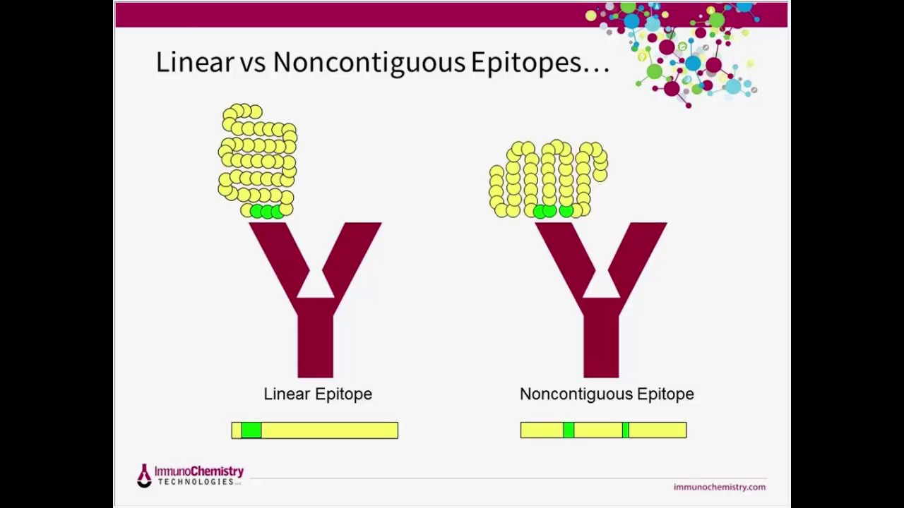
Resolution of Several Common Immunoassay Performance Issues
453
This webinar addresses common performance issues in solid-phase immunoassays (ELISA, lateral flow, beads, ELISPOT) including signal variability, excessive background, and poor dose response, explaining that these problems often stem from improper plate coating (antibodies not oriented correctly, incorrect coating concentration causing hook effects), inadequate blocking (non-specific binding, cross-reactivity), and unstable reagents (denatured enzymes, degraded proteins). The presentation emphasizes that successful assay development requires understanding the fundamental principles of antibody-antigen interactions, proper solid support utilization, and careful optimization of coating buffers, blocking agents, and stabilization protocols to achieve consistent, reliable results.
Click to watch →

Hook effect in ELISA
1.4K
The hook effect is a phenomenon in one-step sandwich ELISA where high analyte concentrations produce falsely low results due to saturation of capture antibody binding sites, which prevents sufficient detector antibody binding to the antigen-antibody complex; this can be identified by comparing undiluted and diluted samples (where diluted samples show higher concentrations) and corrected by performing serial dilutions and multiplying the concentration by the dilution factor.
Click to watch →
Immunochemistry: Basic Concepts
18.3K
Competitive immunoassays involve competition between sample analyte and labeled antigen for limited antibody binding sites, producing inverse proportionality between analyte concentration and signal. Non-competitive (sandwich) assays capture antigen between two antibodies, providing direct proportionality and higher sensitivity/specificity. The hook effect occurs at extremely high analyte concentrations, causing dose-response curves to flatten or invert, potentially producing falsely low readings. Dilution protocols verify result accuracy by distinguishing true high concentrations from assay saturation effects. These design principles fundamentally shape assay performance characteristics and clinical interpretation.
Click to watch →

Armadilhas no tubo de ensaio: interferentes analíticos em dosagens hormonais
213
Hormone laboratories use immunological assays (antigen-antibody reactions) and chromatography-mass spectrometry. Immunological assays include chemiluminescence methods with significant platform variability due to different antibodies, epitopes, and designs. Two-step sandwich assays with wash steps reduce hook effect risk, while one-step assays lack this protection. The hook effect occurs when excess analyte prevents both capture and detection antibodies from binding the same antigen molecule, causing falsely low results. Biotin interferes with assays using biotin-streptavidin reactions, causing falsely low results in sandwich assays and falsely high in competitive assays. Heterophilic antibodies are non-specific antibodies binding assay reagents, causing falsely positive results in sandwich assays. They are present in up to 30% of the population, with interference in 2-3% of tests. When results are incompatible with clinical presentation, the laboratory can: compare with different assays, test linearity by serial dilution, precipitate with PEG to remove large molecules, or use heterophilic antibody blocking reagents.
Click to watch →
Deep Dive
Comparative analysis of lateral flow assays against laboratory standards like ELISA and PCR in clinical diagnostic settings.

Webinar PATELKI 9: Kupas Tuntas Pemeriksaan Serologi COVID-19
35.3K
Lateral flow tests compared to laboratory methods (ELISA, chemiluminescence) show: (1) Lower sensitivity but higher specificity in some tests, (2) Faster results but less quantitative information, (3) Greater accessibility but more potential for human error, and (4) Lower cost but potentially lower accuracy. The choice of test method should be based on the intended application, available resources, and required accuracy.
Click to watch →

COVID-19 Educational Series - Full webinar 2nd of June
207
Rapid diagnostic tests (lateral flow assays) provide qualitative results with turnaround time of just minutes, medium throughput (one patient per test), and are meant for point-of-care use. ELISA assays can be qualitative or semi/fully quantitative, take a couple of hours, have medium throughput (several hundred per day), and are lab-based. Chemiluminescent assays provide qualitative or quantitative results, can be as fast as 18 minutes (Roche assay), have high throughput (up to thousands per day), and are lab-based. Neutralization assays are quantitative tests requiring days to complete, very low throughput (only several per day), and are reference lab-based requiring biosafety level 3 containment procedures. Only neutralization assays actually test for antibody function, making them considered gold standards.
Click to watch →

COVID-19 Recovery Committee - 18 November 2021
139
Initial comparisons between lateral flow tests and PCR tests appeared to show low sensitivity (around 40%) because they compared fundamentally different tests—PCR detects persistent genetic material while lateral flow detects active viral proteins present only during infectious periods. Proper comparison reveals lateral flow tests achieve above 80 per cent sensitivity and potentially 95 per cent for detecting infectious individuals. For public health purposes, lateral flow tests are more reliable than direct PCR comparisons suggested. PCR tests remain essential for variant sequencing and clinical certainty but should not replace lateral flow for routine monitoring.
Click to watch →

Geert Vanden Bossche, "Wave of the Variants", the surge in need for rapid antibody testing (FULL)
5.2K
Lateral flow assays can be used for both serology (antibody) testing and antigen testing, as well as detecting chemical and biological markers. The technology is scalable globally with manufacturers in Taiwan, India, Brazil, and the US. A universal lateral flow assay reader standardizes tests, improves sensitivity, eliminates human error, and collects metadata including vaccination status and disease history. The reading of each assay takes approximately 1-2 seconds, with machines capable of processing 500 samples per hour. Quantitative lateral flow assays can be developed by calibrating against PCR or ELISA results. Nucleocapsid antibodies are the best candidate for serology testing because they are completely discriminative between natural infection and vaccination. The nucleocapsid protein is more stable and does not mutate like spike, making it a reliable marker for natural immunity. Finger prick tests are not sensitive enough; venous blood draw is required.
Click to watch →
PCR vs lateral flow tests | The key differences
3.7K
PCR tests are more accurate laboratory-based tests that detect the presence of the COVID-19 virus and are primarily used for symptomatic individuals, while lateral flow tests are rapid antigen tests that can be done at home and are suitable for asymptomatic screening; PCR tests require laboratory processing and take up to 3 days for results, whereas lateral flow tests provide results in about 30 minutes, and PCR tests are typically required for hospital procedures, care home staff, and international travel, while lateral flow tests are free and can be ordered online or picked up from test collection sites.
Click to watch →
Gratitude
Main Topic
Key Insights
Deep Dive
Audience Engagement
Gratitude
0:08- 1
Expresses thanks to the audience.
- 2
Sets a warm and appreciative tone.
- 3
Acknowledges the viewers' presence and support.
Molecular and Quantitative Diagnostics (PCR/ELISA) vs. Rapid Lateral Flow Assays
While lateral flow assays (LFAs) offer rapid, low-cost, point-of-care results, they are frequently criticized for their lower sensitivity and specificity compared to laboratory-based molecular and quantitative diagnostics. LFAs typically provide qualitative (yes/no) or semi-quantitative results and have a higher limit of detection, meaning they may miss early-stage infections or low concentrations of an analyte, leading to false negatives. In contrast, molecular techniques like Polymerase Chain Reaction (PCR) and enzyme-linked immunosorbent assays (ELISA) offer superior sensitivity, exact quantification, and lower detection limits. Proponents of laboratory-based methods argue that for critical diagnoses—such as systemic fungal infections—molecular assays and quantitative ELISAs remain the gold standards, as they significantly reduce diagnostic errors and allow clinicians to monitor disease progression and treatment efficacy with high precision.
Fundamental principles of antigen-antibody binding specificity and immunological affinity.
The concept of capillary action and chromatography, specifically how fluids migrate through a porous membrane without external pump assistance.
Basic mechanics of conjugation, specifically how antibodies are conjugated to signal-producing markers like colloidal gold or latex beads.
The functional difference between test lines (which capture target analytes) and control lines (which verify test validity) in rapid diagnostics.
Advanced assay design strategies, including multiplexing to detect multiple target analytes simultaneously on a single strip.
Quantitative lateral flow assays, which utilize digital optical readers or smartphone-based detection to measure analyte concentration.
Troubleshooting assay sensitivity and specificity, including how to mitigate issues like the 'hook effect' and non-specific background binding.
Comparative analysis of lateral flow assays against laboratory standards like ELISA and PCR in clinical diagnostic settings.
458.4K views1likes2:10@miravistadiagnosticsOriginal Release: 2023-02-22
[Music] thank you [Music] foreign [Music] foreign [Music] foreign [Music] [Laughter] [Music]
Up Next
47:44
Optimizing Gold Nanoparticle Conjugation: Size, Shape & Stability
@InnovaBiosciences
40.3K views•2014-09-29
14:12
Quantitative Real-Time PCR (qPCR): Principle, Method & Data Analysis
@animatedbiologywitharpan
129.2K views•2023-09-06

23:33
Microbial Degradation of Plastics: Biodegradation Pathways & Sustainability
@majeedhammad
2.9K views•2021-04-11

16:04
CRISPR and Genetic Engineering: How Gene Editing Works and Why It Matters
@kurzgesagt
30.5M views•2016-08-10
Related Study Plans & Knowledge Roadmaps
Structured learning paths in Biotechnology
biotechnology
AAV Production: Transfection, Prep & qPCR
01Introduction to Gene Therapy and AAV Biology
02Mammalian Cell Culture: Suspension HEK293 Cells
03Upstream Bioprocessing: Triple-Transfection Optimization
+ 2 more learning modules
18h5 modules
Explore Roadmap
biotechnology
Building Gut-on-a-Chip: PDMS, Cells & TEER
01Foundations of Microfluidics and Gut Anatomy
02Cleanroom Fabrication & PDMS Soft Lithography
03Epithelial Cell Culture and On-Chip Seeding
+ 2 more learning modules
26h5 modules
Explore Roadmap
biotechnology
Optimizing CFPS: E. coli Extract & Yield
01Fundamentals of Molecular Biology and Translation
02Introduction to Cell-Free Protein Synthesis (CFPS)
03Preparing E. coli Crude Lysate (The Extract)
+ 2 more learning modules
19h5 modules
Explore Roadmap
biotechnology
Haematococcus PBR: Design, Stress & Astaxanthin
01Biology and Life Cycle of Haematococcus pluvialis
02Bench-Scale Photobioreactor (PBR) Design and Engineering
03Culture Media Formulation and Inoculation
+ 2 more learning modules
15h5 modules
Explore Roadmap