
![Anna Fučíková: Letem světem nanosvětem [DOD 27.11.2025]](https://i.ytimg.com/vi/E31VqnYqRnQ/maxresdefault.jpg)















![naukovo.pl [646B] - Dobra ciotka Magda - #naukovlog 646B. Nobel z chemii – wywiad z prof. Proniem](https://i.ytimg.com/vi/D6WtsUajhNA/maxresdefault.jpg)



Quantum dots are semiconductor nanocrystals made from cadmium selenide that can be synthesized by heating cadmium oxide with stearic acid, adding trioctylphosphine oxide and hexadecylamine, removing water under vacuum, and injecting selenium precursor under argon atmosphere; these nanoparticles exhibit size-tunable fluorescence properties and are used in biomedical applications such as cancer cell detection when coated with lipids for water solubility and targeted with antibodies.
Synthesis of Quantum Dots: Cadmium Selenide Nanocrystals | Lab Guide
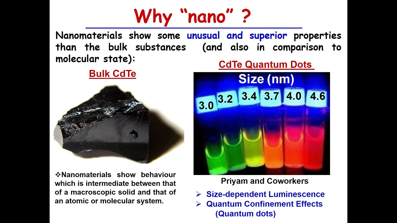
Added:The quantum confinement effect and how a semiconductor material's optical and electronic properties change as its physical size approaches the Bohr exciton radius.
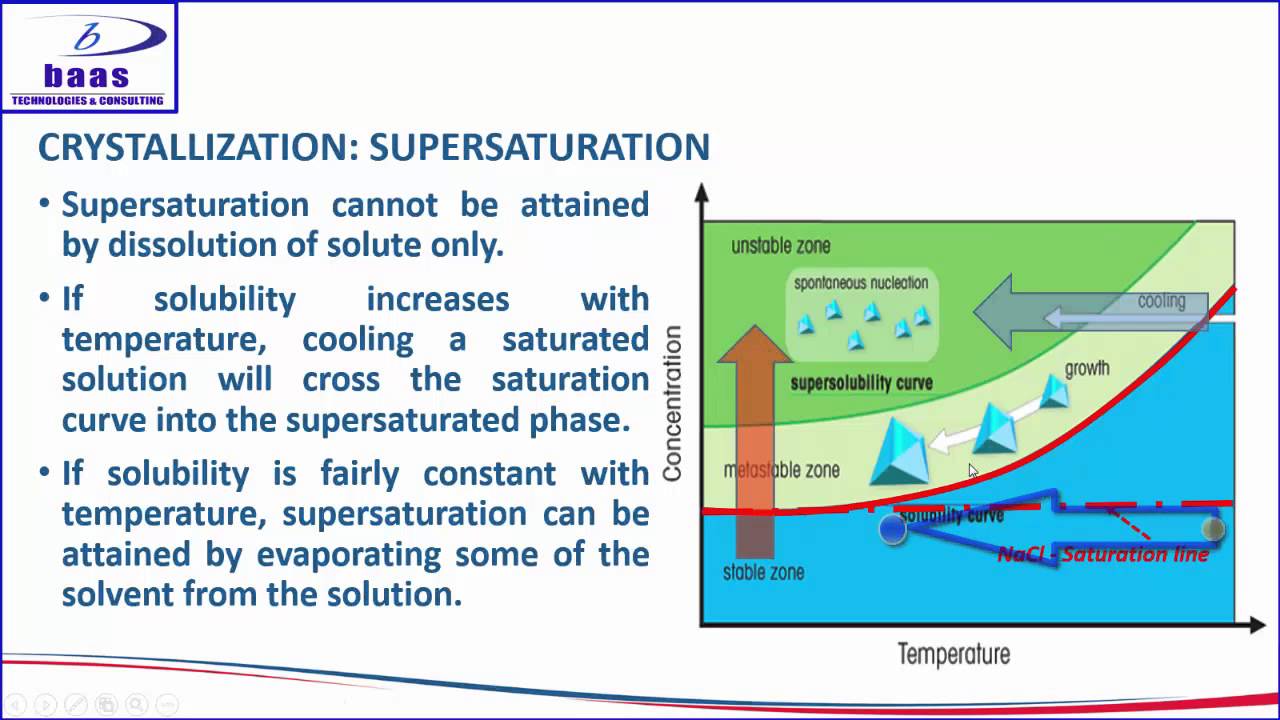
Fundamental principles of colloidal chemistry, specifically the LaMer model of nucleation, supersaturation, and diffusion-controlled nanocrystal growth.
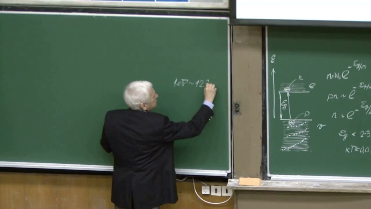
Basic semiconductor physics, including the concepts of bandgaps, valence and conduction bands, and the formation of electron-hole pairs (excitons).
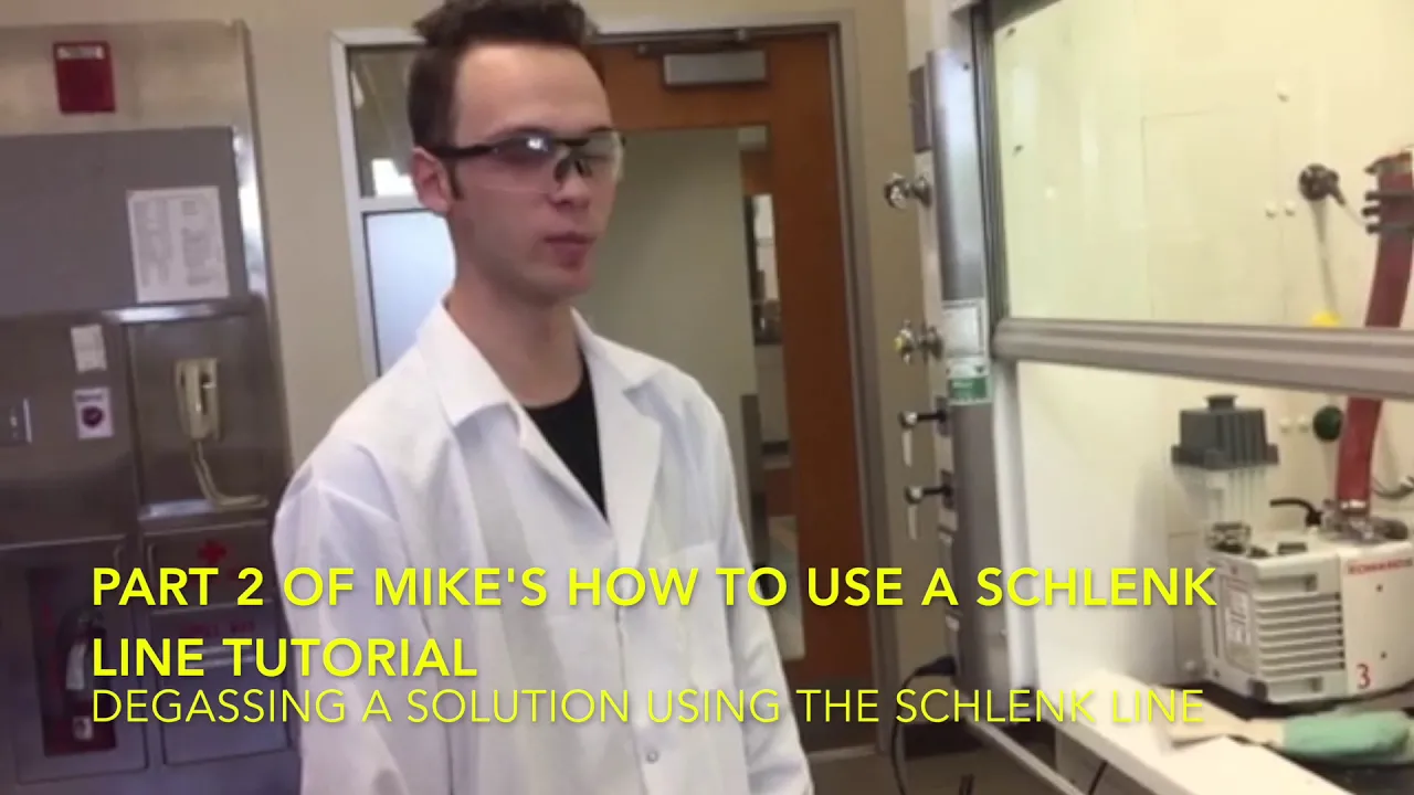
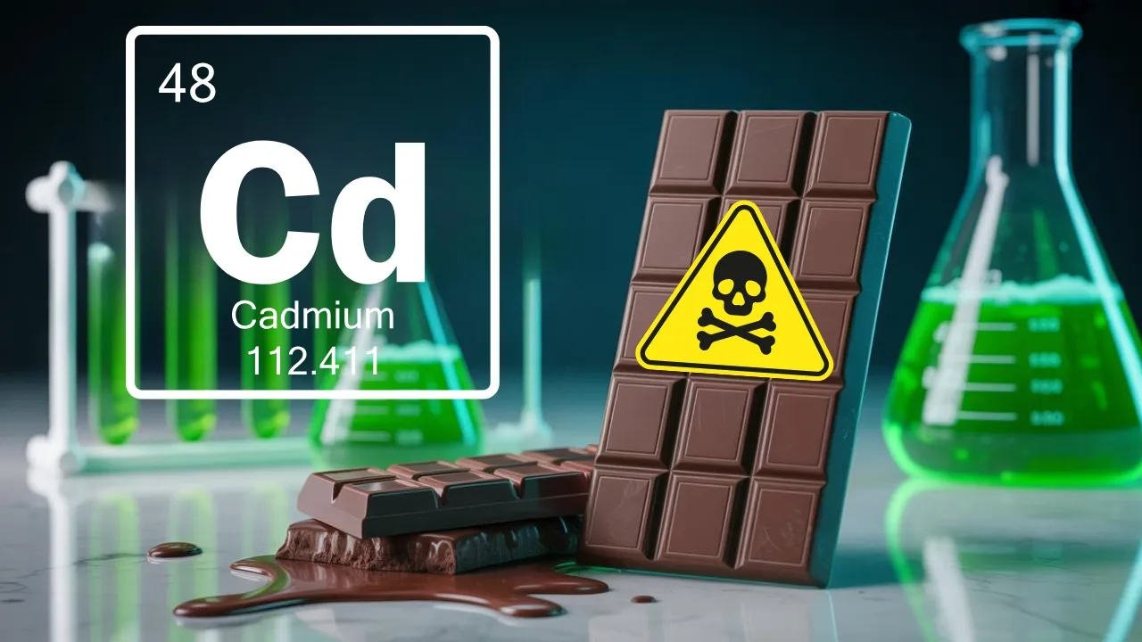
Air-free chemical synthesis techniques (such as using a Schlenk line or glovebox) and safety protocols for handling highly toxic heavy metal precursors like cadmium.
Characterization methodologies for colloidal nanocrystals, specifically UV-Vis absorption spectroscopy, photoluminescence (PL) spectroscopy, and Transmission Electron Microscopy (TEM).
Synthesis of core-shell heterostructures (such as CdSe/ZnS) to passivate surface traps and drastically improve photoluminescence quantum yield.
Surface chemistry and ligand exchange protocols to render hydrophobic quantum dots water-soluble for biological imaging and sensing applications.
Integration of quantum dots into optoelectronic devices, including quantum dot light-emitting diodes (QLEDs), photodetectors, and next-generation solar cells.
100.7K views1.1Klikes7:36Original Release: 2011-09-16
So, in our group, we uh make and use quantum dots, which are cadmium felonide nanop particles. So, today I'm going to start out by showing you how we make them. In here, we have some steeric acid and cadmium oxide that we heat up to dissolve the cadmium oxide in the steeric acid. And then we cooled it back down and added some trictal phosphine oxide and some hexodelamine.
And then we heated that up again until it all melted. And we have been putting it under a vacuum to get rid of all of the water inside. The first step, if you can see, there's uh some accumulation of the cadmium oxide and steeric acid on the wall. So, I just need to heat that up to melt it off so it falls back down. So, I'm just going to use a handy blow torch. And you can see as I'm heating this up, it's falling back in. I don't want to heat it up too much, so try to go slowly. So, now that that's ready, I'm going to go and head over to uh make the selenium for the injection. So, this is a glove box. It's filled with argon, and it serves two purposes. The first is to protect us from the chemicals. Some of them um give off bad odors that could be harmful to the body. So, we keep them in the glove box. And the second is to prevent the chemicals from degrading in the presence of air. So, I'm getting two things in here. The first is selenium, which is in powder form. And then I'm going to dissolve that in tributal phosphine. And then I'm going to take that, split it between two syringes, and head back over to do the injection. So, first I'm just measuring the selenium powder. But the selenium is the component that we add in excess. So now I'm just going to dissolve it in the TBP. And when I dissolve it, it is an exothermic reaction. So it heats up a little, but not to an unbearable amount. It takes a little bit of mechanical stimulation to get this to dissolve. So I just shake it up.
So now I'm going to split this between two syringes uh so that we can inject it faster and so that everything is injected more at the same time and therefore it will grow to be about the same size. So now that these are ready, I'm going to get them ready to be taken out.
Okay, so now we've been vacuuming for about 2 hours and we've gotten rid of all of the excess water in our reaction flask. So, I'm going to go ahead and put it under argon again, uh, the same as was in the glove box. So, I'm putting it under argon in order to prevent reaction with the air. And I just do that by flipping this switch right here. So, now it's off of vacuum and it's on argon.
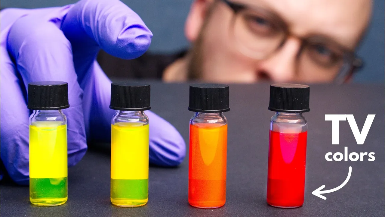
So now I can turn off the vacuum pump and I can set the temperature now to 300° and wait for it to heat up. So now I am injecting the selenium and you can see the color changes immediately. I'm going to lower the temperature to 290° to anneal it. You can see in the beginning it flueses green but as it heats up it turns to a more red color.
So now we just wait about 12 minutes while this anneals. So after this process is over, we have what we call the quantum dot cores. And this is essentially the quantum dot, but it's imperfect. The shell or the exterior of the quantum dot is um not very smooth. And those imperfections mean that it doesn't glow as bright as it potentially can. So we do another process where we put a shell around that to help smooth those imperfections and get very bright quantum dots. And after that whole process is complete, we end up with the finished product. And these we can tailor to be different colors. But these are in chloroform or hexane. They're in an organic solvent. And if we want to ever use them with things in the body, we need to put them in water. Um, in order to put them into water, we end up usually with a much more dilute solution, and we cover them with lipids, which basically are two components. They uh coat the quantum dot with a hydrophobic portion, and then the exterior of the lipid is hydrophilic or water loving. And you end up with something like this. The reason they flues is because they're excited with the UV light. So, when we take away that UV light, they no longer glow.
At this point, the quantum dots have annealed and so now we are able to just cool them down to a temperature where we can clean them. And as they cool, they get brighter. So at this point, the reaction is over. So if I excite them with the black light, you can see that now they flues.
At the lower temperatures, we're able to see the the color much more clearly. Quantum dots are very versatile in their applications. Anything that could be identified or detected with a glowing marker could be identified or detected with a quantum dot. For example, cancer cells are what we identify in this lab. So often cancer cells have uh markers on their membrane on their external membrane and we are able to use that to target those markers with our quantum dots and then we're able to distinguish cancerous cells from non-cancerous cells in the lab. Once we have our quantum dots in water, we can activate them with antibodies, which are what we use to target the cancerous cells. And then we incubate the quantum dots with the cell lines and various different cell lines to see whether or not the quantum dots will uh adhere to the cells. And so here we just when that's all finished we look at them with the microscope and we just put it on the microscope and then we are able to to view the cells um in a variety of different manners both with the phase contrast which is what you see normally with your eye and also with different fluorescents.
Right now, we're looking at images of the cells, cancer cells that have been targeted by quantum dots. And this fluorescent or glowing is the quantum dots attached to the cell membrane. The video is looking at different focal planes. So, it focuses on the bottom of the cell and then up to the top of the cell.
And you can see that it's the glowing is the brightest around the perimeter of the cell where the quantum dots are adhered. I am Charlie D'vorchek, a member in Dr. Sears's lab at John's Hopkins University.
Up Next
6:50
Graphene Exfoliation via Electrochemical Process with Pencil Leads
@JesseSchoch
3K views•2021-01-25

12:54
Triumph of Orthodoxy Icon: Byzantine Art & History Explained
@BenCallan
2.1K views•2024-08-06

9:49
FastAPI vs Flask vs Django: Choosing the Right Python Web Framework
@TechWithTim
302.5K views•2024-05-26

1:42
Game of Thrones Opening Credits: A Cinematic Analysis
@gameofthrones
46.3M views•2011-04-18
Related Study Plans & Knowledge Roadmaps
Structured learning paths in General & Interdisciplinary Studies
general-studies
Elevator Engineering: History, Physics & Cities
01From Hoists to Safety Brakes: The Dawn of Vertical Lift
02Hydraulic vs. Traction: The Engineering Physics of Elevators
03Modern Speed, Safety, and Algorithms
+ 2 more learning modules
12h5 modules
Explore Roadmap
general-studies
Rubber: Material Science, History & Geopolitics
01Origins of Natural Rubber & Mesoamerican History
02The Chemistry of Natural Rubber & Vulcanization
03The Geopolitics of the Rubber Boom & Colonization
+ 2 more learning modules
12h5 modules
Explore Roadmap
general-studies
Containerization: Evolution, Logistics & Trade
01Ancient Trade & Bulk Cargo: Amphorae to Barrels
02The Industrial Revolution & Cardboard Packaging
03The Containerization Revolution: Malcom McLean
+ 2 more learning modules
12h5 modules
Explore Roadmap
general-studies
Textile Tech: Evolution, Labor & Global Impact
01Pre-Industrial Textiles: Handloom & Cottage Industry
02Mechanization: The Spinning Jenny to the Power Loom
03The Factory System & Global Labor Transformation
+ 2 more learning modules
10h5 modules
Explore Roadmap