



![Totipotent, Pluripotent & Multipotent Stem Cells - What is the Difference? [Cell Potency]](https://i.ytimg.com/vi_webp/6vkFU5dWjTU/maxresdefault.webp)







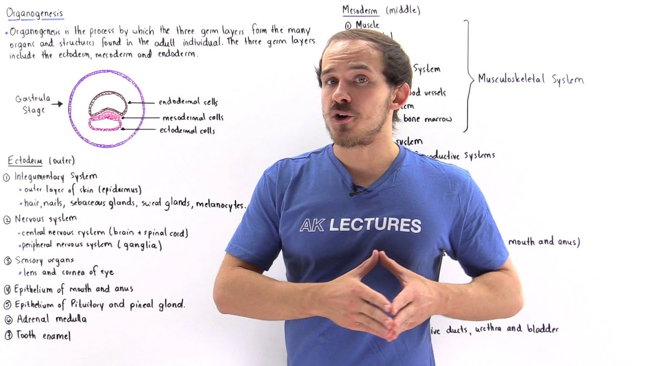

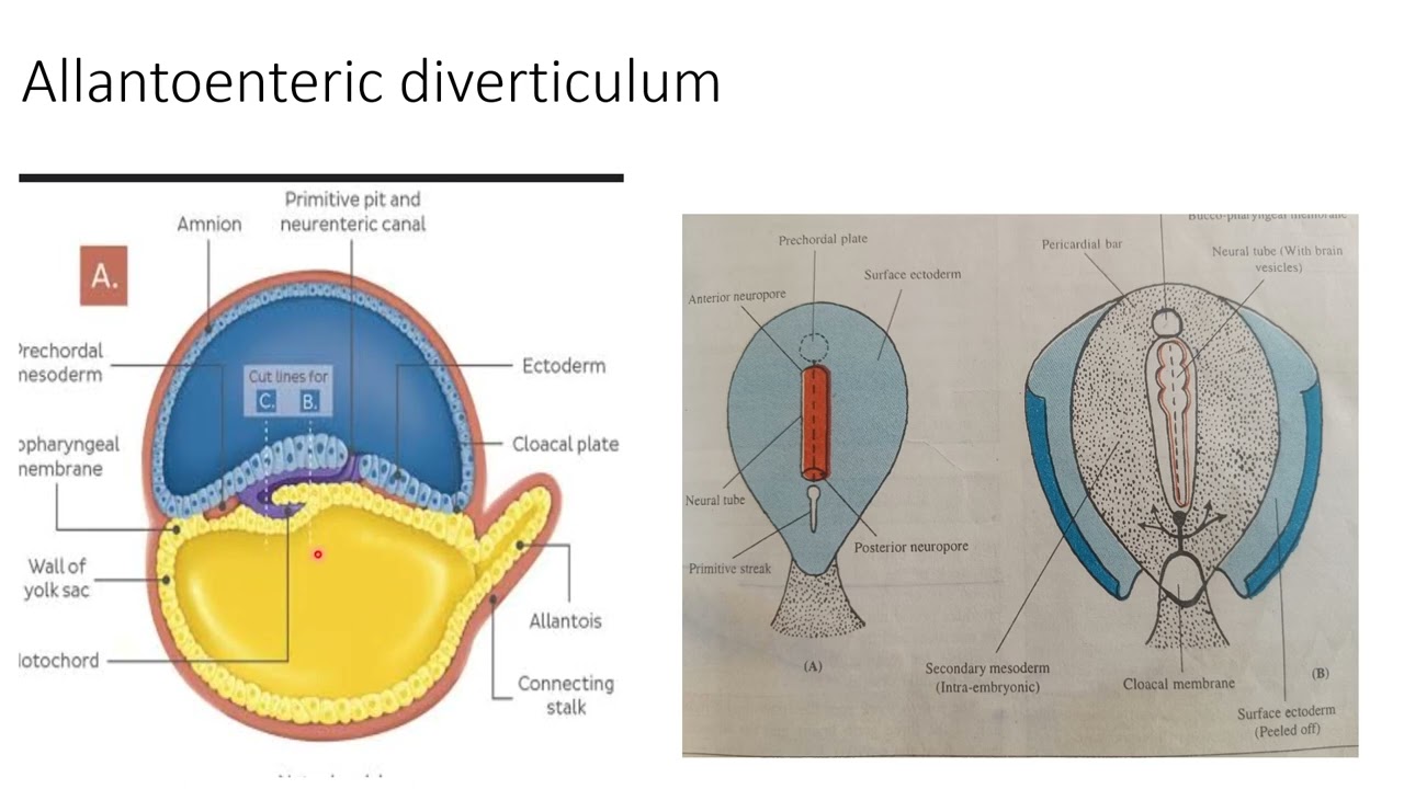




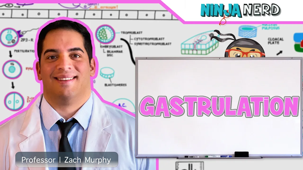
Gastrulation is an early embryological process following blastocyst implantation where the three germ layers—ectoderm (outer layer forming skin, hair, ears, nails, and nervous system including brain and spinal cord), mesoderm (middle layer forming musculoskeletal system, cardiovascular system, excretory system, and reproductive system), and endoderm (inner layer forming digestive system, lungs, liver, pancreas, bladder, thyroid, parathyroid, and thymus)—are formed through a process involving invagination along the primitive streak, ultimately giving rise to all organs and systems in the adult human body.
Gastrulation: Formation of the Three Germ Layers
Added:Understanding of early embryonic development stages, specifically fertilization, cleavage, and the structure of the blastocyst (including the inner cell mass and trophoblast).
Familiarity with the concepts of cellular potency (totipotency vs. pluripotency) and the basics of cell differentiation.
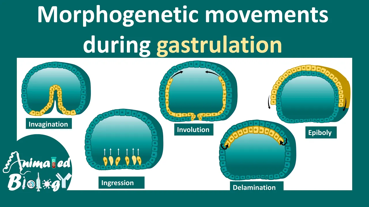
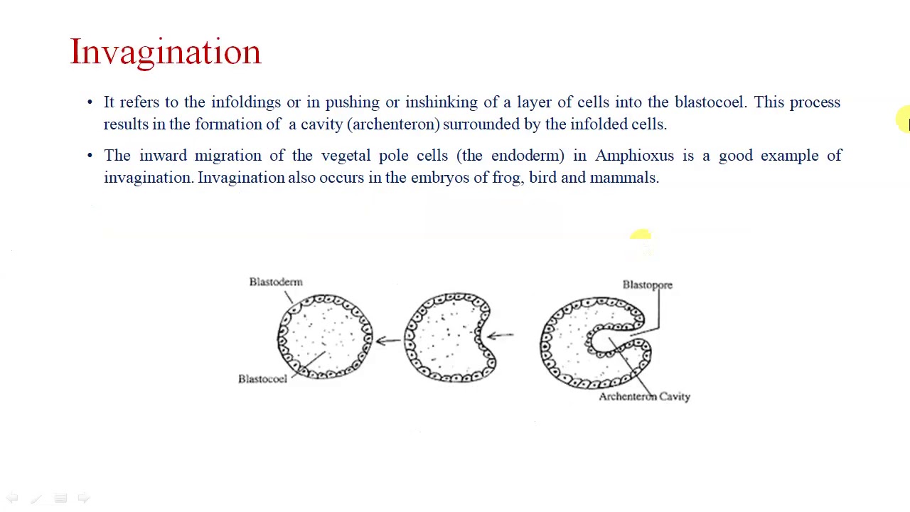
Basic knowledge of cellular movement mechanisms, such as invagination, involution, and epiboly, as well as cell adhesion molecules.
An understanding of basic anatomical axes and planes of symmetry (anterior-posterior, dorsal-ventral) in developing organisms.
The process of neurulation, which is the immediate next step involving the formation of the neural tube and the early nervous system from the ectoderm.
Detailed organogenesis, studying how specific organs and organ systems develop from each of the three germ layers (e.g., cardiogenesis from mesoderm).
Clinical applications and developmental pathologies, such as congenital birth defects (e.g., sacrococcygeal teratoma) that arise from abnormal gastrulation.
Comparative embryology, analyzing how gastrulation patterns differ across different species (amphibians, birds, and mammals).
Applications in regenerative medicine and stem cell research, specifically how scientists mimic gastrulation signals in vitro to differentiate stem cells into specific tissues.
591.5K views7Klikes11:35Original Release: 2014-12-09
Following the implantation of the blastoyst into the endometrium of the uterus, another process known as gastrolation begins. And gastrolation is an early embryological process in which the three distinct germ layers are formed. And these germ layers include the ectoerm, the endoderm, and the mesoderm. Now, as we'll see in just a moment, the cells of each one of these germ layers eventually give rise to specific organs and structures and systems found within the adult human individual. So, let's begin by briefly discussing which structures and which organs and systems are formed by each one of these three germ layers. And let's begin with the ectoerm. Now, the ectoerm is the outer layer. It's the external layer of that developing embryo as we'll see in just a moment. And that ectoerm gives rise to the integimementary system of the human body. And that includes the outer portion of the skin, the ears, the nails, and the hair. Now, because of an invagination process, as we'll see in just a moment, a portion of that ectoderm makes its way into that developing embryo and this forms the nervous system of the human body and that includes the brain and the spinal cord and it also includes all the nerve cells that are part of the peripheral nervous system. We also form the pituitary gland that is found beneath that hypothalamus in the brain. Now let's move on to the middle layer. The actually not the middle layer let's discuss the internal layer. So this is the innermost layer found in that developing organism in that developing embryo and this is called the endoderm.
Now the endoderm layer gives rise to the epithelial layer of the lungs, our digestive system, the pancreas, the bladder, the liver and it also forms the thyroid and the parathyroid gland as well as the thymus. And finally, what about the messaderm? Well, the mesoderm is the middle layer of the developing embryo. It's found between these two layers. And so it makes sense that the messaderm basically creates everything between the skin as well as our digestive epithelium. So we have things like the muscularkeeletal system. So that includes the bone and the cartilage as well and the three types of muscles.
We have the cardiac muscle, we have the smooth muscle and we have the skeletal muscle. Now because we form the cardiac muscle that means we also form the cardiovascular system of the body and that includes the heart as well as the blood vessels. We also form the excratory system as well as the reproductive system. So that includes the gonads of the female the ovaries as well as the gonads of the male the testes. So these are the three different layers that are formed during the process of gastrolation. The question is how does gastrolation actually take place? So let's take a look at the following six diagrams that ultimately describe the process of gastrolation the formation of the three distinct germ layers. So let's begin with diagram one which describes the process of implantation when that blastois the embryo actually develop actually implants itself onto the endometrium the lining of the uterus. So this is the endometrium and this is the blastoyst.
So recall that the blastoyst consists of a region known as the trophoblast and these are the purple cells the dark purple cells found as shown. We have the light purple cells that form the inner cell mass and this entire inner cavity that contains a fluid that provides nutrition to these developing cells is known as the blastoal.
Now the inner cell mass the cells shown in light purple eventually form these three distinct germ layers while the cells of the trophoblast eventually give rise to the corion as well as to the placenta. And remember the placenta is the structure that provides a source of nutrition and oxygen from the mother and to that developing organism that developing fetus. Now let's suppose we move from diagram one to diagram 2. What happens in between these two diagrams?
Well, basically the cells of the trophoblast begin secretreting and releasing digestive enzymes and these digestive enzymes degenerate this section of the endometrium and that allows the entire blast to make its way entirely into the endometrium and that hole is eventually sealed off by blood clots as well as uh as well as by developing epithelial cells. So in diagram 2, the entire embryo is found inside that endometrium. And also notice that the cells of the inner cell mass differentiate into two different types of cells. We have the green cells that make up the hypoblast and we have these blue cells that make up the epibblast.
Now the hypoblast cells eventually give rise to the endoderm while the epibblast eventually gives rise to the ectoerm and as we'll see in just a moment the mesoderm develops in between as a result of a process called invagination when we form that primitive streak as we'll see in just a moment. Now when we go from diagram 2 to diagram 3, what happens is the upper portion of the hypoblast and the upper portion of the epiblast eventually migrates upward towards this pole and we basically form the following diagram. So notice this entire green portion eventually develops into the umbilical vessel. Now in nonhumans it's called the yolk sack and the umbilical vessel uh vesicle eventually becomes part of the umbilical cord system and we also form this cavity that is created by the epibblast cells and this is known as the amniotic cavity and this is where that organism the fetus will actually be found as we'll see in just a moment. Now we also have this extension of the trophoblast. This purple section that will develop into the coron and ultimately into the placenta. Now when we go from diagram 3 to diagram 4, something important takes place. We form something called the mesoderm. So that's the middle layer of that developing embryo. And these are the red cells as shown in this diagram. Now what happens is if we zoom in on this section we get the following diagram. And what happens is we have this invagination process where the blue layer basically invaginates and it forms the primitive streak and along the primitive streak which is basically this axis where invagination takes place. These blue cells essentially invaginate and move inwards. And as they move inwards, these blue cells develop into the red cells that make up the mesoderm. And as this pushing process takes place, all these red cells eventually are pushed around this entire structure. So if we look at this diagram, what happens when we go from diagram 4 to diagram five is there is an inward push that goes into this direction and that pushes all these red cells around the following diagram. And so all these red cells eventually make their way uh around the green structure, around the blue structure, and ultimately around the entire trophoblast, the purple section of this diagram. And so when we go from diagram 4 to diagram 5, this is what we produce.
So this invagination process along the primitive streak, the movement of the ectoderm cells into the red section, the formation of these red cells eventually pushes all these red cells and the masser layer around the green structure, the blue structure and around this entire purple structure to form the following diagram. So this is basically our umbilical vasicle and this is basically the amniotic cavity where that fetus will be found. Now what happens when we go from five to six is this section basically moves inward. So it pushes this way and eventually we form an embryo that looks something like this. And notice that this entire structure here is that developing embryo. It's that developing fetus. So the red portion is the actual embryo along with this green portion and the blue portion. The red portion is basically that middle layer. It's that messed and the messed will basically form the musc the muscularkeeletal system, the cardiovascular system, the excretoryy system, the reproductive system, everything found in between the green layer and that blue layer. Now the blue layer is found on the outside. It's the external layer and it will develop that skin layer as well as the ears, the nails and the hair. And not only that because this blue layer, notice it will eventually move inside. It also forms the nervous system, the brain as well as our spinal cord. And this green layer which is found on the inside will form our digestive system. It will form the epithelial layer of our digestive system as well as the lungs, the liver, the pancreas and the bladder. Now this structure develops into the umbilical vesicle. This entire structure is the corionic cavity and this outside portion is the corion. This extension is what becomes our placenta. This uh uh this structure here that connects the placenta, the coron to the growing embryo is the embryionic stock and this will eventually develop into the embryionic tube. that embryionic um section the embryionic cord that connects that placenta to that developing fetus and this is the amniotic cavity that contains that houses that growing fetus. So this is the process that we call gastrolation.
It it is basically an early phase of embryological development in which following implantation the three germ layers the ectoerm the endoderm and the mesoderm begin to form and ultimately form the different types of structures organs and systems that are found inside the human Body.
Up Next

11:43
Neural Tube Patterning: Early Nervous System Development (Part 2)
@thehistologywizard5783
8.6K views•2020-08-02

50:12
Bacterial Communication: Quorum Sensing and Biofilm Formation Explained
@JHUAAP
12.6K views•2011-06-28

7:47
Post-Translational Modifications Explained: Types & Functions
@AKLECTURES
136.8K views•2014-09-11

7:08
Bacteriophages: Earth's Deadliest Killers and Future Antibiotics
@kurzgesagt
34.6M views•2018-05-13
Related Study Plans & Knowledge Roadmaps
Structured learning paths in Biology
biology
RNAi: Biogenesis, RISC & Gene Silencing
01Molecular Biology Foundations: DNA to Protein
02Introduction to RNA Interference (RNAi)
03Molecular Biogenesis of miRNA and siRNA
+ 2 more learning modules
10h5 modules
Explore Roadmap
biology
Mycorrhizae: Signaling, Biophysics & Ecology
01Foundations of Mycorrhizal Symbiosis
02Molecular Signaling and Root Colonization
03Biophysics and Bioenergetics of Nutrient Exchange
+ 1 more learning modules
11h4 modules
Explore Roadmap
biology
Circadian Clocks: TTFL, SCN & Periphery
01Foundations of Molecular Biology & Gene Expression
02Introduction to Chronobiology and Actograms
03The Core TTFL: Molecular Architecture of the Clock
+ 3 more learning modules
14h6 modules
Explore Roadmap
biology
Population Genetics: Math, Drift & Selection
01Genetic & Probability Foundations
02The Hardy-Weinberg Principle and Baseline Models
03Mathematical Modeling of Natural Selection
+ 3 more learning modules
18h6 modules
Explore Roadmap