


















![🧠👀Cuánta CAFEÍNA debes tomar (y a Qué HORA) para *Maximizar sus Beneficios*⚠️[Lo estás haciendo Mal]](https://i.ytimg.com/vi_webp/O4CnRSTS0zA/maxresdefault.webp)


Circadian clocks are self-generated biological rhythms that evolved to anticipate daily environmental changes, controlled by a core molecular feedback loop where CLOCK and BMAL1 proteins activate transcription of period and cryptochrome genes, which then repress their own activators to create rhythmic oscillations; these clocks exist in nearly all body cells and tissues, with the suprachiasmatic nucleus serving as the master coordinator that synchronizes peripheral clocks through temperature signals and other molecular pathways.
Circadian Clocks: Clock Genes, Cells, and Circuits Explained
Added:Basic gene expression mechanisms, specifically transcription, translation, and negative feedback loops.
Fundamental neuroanatomy, including the structure and function of the hypothalamus and the visual pathway.
Cellular signaling and transduction, particularly how external stimuli (like light) trigger intracellular changes.
The general concept of homeostasis and physiological rhythms in living organisms.
The distinction and synchronization between the central master clock (Suprachiasmatic Nucleus) and peripheral clocks in organs like the liver and gut.
Pathologies associated with clock disruption, such as shift-work sleep disorder, metabolic syndrome, and mood disorders.
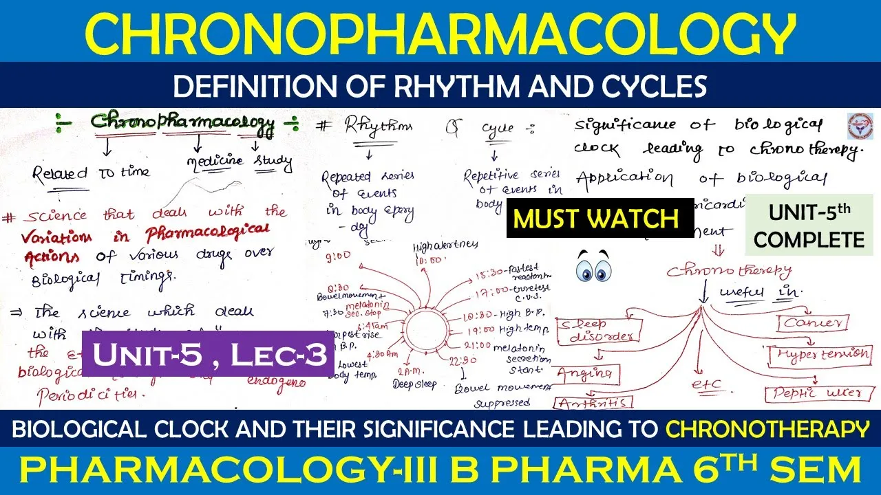
Chronopharmacology and chronotherapy, which study how the timing of drug administration affects efficacy and side effects.
The evolutionary biology of circadian systems across different species, from cyanobacteria to mammals.
42.7K views496likes33:17Original Release: 2014-03-24
Hello, my name is Joe Takahashi. I'm at uh UT Southwestern uh medical center in Dallas and the Howard Hughes Medical Institute. And today I'll be talking about uh circadian clocks in uh three different lectures.
So as all of us know we live on a planet that is uh governed by the dal cycle which is caused by the earth's rotation.
Uh and this has led to uh the evolution of biological clocks in virtually all living organisms to anticipate uh the changes in the environment. And what I'd like to do today is really to uh give you an introduction to biological clocks. First talk about the genes and then about the clocks and cells and then how they're organized uh in the body. So to get us all oriented uh perhaps the most familiar uh biological rhythm to us is our sleepwake cycle. And so this is an example of a sleepwake record from a young German medical student who volunteered for an experiment in the 1960s uh in a Mox plank institute in Andex, Germany where uh they have two underground bunkers where uh people can go in to what we call temporal isolation. And so in this record here, uh, the blue bars show when the German medical student is awake and the yellow bars show when he's sleeping. And then there's a little white arrow that shows the low point in the body temperature rhythm of our circadian rhythm uh, which occurs uh, about 3 hours before we wake up. And so this experiment which actually lasted about 40 days um shows two really important features of circadian rhythms. So in the first week um the student is living in the apartment. The door is open so he's exposed to the 24-hour light schedule.
Uh and what you can see is that he's waking up at about 8 o'clock um in the morning real time shown at the uh bottom of this graph. Uh and then he uh goes to sleep around midnight each day for about a week. But then at this point right here, the door to that apartment is closed and he goes into temporal isolation uh shown in this green area here. uh and this shows the first feature of his circadian rhythms and that is you can see that his sleepwake pattern continues but there's a difference. The timing of that pattern drifts to later and later times each day. So down at the bottom of the record he's waking up every 25.4 hours instead of every 24 hours. Okay.
So that shows the first feature of circadian rhythms. They are selfgenerated. they're endogenous to our bodies. Uh, and they're not precisely 24 hours. The second feature is actually shown in the bottom here where he continues to live in the apartment, but the door is open again and he's exposed to the 24-hour environment. Uh, and he then goes back to a very regular schedule where he wakes up actually about 8:00 in the morning and goes to sleep at midnight.
Um, and this shows the second feature of rhythms, and that is that even though they're endogenous, they're still regulated or synchronized by the external environment. So now the period of his rhythm matches the period of the earth's rotation 24 hours. And that process is called entrainment. Uh, which involves two features. One is period control. So, our rhythms adopt a 24-hour cycle. And the second is phase control where he wakes up at 8:00 in the morning again uh with the same phase as he had before. Now, there is one curious feature in this record and that is on this graph his uh phase plot is actually on the second 24-hour cycle of the graph. uh instead of waking up at 8:00 over here, he's waking up at 8:00 over here. Uh so why did that happen? Well, it turns out that while he was in isolation, he went through one fewer biological cycles than the number of real days that actually uh passed during that time period. So he lost a day. So anyway, just an example uh of our most familiar biological rhythm.
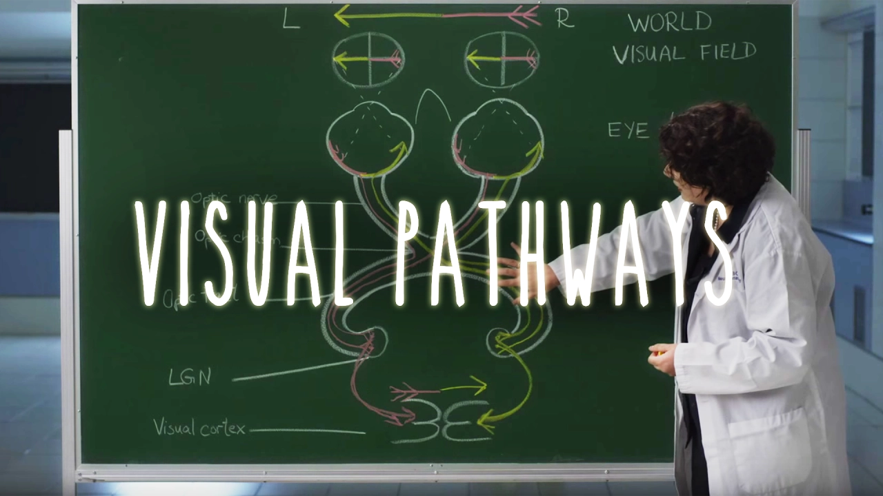
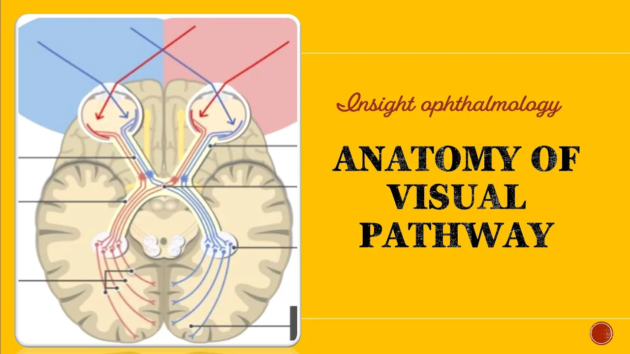
Now in humans the principal synchronizer of our rhythms is light. Light cycle mediated by special photoceptors in our eyes in cells that are called intrinsically photoceptive gangalene cells which project down the optic nerve into these two yellow wing-like structures in the hypothalamus which are called the supercismatic nuclei. And within each nucleus are about 10,000 neurons, each of which is capable of generating circadian rhythms in a solutonomous manner. And so even in humans and mammals, the fundamental unit for generation of circadian rhythms is the cell.
Now the uh origin of what we would call the modern era of the genetics of circadian rhythms really was started by these two individuals, Ron Kopka and Seymour Benzer. Uh and this is a picture of Ron and Seymour uh back in 2000.
And it turned out what Ron did in the 60s was he undertook a genetic screen in fruit flies and isolated mutations that changed the circadian rhythms of those of those flies. U he isolated three mutants, one that abolished the rhythm, one that shortened the rhythm and one that lengthened the rhythm. Um and incredibly these three mutants uh were uh mapped to the same locus the same gene and they named that gene the period gene.
Now since uh that discovery which was made in 1971 uh many different groups tried to isolate genes uh of a similar manner in mammals but we all failed until the late 90s.
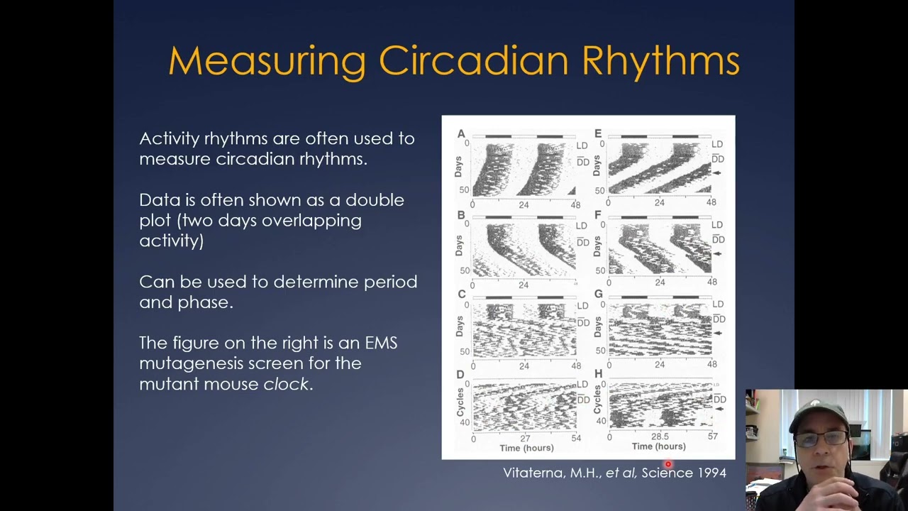
uh and so it wasn't possible in those early days to go from the Drosophila period gene to the mouse or human period gene very easily using DNA sequence homology. And so uh in the early 90s instead we decided to take a step back uh and to not look for similar genes but instead to look for clock mutants uh but instead of using fruit flies uh we would do the same kind of experiment that Kopk and Benzer did uh but we would do those experiments in the mice in mouse um and so uh using the mouse house, we were able to conduct uh a genetic screen which was done by Martha V. Turna and this illustrates uh the products of that very first screen we conducted in the early 90s where we used the activity rhythm of a mouse shown here. This is the record of a normal mouse. The black bars indicate when the mouse is active.
Uh and uh in this case the plot is double plotted. So you can see the pattern better. Uh the green and white bar at the top indicates the light cycle to which the mouse was exposed at the beginning of the record. But then uh in the bottom of this record right here, the mouse is in constant conditions. And so we can measure the period of its biological clock. Uh and in this screen, um Vita Turner found this particular mouse shown here in the bottom right. uh which has a period of 25 hours. It was mouse number 25.
Uh and it turned out this period lengthening was caused by a single gene mutation. And so uh we named this mutation clock and this shows uh the effect of that mutation on the periodicity of the rhythm. So these are actually grandchildren of that founder mouse. This is a wild type normal mouse.
two mice that carry uh one copy of the mutant gene. And then at the bottom, this is the record of a mouse that has two mutant copies of the gene or a homozygous mutant clock. Uh and this mouse is very unusual because it has a period of its rhythm that's 28 hours long. It wakes up four hours later each day.
uh and so um using this mouse uh we were then able to try to identify the gene.
Now back then in the '9s uh this was still a difficult task. The mouse genome is comparable to the human genome. There are about 20 chromosomes. Uh in the early 90s we had only about 300 genetic markers. Uh, and of course today we have genome sequence, but back then there was no genome sequence. And so we're working in the blind, uh, if you will. And so my laboratory uh shown here in this picture taken around 1997 uh worked together as a team over a three-year period uh to actually isolate the gene using a method that we call positional cloning which today I sort of call as the geneticist genome positioning system or GPS.
uh where we just try to uh use markers in the genome and hone in and try to find the location of that gene.
And so the first step in this process is what's called genetic mapping. And this is shown at the very top here in these blue bars which represent a region of mouse chromosome 5 uh where we use DNA markers and find uh whether or not they're associated with the mutant phenotype or not. And using this process which is called genetic mapping we can estimate the position of the gene in in a chromosomeal location. Now, back then, of course, unfortunately, none of the genes here were known. And in fact, the DNA in this region wasn't even cloned.
And so, shown below are what are called physical maps, which are large fragments of DNA shown in green and yellow, uh, which represent clones of DNA that we had to isolate to, um, find this region.
Um and so uh we were able to um cover the region in both the green clones which are called yeast artificial chromosomes or bat clones which are called bacterial artificial chromosomes in yellow.
Um now the breakthrough for us um came through a functional approach where we took a mutant mouse okay and we were able to then inject DNA large fragments of DNA from that region to test whether they might be able to rescue the mutation in the clock mutant mouse. So this shows you the general idea of the experiment. We're injecting DNA into eggs and then we uh recover mice that are carrying these large bacterial artificial chromosome clones uh and we then see whether they can rescue or fix or repair the mutation. And these are the results of that experiment. On the left are shown four activity records of clock mutant mice as as you've seen before that have the long period and loss of rhythm phenotype. And on the right are four clock mutant mice that are carrying this very large transgenic fragment that completely rescues the behavior. So this is an a very important result for us because it told us the gene was contained in that piece of DNA.
Uh, and so, uh, going back to that physical map, uh, the yellow clones here are the pieces of DNA that could rescue the mutant phenotype in a mutant mouse.
Uh, they're pretty large. Back 54, the top one is 140 kilobases in size. And then these two orange fragments above were also um fragments that we made into mice but they failed to rescue. And so this told us that the gene had to be located in this region where the yellow bars are located. Uh and ultimately we found that there was a very large gene in this region shown here at the top uh which eventually we named the clock gene. Okay. It's a very interesting gene. It's about 100,000 base pairs in size. It's very big and it has 24 exxons. Uh we say facitiously one exxon for every hour of the day.
Um now in that mutant mouse there was only a single DNA change between the mutant and the wild type and that was an A to T transversion shown here. uh in a splice donor site that caused skipping of a single exxon in the clock protein.
Um now the clone the cloning of this gene revealed that the clock protein the predicted protein uh was very interesting because it had some clear protein motifs indicating that its function. So it had a basic helix loop helix uh domain shown in green which is a DNA binding domain. It then had a second domain called PAS or PASS uh which is a protein interaction domain.
Incredibly that's a sequence that was found in the original period protein.
uh and then finally in the C terminus of the protein there's a glutamine or Q-rich uh region which is characteristic of activation domains of transcription factors or proteins that activate transcription. So clock turned out to be very interesting because the amino acid sequence gave us very strong indications of what the function of this protein might be. Uh this is really not the case for the period protein, the original first clock protein to be found uh because period only had this PAS domain uh which at the time really had no known function. Uh so what is clock doing? So um shortly after isolating clock uh we found that it interacted with a second protein called BAL1 uh in work that we did with Char Charles Whites at Harvard. Uh and it turned out that BAL and clock form what's called a transcriptional activator. They bind to regulatory DNA sequences in the promoters of genes. One of those genes turned out to be the period gene itself.
Uh, and it turns out that the clock protein uh can still interact with BMAL, can still bind DNA, but it's deficient in uh activating transcription of many genes.
So this uh animation then shows you uh how we think clock be period and cryptochrome work together to form a molecular clock in mammals. So there are three period genes per one two three and two cryptochrome genes cry 1 and two and these genes are both activated by clock and bell in the daytime. their RNAs are transcribed. The proteins are translated in the cytoplasm.
Uh the purr and cry proteins as we call them for short can interact with each other and then they transllocate back into the nucleus at night. uh and as their levels increase at night they then interact directly with clock and be and repress the activation potential of clock BL which thereby turns off their own transcription.
uh once their transcription goes down their protein products go down and eventually at the end of the night the purr and cry proteins are turned over and disappear and the next morning uh clock and be can then activate a new round of transcription. So this is a very simple uh description of what we call the core feedback loop which uh makes up the mechanism of the circadian clock in mammals.
So uh incredibly uh work done by Louis Pachek uh showed that there was a sleep disorder that uh is caused by a change in the timing of sleep preference in humans which is called advanced sleep phase syndrome. And this is an ASPS pedigree um from Pachek.
And when they found the causitive gene in this family, what they found was it was caused by mutation in the human period 2 gene, one of the clock genes that we're just talking about. So this pathway that uh we just described, we now know is conserved uh from mouse all the way to humans. uh and distant relatives of these genes also uh are conserved in fruit flies.
So this is a uh a more modern view of the clock gene network. In the middle here is that core transcriptional feedback loop with clock purr and cry.
But in addition at the top is a second loop discovered in about 2002 by Ulie Shibler's lab that uh involves the nuclear receptors uh reverb alpha and roar. Uh these form a second feedback loop and it turns out that they regulate the transcription of be in most tissues and clock in some tissues to cause a second oscillation.
Uh also shown in this diagram are two very important um pathways that regulate the stability of the cry protein and the period protein. Turns out that the half- livives of these two repressor proteins are very important in regulating the periodicity of the clock.
uh and then finally at the bottom um clock and be turn out to have uh many other target genes uh in the genome which we'll discuss later.
So how is it that we study the clock? Uh and so one of the important tools that we use is to use reporter genes such as luciferase or firefly luciferase um an enzyme that can generate light from a substrate. We use luciferase and we fuse it to the period 2 protein uh and then uh replace the period 2 gene in a mouse with a period 2 luciferase fusion gene. Um and what this allows us to do is to visualize the uh amount of per two protein. So, this time-lapse movie shown here on the right uh is a 7-day time-lapse recording of a brain slice that contains the supercismatic nucleus of a mouse uh in which we can see luciferase levels oscillating uh in level but also spatially we can see a pattern of luciferase that progresses as a wave across the nucleus.
Um, using this kind of reporter, uh, we can even go to the cellular level. So, this, uh, movie here shows a time-lapse movie recorded by David Welsh for a six-w week period in time, uh, in which it's possible to follow the circadian rhythm of individual fibroblast cells. So in this movie, each of these twinkling stars is a single cell that is oscillating over a six week period. And on the right here, I've given you six examples of individual cells that are showing circadian oscillations that persist for the duration of this experiment.
Now, this uh experiment really changed our view completely about how the clock works. So, I told you there's a clock in the hypothalamus, the supercismatic nucleus, and we've known about that clock since the 1970s, but we really did not realize that the body also contained such robust oscillators, especially at the single cell level. And it really wasn't until we could literally see the single cell rhythm that we could appreciate how robust or strong the cell autonomous clock is in our bodies.
So we now know that virtually all of our major organ systems in the body contain circadian clocks. They're driven by these cell autonomous clocks.
uh the brain SCEN clock we think is still in charge but many different tissues throughout the body the liver pancreas lung even skin have the capability of generating circadian rhythms. uh and this has really led to a whole new set of questions having to do with what is the function of these clocks in the body and how might they be controlled uh in the organism. So one example of a what we would call a peripheral clock are clocks in the pancreas. So these experiments done by my colleague at at Northwestern Jabass shows that pancreatic eyelet cells have beautiful circadian rhythms as you can see here with per luck recording.
Uh and we then asked what happens if we disrupt the gene for clock in this case the BL1 gene specifically in pancreatic beta cells.
uh and this shows uh two photo microraphs of pancreatic eyelets. The red indicates BMAL protein which has been deleted in the BMAL pancreatic specific knockout shown here. The green and blue show insulin and um uh the rest of the eyelet cell anatomy here. What we find is by deleting BAL uh the mouse has a loss of glucose regulation as shown here compared to control mice that have intact uh BAL in their pancreas. And this loss of glucose regulation uh is due to insufficient insulin production as shown in this bottom graph here. So this is a very nice example of the function of a clock gene in a peripheral tissue uh which can lead to uh diabetes and uh insufficient insulin production.
So how is it that timing information might be integrated in the SCN and throughout the whole body?
So um if we do a thought experiment, this is actually an experiment uh proposed by the philosopher Derek Parett at Oxford uh in which he had a scientist gradually replace the cells in his body uh with the cells from Greta Garbo. And he asks as uh you increase the number of cells that were placed in your body, at what time uh during that process do you cease to be yourself and become Greta Garbo?
Um, now for those of you who aren't familiar with Greta Garbo, of course you might just think of Angelina Jolie as uh a substitute or modern-day version of Greta Garbo. Now, interestingly, that very pro provocative kind of experiment can be done uh in in the laboratory. We can do that in mice. So the way we do that is that we can mix the cells uh from two different strains of mice together and create a single mouse that contains cells derived from two different types of mice. Uh and these mice are called chimeas.
And so the way these mice are made is you um prepare embryos from the two strains of mice indicated in blue and yellow. You can mix these embryos at an early stage and they will form a chimeriic embryo uh which you can then transplant into mice uh and then produce chimeic mice.
So this kind of experiment was done by Sharon Lozettes.
Uh she made the mice, tested their circadian behavior and then uh using anatomical methods uh looked at the proportion and distribution of those cells uh in the brain. So in this case the normal cells were marked uh by the laxi gene which we could visualize with blue dye. So this slide shows the two strains of mice we used. On the left are the wild type strain which is pigmented has normal behavior and is laxi positive. So the cells are blue and on the right is the mutant strain which is albino has clock mutant behavior and the cells are laxi negative and in the middle is shown the genetic cross of these two strains the F1 which confirms that uh the behavior is intermediate in phenotype. Sharon made a huge number of chimeas. These are a set of them. And what you can see already is that mice at the top are carrying pigmentation in their coat colors and the mice at the bottom are generally albino and that's because they were ordered by their phenotype.
If we look at their SCNs, you can also see that the mice at the top have more blue cells. Those are wild type cells.
and the mice at the bottom have more mutant cells which are white. Okay. And then finally, this uh illustration here uh shows you the activity record of each one of these mice as little postage stamps. But what's important, it's a little hard to see, but what's important is at the top uh the mice have wild type or normal behavior. At the bottom, they have clock mutant behavior. But in the middle they show something very interesting. They show an intermediate phenotype.
Uh and so here's a better picture of this this idea. The uh left shows the mice that have mainly blue or wild type cells. And then in the middle are mice that are carrying about a 50/50 mixture of mutant and normal cells. uh and then here are clock mutants which are predominantly u blue negative which have mutant behavior. What's interesting is that the behavior is intermediate when we have a 50/50 mixture of cells. Okay, this is shown here uh for three examples of mice carrying about 50/50 mixtures of cells. And what is really amazing here is that these mice have behavior that's identical to a clock hetererozygate mouse. Uh but no cell in the body of those mice is clock hetererozygous.
Uh they're either homozygous clock or wild type mixture. So we call them clock hetererozygot phenocopies because they uh have the same behavior but genetically they're not the same. Okay.
And what this tells us is that the system that controls circadian behavior in the mouse has some way of integrating information uh across wild type and mutant cells to in some kind of additive way produce an intermediate phenotype uh that actually is equivalent to what you would see in a genetically intermediate cell.
So this uh kind of experiment shows the circadian system really is integrating uh quantitative information in a very uh additive or almost mathematical way uh to generate behavior in the mouse.
So in summary um I've tried to uh summarize for you the uh really the discovery of the clock gene network and then how we study those genes in a cellular context uh both at the cell autonomous level and at the organismal level looking at tissue organization.
And so what we can see from this work is that clock cells play a very important role throughout the body, but how they're organized uh is fundamentally different in the brain and the periphery. And in the next section, we'll go into the actual details of how those differences occur.
Up Next
8:18
How Our Cells Track Time: Circadian Rhythms Explained
@QuantaScienceChannel
85.7K views•2023-10-10

50:12
Bacterial Communication: Quorum Sensing and Biofilm Formation Explained
@JHUAAP
12.6K views•2011-06-28

42:45
MicroRNAs: Gene Regulation Explained by David Bartel | iBiology
@scicommlab
233.1K views•2014-03-24

7:08
Bacteriophages: Earth's Deadliest Killers and Future Antibiotics
@kurzgesagt
34.6M views•2018-05-13
Related Study Plans & Knowledge Roadmaps
Structured learning paths in Biology
biology
RNAi: Biogenesis, RISC & Gene Silencing
01Molecular Biology Foundations: DNA to Protein
02Introduction to RNA Interference (RNAi)
03Molecular Biogenesis of miRNA and siRNA
+ 2 more learning modules
10h5 modules
Explore Roadmap
biology
Mycorrhizae: Signaling, Biophysics & Ecology
01Foundations of Mycorrhizal Symbiosis
02Molecular Signaling and Root Colonization
03Biophysics and Bioenergetics of Nutrient Exchange
+ 1 more learning modules
11h4 modules
Explore Roadmap
biology
Circadian Clocks: TTFL, SCN & Periphery
01Foundations of Molecular Biology & Gene Expression
02Introduction to Chronobiology and Actograms
03The Core TTFL: Molecular Architecture of the Clock
+ 3 more learning modules
14h6 modules
Explore Roadmap
biology
Population Genetics: Math, Drift & Selection
01Genetic & Probability Foundations
02The Hardy-Weinberg Principle and Baseline Models
03Mathematical Modeling of Natural Selection
+ 3 more learning modules
18h6 modules
Explore Roadmap