Yeast transformation is a fundamental genetic technique that allows researchers to introduce foreign DNA (either as plasmids or PCR products) into yeast cells to modify their genotype through integration or replication, enabling applications such as gene mutation, deletion, overexpression, or suppression of lethality; the standard protocol involves a six-day workflow including strain thawing and growth, liquid culture inoculation, cell harvesting and washing with TEL buffer, transformation reaction setup with PEG mix, heat shock treatment, plating on selective media, replica plating for selection verification, and final strain freezing after PCR confirmation.
Yeast Transformation Lab Tutorial: A Step-by-Step Protocol
Added:Deep Dive
Fundamental understanding of yeast (Saccharomyces cerevisiae) biology, including its eukaryotic cellular structure and life cycle.

SACCHAROMYCES BSc 1st year classification,life cycle, reproduction
16.4K
Saccharomyces cerevisiae is the only unicellular fungus in the kingdom Fungi, belonging to Ascomycota division, with approximately 2016 species; it has a plasma membrane, cell wall containing chitin, nucleus, and large vacuole, and reproduces through budding (vegetative reproduction under favorable conditions) and sexual reproduction (fusion of haploid cells forming diploid zygote that undergoes meiosis to produce ascospores under unfavorable conditions).
Click to watch →
![Structure of Yeast [Fungi] Life Cycle of Saccharomyces | Reproduction Budding in Yeast Microbiology](https://i.ytimg.com/vi_webp/FzuKH5Sz1R4/maxresdefault.webp)
Structure of Yeast [Fungi] Life Cycle of Saccharomyces | Reproduction Budding in Yeast Microbiology
49.1K
Saccharomyces yeast cells are eukaryotic fungi with a chitin cell wall containing ectoplasm and endoplasm, possessing enzymes invertase and zymase for sugar metabolism; they reproduce vegetatively through budding (forming unequal cells that detach as pseudomycelium) or fission (producing equal daughter cells), and sexually through three life cycle types: haplobiontic (dominant haploid phase with diploid zygote forming eight ascospores), diplobiontic (prominent diploid phase with ascospores functioning as gametangia), and haplodiplobiontic (equal duration of haploid and diploid phases); economically, yeast is vital for baking (bread leavening), brewing (alcoholic beverages), producing invertase enzyme, synthesizing proteins and vitamins, generating biofuel ethanol, and treating skin diseases.
Click to watch →
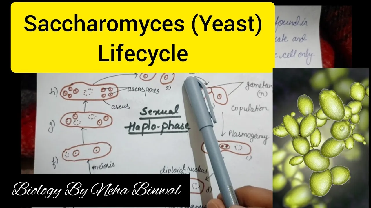
Saccharomyces (Yeast) Lifecycle l Biology By Neha Binwal l UPSC Botany l NEET l
20.4K
Saccharomyces belongs to Kingdom Fungi, Division Ascomycota, Class Hemiascomycetes, Order Saccharomycetales, and Family Saccharomycetaceae. Commonly known as yeast, it is a saprophytic fungus with approximately 40 species worldwide, thriving in sugar-rich environments. The body is unicellular but can form pseudomycelium when cells attach in short chains. The cell can be oval, elongated, or rectangular in shape. Saccharomyces reproduces through vegetative methods (budding, fission, endospore formation) and sexual reproduction. Endospores serve as survival structures during adverse conditions. Three life cycle patterns exist: haplobiontic (haploid phase dominant, diploid confined to zygote), diplobiontic (diploid phase dominant, haploid confined to ascospores), and haplodiplobiontic (both phases equally present). The haplodiplobiontic cycle is most common in Saccharomyces cerevisiae (baker's yeast), where ascospores behave as gametes, fuse to form diploid cells, which undergo budding and meiosis to continue the cycle.
Click to watch →
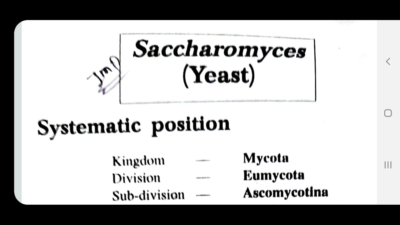
Yeast or Saccharomyces Part 1st(B.Sc. 1st Botany,Microbiology and Fungi)
5.8K
Yeast (Saccharomyces) is a haplobiontic fungus belonging to Kingdom Fungi, Division Ascomycota, with approximately 40 species; it has significant economic importance in baking and alcohol production, and its cell structure includes a three-layered cell wall (mannoprotein outer layer, glucan middle layer, and chitin inner layer), a large central vacuole containing hydrolytic enzymes, and a single nucleus that divides through mitosis with spindle body formation at opposite poles, followed by cell division into daughter cells.
Click to watch →

Life Cycle of Yeast (Saccharomyces) | Why called a Haplodiplobiontic life cycle? #yeast #fungi
16K
Saccharomyces cerevisiae (baker's yeast) exhibits a characteristic haplo-diploid life cycle essential for understanding eukaryotic biology. The organism exists in two equally dominant phases: haploid and diploid. The cycle begins when haploid cells of mating types 'a' and 'alpha' fuse to form diploid cells under pheromone influence. Diploid cells reproduce asexually through budding, doubling approximately every 100 minutes. During starvation, diploid cells function as asci, undergoing meiosis to produce four haploid ascospores (two of each mating type). Upon ascus rupture, haploid cells are released and continue the haploid phase through budding. Eventually, haploid cells fuse again to restart the cycle. This alternating pattern between haploid and diploid phases defines the haplo-diploid life cycle, making yeast an ideal model organism for studying eukaryotic cell biology.
Click to watch →
Deep Dive
Basic principles of molecular cloning, including the structure and function of plasmids, promoters, and selectable markers.

Plasmid design (bacterial expression vector)
33.5K
Plasmids are circular DNA molecules naturally found in bacteria that serve as essential cloning vectors in genetic manipulation. They function by moving DNA between different species, similar to how mosquitoes transmit malaria parasites. Bacteria contain both chromosomes with essential genes and plasmids with optional genes like antibiotic resistance. Plasmids are self-replicating and range from 1,000-20,000 base pairs, making them ideal for inserting typical genes of 2,000-4,000 base pairs. Three fundamental components are required for functional plasmid vectors: (1) Origin of replication (ori) - a specific DNA sequence enabling DNA replication machinery to recognize and copy the plasmid, controlling copy number per cell (2-3 to hundreds); (2) Selectable marker - provides visible phenotype (like antibiotic resistance) to identify cells that have taken up the plasmid, since transformation efficiency is very low (~1 in 10,000 cells); (3) Multiple cloning site (MCS) - contains multiple restriction enzyme recognition sites for inserting genes of interest. The standard cloning workflow involves: (1) amplifying the gene of interest via PCR; (2) digesting both plasmid and gene with restriction enzymes; (3) purifying and ligating the fragments using DNA ligase; (4) transforming the recombinant plasmid into host cells (typically E. coli).
Click to watch →

Plus Two Biology Christmas Exam | Biotechnology:Principles And Processes | Chapter 9 | Exam Winner
229K
Cloning vectors are DNA molecules used to carry foreign genetic material into host cells. The cloning process involves inserting DNA fragments into vectors, introducing vectors into host cells, and allowing replication to produce multiple identical copies. Plasmids contain essential features including the origin of replication (ori) for independent replication, selectable markers (such as antibiotic resistance genes) for identifying transformed cells, and cloning sites (multiple cloning sites) where foreign DNA can be inserted. Plasmid copy number refers to the number of plasmid molecules per bacterial cell, affecting DNA production capacity.
Click to watch →

Biotechnology - Principles and Processes | Superfast Revision with Handwritten Notes and PYQs
711.4K
Plasmids are circular, double-stranded DNA molecules that replicate independently and are not essential for bacterial survival. Good cloning vectors require: origin of replication (ori) for independent replication, multiple cloning sites (MCS) for various restriction enzymes, and selectable markers (antibiotic resistance genes). pBR322 is a widely used plasmid with ampicillin and tetracycline resistance genes, ori, rop gene, and multiple cloning sites. The rop protein aids plasmid replication. Vectors enable transport of foreign DNA into host cells for cloning.
Click to watch →

Plus Two Biology | Chapter 9 |Biotechnology : Principles And Processes | Full Chapter | Exam Winner
414.5K
Plasmid vectors are circular DNA molecules used to carry foreign DNA into host cells. Essential vector components include the origin of replication (ori) for plasmid maintenance, selectable markers (such as antibiotic resistance genes) for identifying transformed cells, and cloning sites for inserting foreign DNA. The PBR322 plasmid is a widely used cloning vector containing multiple selectable markers including ampicillin and tetracycline resistance genes.
Click to watch →

Biotechnology: Principles and Processes Mega-Marathon | BioBali Series for NEET ft. Vipin Sharma
122.5K
Effective cloning plasmids must contain several features: (1) Origin of replication (ori) for plasmid maintenance, (2) Selectable markers (like antibiotic resistance genes) for identifying transformed cells, (3) Cloning sites (multiple cloning sites) for inserting foreign DNA, and (4) Promoters for gene expression in host cells.
Click to watch →
Deep Dive
The concept of auxotrophy and selection media (e.g., synthetic defined amino acid dropout media).

Understanding Selection and Drop-out Media
269
Selection media in molecular biology use either antibiotics or nutrient dropout to identify cells that have successfully taken up plasmids, with antibiotic-based selection relying on resistance genes expressed from the vector to kill non-transformed cells, while dropout media exploits auxotrophic host strains that cannot synthesize certain nutrients unless the vector provides the necessary biosynthetic genes; however, both methods only identify transformed cells and require additional techniques like colony PCR or blue-white screening to distinguish between recombinant plasmids (with inserted genes) and non-recombinant plasmids (empty vectors), with phenotypic lag being an important consideration as transformed cells take approximately one hour to express antibiotic resistance.
Click to watch →
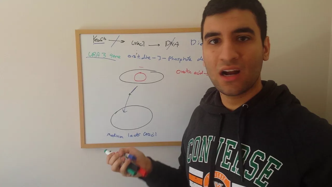
Auxotrophy In Yeast (Auxotrophic Complementation technique)
5.9K
To perform auxotrophic complementation, researchers use selective growth media that lack the essential compound the mutant strain cannot produce. For example, when working with URA3 mutants, the medium is supplemented without uracil. Only yeast cells that have acquired the ability to produce uracil through transformation with a plasmid containing the functional URA3 gene can grow on this selective medium. This creates a direct link between the presence of the marker gene and the ability to grow, enabling efficient selection of successfully transformed cells.
Click to watch →

Bacterial growth media - terms, types (LB, TB, SOC, etc.), and usages in biochemistry
987
Bacteria are classified as autotrophic (synthesize required nutrients) or auxotrophic (require external supply). The Ames test exploits histidine auxotrophs: mutagenic compounds increase revertant frequencies restoring histidine synthesis. Selection allows only bacteria with desired traits (antibiotic resistance) to grow. Screening reveals differences without growth restriction (blue-white screening using lacZ disruption). These principles underlie bacterial-based assays for detecting mutagens and identifying genetically modified organisms.
Click to watch →

Types of Culture Media.mp4
13.2K
Minimal media contain the minimum ingredients required to support bacterial growth. These media lack one or two specific amino acids and are used to select for auxotrophic bacteria (organisms that cannot synthesize certain compounds and must obtain them from the environment). This helps study natural recombinations occurring in nature, as only bacteria capable of synthesizing the absent amino acids will grow on minimal media.
Click to watch →

Sinh học phân tử - Buổi 5 - Đột biến gen_2
275
Microbiologists use multiple methods to isolate mutants: antibiotic resistance selection uses media containing antibiotics to isolate resistant bacteria; phage resistance selection uses media containing bacteriophages; replica plating transfers colonies from complete to minimal media to identify auxotrophic mutants that fail to grow without specific nutrients; penicillin enrichment selects auxotrophic mutants that survive penicillin treatment but cannot grow. Auxotrophic mutants cannot synthesize specific nutrients (amino acids, vitamins) and require supplementation for growth. Fungal auxotrophic mutants are selected by growing fungi on minimal media, where only prototrophic fungi grow and form fluffy colonies. The mixture is filtered to separate growing from non-growing fungi, with the non-growing fraction containing auxotrophic mutants. These mutants are verified by growing on minimal media supplemented with specific nutrients; colonies that grow on supplemented media but not on minimal media are auxotrophic.
Click to watch →
Deep Dive
Standard aseptic laboratory techniques for culturing and handling microorganisms without contamination.

Aseptic Techniques Required Practical 6 | A-Level Biology
1.6K
Aseptic techniques are essential methods for culturing bacteria that prevent contamination. Key techniques include: using sterile pipettes (pre-packaged, single-use, discarded in disinfectant); flame sterilizing inoculating loops until they glow red; flaming the neck of McCartney bottles before use; working near a lit Bunsen burner to create upward convection currents that carry airborne microorganisms away; boiling agar to kill contaminating microorganisms; keeping petri dish lids at an angle during transfer to minimize exposure time; taping lids securely but not sealing completely to allow oxygen for bacterial respiration. These techniques ensure only the intended bacterial strain grows without environmental contamination.
Click to watch →

Técnicas Assépticas
3.5K
Aseptic techniques are essential laboratory methods for manipulating microorganisms while minimizing contamination, involving the sterilization of all materials (Petri dishes, culture media, test tubes) before use and the use of flame sterilization to create a sterile environment around the work area; key practices include working alone with materials within reach, opening Petri dishes minimally with one hand while holding the base with the other, sterilizing the platinum loop by passing it through the flame before and after each use, and performing all manipulations behind the flame to protect both the material and the operator from contamination.
Click to watch →

A Level Biology - 6.2.1.7 - Microorganism Cultures
808
Aseptic techniques are essential methods to prevent contamination when culturing microorganisms, including hand washing or wearing gloves, disinfecting work surfaces, working near a Bunsen burner to create a protective heat current, flame sterilizing bottle necks, and minimizing air exposure when handling Petri dishes; microorganisms require carbon compounds for respiration, nitrogen compounds for proteins, and appropriate temperatures to grow, so all equipment and nutrient agar (either solid or liquid broth) must be sterilized in an autoclave at 121°C for 15 minutes before use; inoculation methods include streaking with a wire loop, seeding liquid culture onto agar, spreading with a glass spreader, or using a swab, followed by incubation at 24-36 hours (not 35°C to avoid pathogen growth) and observation of colony characteristics without opening the dish, with control plates included to verify aseptic technique effectiveness.
Click to watch →
GCSE Biology Revision "Required Practical 2: Culturing Microorganisms" (Triple)
864.1K
Aseptic technique is essential when preparing bacterial cultures to prevent contamination by unwanted microorganisms naturally present in the environment. Key steps include: sterilizing petri dishes, bacterial nutrient broth, and inoculating loops by passing them through a Bunsen burner flame; using adhesive tape to secure agar plate lids; and placing plates upside down in an incubator at 25°C. This prevents moisture from dripping onto colonies and reduces the risk of harmful bacteria growing.
Click to watch →

TECNICA ASEPTICA
28K
Aseptic technique is a set of procedures designed to prevent contamination during the handling of microbial cultures by creating a sterile working area (approximately 12 cm radius) using a Bunsen burner, sterilizing instruments like inoculation loops and pipettes before and after use, and maintaining all materials within the sterile zone throughout the procedure.
Click to watch →
Prerequisite Knowledge
- Concept 01Fundamental understanding of yeast (Saccharomyces cerevisiae) biology, including its eukaryotic cellular structure and life cycle.
- Concept 02Basic principles of molecular cloning, including the structure and function of plasmids, promoters, and selectable markers.
- Concept 03The concept of auxotrophy and selection media (e.g., synthetic defined amino acid dropout media).
- Concept 04Standard aseptic laboratory techniques for culturing and handling microorganisms without contamination.
Subsequent Learning
- Step 01Advanced gene editing techniques in yeast, such as CRISPR-Cas9 genome editing and homologous recombination for gene knockouts.
- Step 02Yeast Two-Hybrid (Y2H) assays to screen and study protein-protein interactions in vivo.
- Step 03Industrial scale-up applications of engineered yeast in metabolic engineering, synthetic biology, and biofuel production.
- Step 04Troubleshooting methods for low transformation efficiency and optimization of expression protocols for recombinant protein production.
Experiment Intro
Culture Prep
Cell Harvest
PEG Mix Step
Heat Shock
Replica Plating
Test Positives
Experiment Intro
0:10Playing Section
- 1
Yeast transformation changes strain genotype via DNA integration or plasmid replication.
- 2
Purpose includes gene mutation, deletion, overexpression, or lethality suppression.
- 3
Plan starts with fresh cultures and proceeds through five-day workflow.
Rapid Single-Day Protocols and Cell-Free Alternatives
While the traditional six-day yeast transformation protocol is a foundational teaching tool, modern synthetic biology and high-throughput research increasingly critique it as slow, labor-intensive, and inefficient. Opposing methodologies advocate for "one-step" or "rapid" transformation protocols that bypass the lengthy strain revival and multi-day preparation phases, reducing the timeline from nearly a week to a single day. Furthermore, advanced research is moving toward cell-free protein synthesis (CFPS) systems. Cell-free technology eliminates the need for living host cells and transformation entirely, allowing researchers to transcribe and translate DNA in vitro in a matter of hours. This shifts the focus from tedious cell-culture maintenance to rapid, automated genetic prototyping, presenting a major alternative to classic in vivo yeast transformation.
Deep Dive
Advanced gene editing techniques in yeast, such as CRISPR-Cas9 genome editing and homologous recombination for gene knockouts.
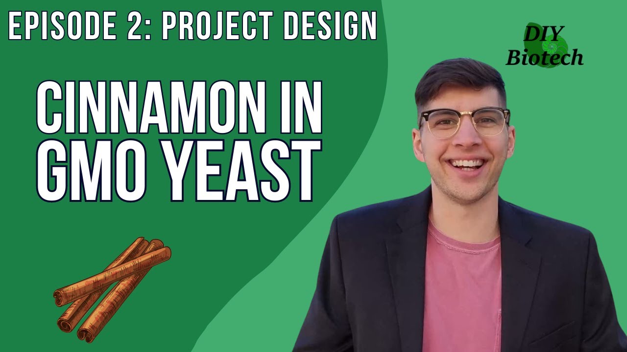
Writing in DNA | How to Design CRISPR GMO Yeast
2.8K
This video demonstrates how to design and construct CRISPR-GMO yeast capable of producing cinnamon compounds by integrating specific genes using a dual plasmid transformation system. The process involves finding the necessary genes from scientific literature, codon optimizing them for the host organism (Yarrowia lipolytica), designing Gibson assembly arms for seamless integration, and using CRISPR-Cas9 to make precise cuts in the yeast genome where the new genes will be incorporated through homologous recombination. The dual plasmid system uses auxotrophic markers to ensure the yeast retains both plasmids temporarily, allowing the CRISPR system to function before the plasmids can be cured away, leaving only the integrated cinnamon-producing genes.
Click to watch →
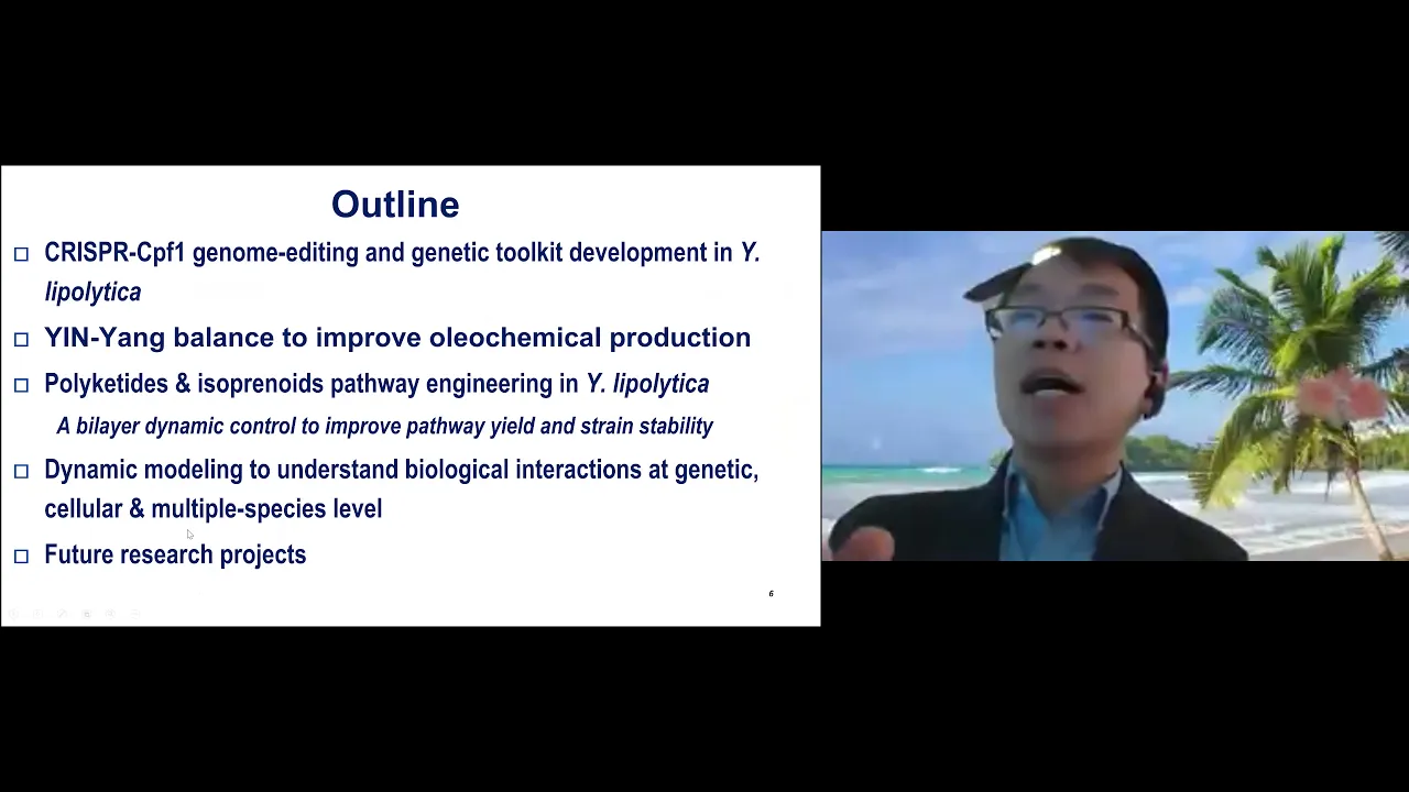
Encoding decision-making functions into cell metabolism
242
Living cells function as intelligent systems capable of organizing information, responding to stimuli, and maximizing their own benefit. This perspective frames cells as computational entities rather than passive chemical reactors. The intersection of synthetic biology, metabolic engineering, and intelligent control aims to discover metabolic costs, stability, and social interactions at genetic, cellular, and multi-species levels. By rewriting genetic code, researchers can control both mass flow and information flow in living systems, enabling cells to be reprogrammed for energy production and biomedical applications. Traditional CRISPR-Cas9 systems face limitations in yeast due to high genome GC content (~65%), causing off-target effects. Cpf1 overcomes these limitations by recognizing TTNTN PAM motifs with lower mismatch probability. Researchers enhance CRISPR efficiency through self-cleaving ribozymes, optimized promoters, and tRNA-based expression systems. Multiplex genome editing achieves 80% efficiency for three genes simultaneously. Reporter systems using housekeeping genes enable visual screening of successful edits, achieving 45-75% mutation rates. These advances enable precise genetic modifications essential for metabolic engineering and synthetic biology applications in yeast platforms.
Click to watch →
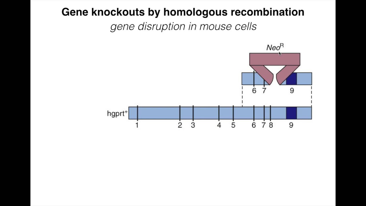
Homologous Recombination IV
14K
Homologous recombination is a molecular biology technique that allows scientists to precisely modify genes by using PCR-generated DNA constructs containing a gene of interest (such as antibiotic resistance genes like kanamycin or neo R) flanked by homologous sequences (~50 base pairs) that match the target gene's genomic regions; this enables targeted gene knockouts or insertions by exploiting the cell's natural recombination machinery, with selection markers allowing identification of successfully modified cells, and in mice, this process involves embryonic stem cell manipulation followed by injection into early embryos to generate transgenic animals.
Click to watch →
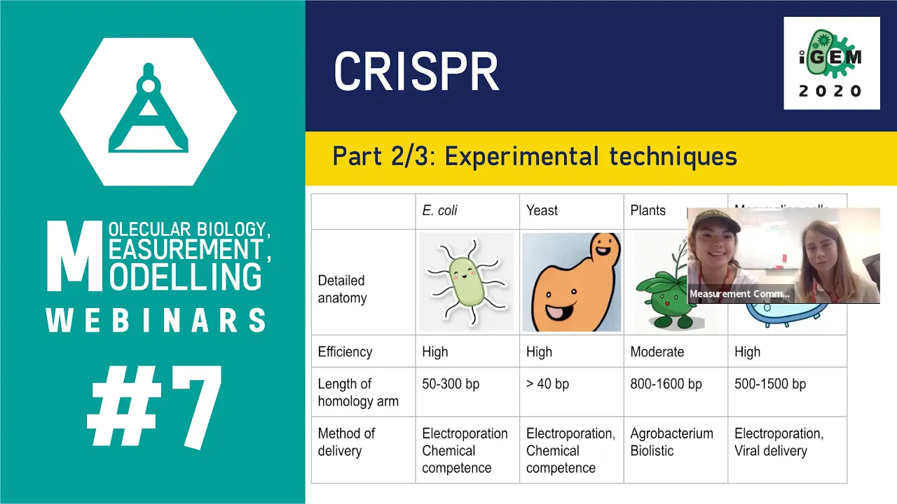
Week #7: CRISPR 2/3: Experimental techniques
988
CRISPR systems have evolved from natural bacterial defense mechanisms into versatile gene editing tools. Cas9 produces blunt cuts with G-rich PAM sequences, while Cas12a creates sticky cuts with T-rich PAM sequences. Beyond DNA cutting, CRISPR enables gene repression using catalytically dead Cas9 fused to repression domains, and gene activation through activation domain fusions. A functional CRISPR system requires three essential elements: the Cas protein (nuclease), guide RNA, and donor DNA for precise editing. Two repair mechanisms exist: Non-Homologous End Joining (NHEJ) creates indels for knockouts through error-prone repair, while Homology-Directed Repair (HDR) uses donor templates for precise gene knock-ins. For yeast, a toolkit consists of linearized Cas9 plasmid, guide RNA plasmid, and donor DNA. Guide RNA is designed in silico targeting ~20 bp sequences, constructed by annealing complementary oligos, and assembled via Golden Gate assembly with BsmBI.
Click to watch →
Blonde Ale Yeast Experiment | Sundew Vs. Bananza Vs. Kaiser | New Omega Yeast's
5.7K
CRISPR-Cas9 gene editing technology enables precise modification of brewing yeast by removing specific genes, such as the one that produces 4-vinyl guaiacol (clove/fennel flavor), allowing brewers to create new flavor profiles by enhancing other ester-producing characteristics like banana (from German hefeweizen strains) or strawberry/melon (from Belgian strains).
Click to watch →
Deep Dive
Yeast Two-Hybrid (Y2H) assays to screen and study protein-protein interactions in vivo.

Yeast Two-Hybrid Assay
4K
The yeast two-hybrid assay is a powerful biochemical technique developed in 1989 by Stanley Fields and Ok-Kyu Song at SUNY Stony Brook that enables researchers to study protein-protein interactions in vivo. The method exploits the modular nature of transcription factors like GAL4, where the DNA binding domain and activation domain can be separated and fused to different proteins. When two proteins of interest interact, they bring these domains together to reconstitute the transcription factor, activating reporter genes and indicating a protein-protein interaction. This technique has been widely adopted in research labs worldwide and has spawned various derivatives including yeast one-hybrid, three-hybrid systems, and reverse versions for screening interaction-deficient alleles, making it applicable for studying protein-DNA, protein-RNA, and protein-ligand interactions.
Click to watch →

Yeast Two-Hybrid Screen (Y2H) | Protein-Protein Interaction| Genomics Lab
578
The Yeast Two-Hybrid (Y2H) system is a molecular biology technique that detects protein-protein interactions by fusing two proteins of interest to different domains of the GAL4 transcription factor; when the proteins interact, they bring together the DNA-binding and activation domains, enabling transcription of reporter genes (such as those encoding histidine biosynthesis enzymes) that allow yeast cells to grow on selective media lacking histidine, thereby indicating a successful protein interaction.
Click to watch →
3.2. Interacción entre proteínas (Y2H)
10.6K
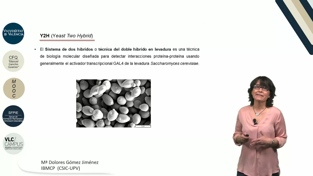
The yeast two-hybrid (Y2H) technique is a powerful molecular biology method that detects protein-protein interactions in vivo by exploiting the transcriptional activation properties of the Gal4 factor in Saccharomyces cerevisiae; it works by fusing two proteins of interest—one to the DNA-binding domain (bait) and another to the activation domain (prey)—so that when they interact, the activation domain approaches the regulatory regions of reporter genes, triggering transcription and allowing researchers to identify interacting protein pairs through phenotypic selection on selective media.
Click to watch →
Yeast two hybrid assay || protein-protein interaction #yeast two hybrid assay
106
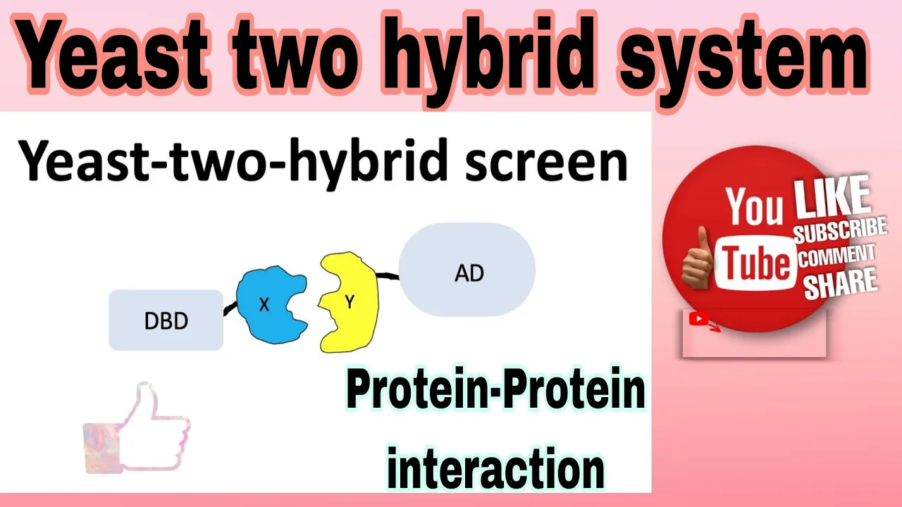
The yeast two-hybrid assay is a molecular biology technique that detects protein-protein interactions by utilizing a transcription factor with two separable domains: the DNA-binding domain (DBD) and the activation domain (AD). In this system, one protein of interest (the 'bait') is fused to the DBD, while another protein (the 'prey') is fused to the AD. When the bait and prey proteins interact, the DBD and AD come together to form a functional transcription factor that activates reporter gene expression; if no interaction occurs, transcription remains inactive. This allows researchers to identify and study protein-protein interactions in vitro.
Click to watch →

Yeast Two-Hybrid Assay (Y2H Assay) | Y3H and Y1H | Methods in Biology
376
The Yeast Two-Hybrid (Y2H) assay is a powerful molecular biology technique used to detect protein-protein interactions by splitting a transcription factor into two separate domains: a DNA-binding domain (BD) fused to one protein (bait) and an activation domain (AD) fused to another protein (prey). When the bait and prey proteins interact, the BD and AD domains are brought into proximity, reconstituting a functional transcription factor that activates a reporter gene (such as histidine synthase), allowing yeast cells to grow on selective media lacking histidine. This technique can be adapted into variants like Y1H (protein-DNA interactions), Y3H (protein-protein interactions mediated by RNA), and split ubiquitin Y2H (for membrane proteins).
Click to watch →
Deep Dive
Industrial scale-up applications of engineered yeast in metabolic engineering, synthetic biology, and biofuel production.
Synthetic Biology: Engineering Microbes to Solve Global Challenges - Jay Keasling
36.5K
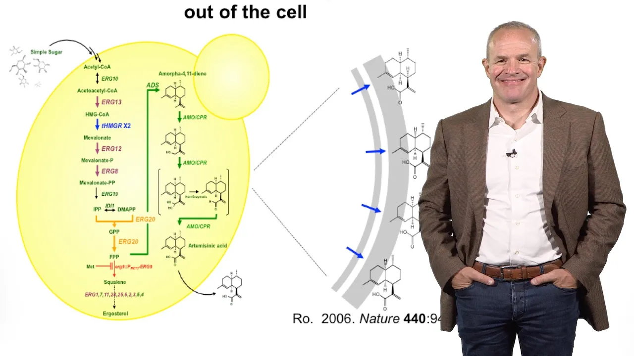
Ambrx optimized engineered yeast for high productivity, achieving industrial-scale production in Italy. Artemisinic acid is converted to artemisinin through light-catalyzed processes mimicking plant chemistry, with tablets manufactured in Morocco. By May, 16 million treatments had been delivered to Africa, with capacity for 100-150 million annually—addressing half global needs. The same engineering principles apply to biofuels: microbes produce hydrocarbons (gasoline, diesel, jet fuel) that excrete directly, floating to broth surfaces for simple purification. Similar approaches can produce specialty chemicals (mouthwash, perfumes, carpet, paint) currently derived from petroleum, representing 15% of oil value but significant profit margins.
Click to watch →

Engineering Yeast for Industrial Biotechnology
2.2K
This webinar presents advances in engineering yeast for industrial biotechnology, covering two complementary approaches: (1) CRISPR-based genome editing combined with recombination-based DNA assembly enables rapid strain engineering, reducing development time from years to weeks; and (2) systems biology approaches using genome-scale metabolic models with proteome allocation constraints provide predictive frameworks for understanding and improving yeast metabolism. The speakers demonstrate how these technologies are transforming yeast from a platform for biofuels to a versatile host for pharmaceuticals, cosmetics, and nutrition products.
Click to watch →
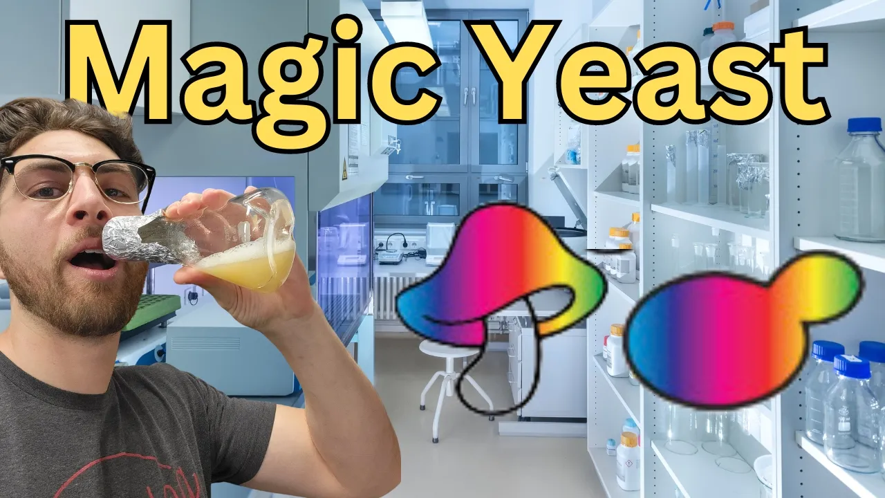
How to Make Psilocybin with Yeast | Journal Club
237.5K
This section covers translating laboratory achievements to industrial production scales. The video explains fed-batch fermentation, a cultivation strategy where nutrients are added gradually to maintain optimal growth conditions while preventing substrate inhibition. This approach increased psilocybin titers from milligram to gram-per-liter levels. The section also discusses aromatic amino acid pathway engineering, including overexpression of ARO1 and ARO2 enzymes, knockout of competing branches, and introduction of beneficial mutations like K229L in ARO4. The research demonstrates how combining multiple engineering strategies—promoter optimization, pathway redirection, and fed-batch cultivation—can achieve commercial-scale production. This illustrates the progression from basic pathway reconstruction to sophisticated industrial bioprocess development.
Click to watch →
Bioengineer Sarah Reisinger - 2014 Nelson Series
482
Native yeast metabolism for farnesene achieves only 23.8% theoretical yield from glucose. Amis engineered synthetic metabolic pathways achieving 28.6% glucose yield (20% improvement) and 170% oxygen utilization efficiency improvement. Implementation delivered 15% glucose yield improvement and over 70% oxygen consumption improvement. Remarkably, engineered strains exceeded theoretical limits of native metabolism, demonstrating that rational pathway redesign can surpass evolutionary constraints. CRISPR-Cas9 further accelerated gene editing from six weeks to one week per modification. This represents the future of industrial biotechnology: systematically redesigning metabolic networks to achieve performance impossible through natural evolution, enabling sustainable production of chemicals, fuels, and materials at commercial scales.
Click to watch →

Expanding yeast's diet to improve biofuel production
1.7K
Researchers at QB3, UC Berkeley, have successfully transferred transporter genes from the fungus Neurospora crassa to Saccharomyces cerevisiae yeast, enabling the engineered yeast to simultaneously import and ferment both glucose and xylose—the two primary sugars in plant cell walls—thereby improving biofuel production efficiency through synthetic biology approaches.
Click to watch →
Deep Dive
Troubleshooting methods for low transformation efficiency and optimization of expression protocols for recombinant protein production.

PDD-UKTPT SERDOS2026 Lidya Kristiani, S.Si, Ph.D. Advance Cellular Oncology Laboratory i3LUniversity
120
Effective troubleshooting requires addressing issues one at a time. When one problem is resolved but other groups still show issues, multiple underlying problems exist. Using positive controls (known working plasmids) distinguishes between technique errors and scientific problems. If a positive control fails, the issue is with technique or reagents. If it works but experimental samples fail, the problem lies with experimental conditions or plasmid quality. Transformation efficiency measures colonies obtained per microgram of plasmid DNA. Low efficiency results from low plasmid concentration, poor cell competency, or suboptimal conditions. Competent cell quality can be assessed by comparing colony counts between different cell preparations.
Click to watch →

Troubleshooting troublesome recombinant protein expression...
2.9K
When recombinant protein expression fails, systematically troubleshoot by first verifying if the protein is actually being expressed using Western blotting; if not expressed, check cloning accuracy and transformation efficiency; if expressed but absent from the supernatant, test for solubility issues by checking the pellet for inclusion bodies; if the protein is in the pellet, optimize conditions by lowering expression temperature, reducing inducer concentration, using fusion partners like SUMO or GST, or switching to different cell types; if the protein is soluble but purification fails, verify antibody specificity and optimize purification parameters.
Click to watch →

Recombinant protein expression systems & trying to get expression to work in bacterial cells
315
When optimizing recombinant protein expression in bacterial cells, first determine whether the issue is lack of expression or improper folding by comparing pre- and post-induction SDS-PAGE samples; if the protein is expressed but insoluble, try lowering the temperature (e.g., 16°C), reducing induction strength (e.g., lower IPTG concentration), using different cell strains (like Rosetta for rare codons), or adding fusion partners (SUMO, GST, MBP) to aid solubility and folding; if these simple optimizations fail, consider switching to more complex expression systems like yeast, insect cells, or mammalian cells (CHO, 293) for proteins requiring post-translational modifications or proper folding assistance.
Click to watch →

Recombinant protein production
1.4K
Common challenges in recombinant protein production include: (1) No or low expression: Caused by incorrect reading frame (addressed by bidirectional sequencing), protein toxicity (addressed by low-copy plasmids and tunable promoters), or codon bias (addressed by codon-optimized strains like BL21(DE3) RIL); (2) Inclusion body formation: Occurs when proteins cannot fold properly, addressed by directing proteins to periplasm, inducing at lower rates, or using chaperone-expressing strains; (3) Protein inactivity: Often due to lack of proper PTMs or improper folding, addressed by changing expression systems or inducing at lower temperatures.
Click to watch →

Engenharia Genética Aplicada à Expressão Heteróloga de Proteínas (Dia 26/09/2025)
129
One common problem in protein expression is complete lack of protein production, which can occur if the protein is toxic to the host bacteria. Toxic proteins can kill bacteria before induction, preventing expression. The problem may also result from leaky expression (protein production before induction), which can be fatal for toxic proteins. For lac-based promoters, adding glucose (0.2-2%) to the growth medium inhibits expression by repressing the lac promoter. For T7-based systems, using BL21(DE3) pLysS bacteria, which express T7 lysozyme, can reduce leaky expression. When proteins are toxic to host bacteria, several strategies can improve expression. Using C41 or C43 bacterial strains, which have additional membrane proteins, can help bacteria survive toxic protein expression. Adding signal peptides can direct proteins to the periplasm, reducing toxicity. Using lower copy number plasmids reduces protein concentration. Codon optimization involves changing rare codons in a gene sequence to improve expression in the host organism. Rare codons can slow translation and reduce protein yield. The software can optimize codons for specific host organisms. However, codon optimization must be balanced with the need to maintain protein structure, as some rare codons may be important for proper protein folding. Different host strains have different codon pools, and choosing the appropriate host can improve expression without codon optimization. For protein expression in Erlenmeyer flasks, the culture volume should not exceed 50% of the flask capacity (50 ml in a 250 ml flask). Excessive volume reduces aeration, which is critical for bacterial growth and protein expression. The flask should be shaken to provide adequate oxygenation. Excessive agitation can cause foam formation, which may require antifoaming agents. Disulfide bonds form between cysteine residues and are important for proper protein folding. In the reducing environment of the bacterial cytoplasm, disulfide bonds cannot form correctly. To express proteins with disulfide bonds, researchers should use periplasmic expression (with signal peptides) or specialized strains like Origami, which have a more oxidizing cytoplasm. Proper disulfide bond formation is essential for protein stability and function. Chaperone-assisted protein folding can be achieved by co-transforming with chaperone plasmids or adding benzyl alcohol to the growth medium. Lower expression temperatures (15-25°C) can improve folding by reducing aggregation. Chromatography separates proteins based on their interactions with a stationary phase and a mobile phase. Affinity chromatography using His-tag exploits the nickel-binding property of histidine residues. The protein binds to nickel ions in the stationary phase. The column is washed to remove unbound proteins. The target protein is eluted using imidazole, which competes for nickel binding. Size exclusion chromatography separates proteins based on size. The column contains porous beads that proteins cannot enter. Large proteins pass through the column faster (elute first), while small proteins enter the pores and elute later. Ion exchange chromatography separates proteins based on charge. The stationary phase contains charged groups that bind proteins of opposite charge. A salt gradient (like NaCl) is used to elute proteins by competing for binding sites. Hydrophobic interaction chromatography separates proteins based on hydrophobicity. The stationary phase is hydrophobic, and proteins bind through hydrophobic interactions. A gradient of organic solvent (like acetonitrile) is used to elute proteins by reducing hydrophobic interactions. After purification, protein activity must be verified. For enzymes, zymography (zymogram) can be used, where the gel contains substrate. Active enzymes will degrade the substrate, creating clear zones on the gel. This assay confirms both protein presence and activity. Inactive proteins may indicate improper folding or degradation. Zymography is particularly useful for verifying enzyme function after purification. Western blotting detects specific proteins in a sample. Proteins are separated by SDS-PAGE, then transferred to a membrane (nitrocellulose or PVDF). The membrane is blocked with BSA or milk to prevent non-specific binding. Primary antibodies specific to the target protein are applied, followed by secondary antibodies conjugated to enzymes. The enzyme-substrate reaction produces a visible signal. Western blotting requires antibodies specific to the target protein. These can be purchased commercially or produced in animals. To produce antibodies, the target protein is injected into an animal (like a rabbit), and the animal's immune system produces antibodies against it. The antibodies are then purified from the animal's blood.
Click to watch →
Experiment Intro
Culture Prep
Cell Harvest
PEG Mix Step
Heat Shock
Replica Plating
Test Positives
Experiment Intro
0:10- 1
Yeast transformation changes strain genotype via DNA integration or plasmid replication.
- 2
Purpose includes gene mutation, deletion, overexpression, or lethality suppression.
- 3
Plan starts with fresh cultures and proceeds through five-day workflow.
Rapid Single-Day Protocols and Cell-Free Alternatives
While the traditional six-day yeast transformation protocol is a foundational teaching tool, modern synthetic biology and high-throughput research increasingly critique it as slow, labor-intensive, and inefficient. Opposing methodologies advocate for "one-step" or "rapid" transformation protocols that bypass the lengthy strain revival and multi-day preparation phases, reducing the timeline from nearly a week to a single day. Furthermore, advanced research is moving toward cell-free protein synthesis (CFPS) systems. Cell-free technology eliminates the need for living host cells and transformation entirely, allowing researchers to transcribe and translate DNA in vitro in a matter of hours. This shifts the focus from tedious cell-culture maintenance to rapid, automated genetic prototyping, presenting a major alternative to classic in vivo yeast transformation.
Fundamental understanding of yeast (Saccharomyces cerevisiae) biology, including its eukaryotic cellular structure and life cycle.
Basic principles of molecular cloning, including the structure and function of plasmids, promoters, and selectable markers.
The concept of auxotrophy and selection media (e.g., synthetic defined amino acid dropout media).
Standard aseptic laboratory techniques for culturing and handling microorganisms without contamination.
Advanced gene editing techniques in yeast, such as CRISPR-Cas9 genome editing and homologous recombination for gene knockouts.
Yeast Two-Hybrid (Y2H) assays to screen and study protein-protein interactions in vivo.
Industrial scale-up applications of engineered yeast in metabolic engineering, synthetic biology, and biofuel production.
Troubleshooting methods for low transformation efficiency and optimization of expression protocols for recombinant protein production.
14.3K views337likes13:31@bioscienceinsightOriginal Release: 2021-03-19
[Music] good day fellow scientists and other curious minds today's bread and butter experiment is yeast transformation so you can use a transformation to change the genotype of a strain the dna we transform is either going to integrate into the genome as a pcr product or replicate as a dna plasmid we can use this transformation to mutate a gene delete a gene over express a gene or try to suppress lethality so we're going to do is we're going to get some strains out fresh make a fresh inoculation grow up some cells for the next day do the transformation and then we will test the strains before freezing them day one thaw and grow strains get fresh cells out of the minus 80 for the transformation streak them onto ypd or selective media incubate at 30 degrees for about 24 hours day two inoculation fill a flask with sterile media inoculate a small amount of cells to start a liquid culture prepare some dilutions if you want to avoid over inoculation incubate the cells overnight at 25 to 30 degrees day three transformation take the liquid cultures out of the incubator measure the od of cultures that are not obviously saturated an od 600 value of 0.6 to 0.8 is ideal for exponentially growing cultures harvest the cells by centrifugation pour off the media and wash with tell buffer tel buffer is just tris edta and lithium acetate diluted in water add tail buffer to the tube and vortex or invert to wash harvest the cells again and remove the wash buffer at some point during the harvest thaw salmon sperm dna at 100 degrees add 35 microliters of salmon sperm to each tube after it has cooled again add 5 to 10 microliters of water to the negative control add 5 to 10 microliters of the plasmid or pcr product to be transformed resuspend cells in tel buffer and split into each tube roughly 100 microliters each surprised your voice that is add 900 microliters of peg mix to each tube and mix well by vortexing peg mix is polyethylene glycol diluted intel buffer pegmix is thought to improve transformation efficiency by altering the chemical environment around the cell to promote dna uptake incubate the transformation reaction at 30 degrees for 30 to 90 minutes get some fresh plates ready for plating the transform cells after the incubation time expires add 60 microliters of dmso to each tube vortex well and incubate at 42 degrees for 15 minutes harvest cells by centrifugation and carefully aspirate away the peg mix [Applause] be very careful not to suck up the gloopy mass near the cell pellet because the cells will likely come with it resuspend the cells until buffer which can be tricky if there's a lot of solidified peg mix plate the full volume of resuspended cells on ypd and spread with glass beads [Music] after the volume soaks in remove beads and incubate at 30 degrees overnight day 4 replica plating place the sterile felt over the replica block and make a stamp on the felt with the lawn of cells from the transformation there's no need to apply significant pressure to the plate just ensure that the agar is making contact with the felt lightly press the new selection plate onto the cell covered felt incubate the new selection plates at 30 degrees for one to two days day five patching positives the moment of truth look into the incubator and see if there are colonies on the positive plate there should be theoretically none on the negative control plate streak a bit of each positive colony onto a new plate with the same selection allow the cells to grow into a patch for one to two days day six freezing and testing the strains after a day or two the patches should be grown and ready to test any false positive colonies will not form a healthy patch a small amount of cells go through the crude genomic prep a tiny amount of dna gets tested by pcr after screening for confirmed positive strains they are frozen check out the follow-up video to see which transformers were positive and how they were tested
Up Next

53:56
Ghost and Stray Light Analysis with TracePro Tutorial
@Lambdaresearchcorporation
2.7K views•2014-09-17

12:54
Triumph of Orthodoxy Icon: Byzantine Art & History Explained
@BenCallan
2.1K views•2024-08-06

9:49
FastAPI vs Flask vs Django: Choosing the Right Python Web Framework
@TechWithTim
302.5K views•2024-05-26

1:42
Game of Thrones Opening Credits: A Cinematic Analysis
@gameofthrones
46.3M views•2011-04-18
Related Study Plans & Knowledge Roadmaps
Structured learning paths in General & Interdisciplinary Studies
general-studies
Elevator Engineering: History, Physics & Cities
01From Hoists to Safety Brakes: The Dawn of Vertical Lift
02Hydraulic vs. Traction: The Engineering Physics of Elevators
03Modern Speed, Safety, and Algorithms
+ 2 more learning modules
12h5 modules
Explore Roadmap
general-studies
Rubber: Material Science, History & Geopolitics
01Origins of Natural Rubber & Mesoamerican History
02The Chemistry of Natural Rubber & Vulcanization
03The Geopolitics of the Rubber Boom & Colonization
+ 2 more learning modules
12h5 modules
Explore Roadmap
general-studies
Containerization: Evolution, Logistics & Trade
01Ancient Trade & Bulk Cargo: Amphorae to Barrels
02The Industrial Revolution & Cardboard Packaging
03The Containerization Revolution: Malcom McLean
+ 2 more learning modules
12h5 modules
Explore Roadmap
general-studies
Textile Tech: Evolution, Labor & Global Impact
01Pre-Industrial Textiles: Handloom & Cottage Industry
02Mechanization: The Spinning Jenny to the Power Loom
03The Factory System & Global Labor Transformation
+ 2 more learning modules
10h5 modules
Explore Roadmap