A lateral flow immunoassay is a rapid diagnostic test that uses capillary action to move a liquid sample across a nitrocellulose membrane containing capture antibodies; in a sandwich (non-competitive) format, target molecules in the sample bind to capture antibodies at the test line and to labeled detection antibodies, producing a visible signal that indicates the presence of the target analyte.
How Lateral Flow Immunoassays Work: Sandwich Format Explained
Added:Deep Dive
Basic immunology, specifically the concept of antigen-antibody binding, epitopes, and binding specificity.

Anticorpo / Imunoglobulina
114.9K
Antigens contain multiple specific regions called epitopes (also called antigenic determinants) where antibodies can bind. Each antibody recognizes and binds to only one specific epitope with extremely high specificity. A single pathogen may have many different epitopes, and different B cells produce different antibodies that recognize different epitopes on the same antigen, creating a diverse immune response.
Click to watch →

Imunoglobulinas / Anticorpos
35.3K
Antibodies bind to specific regions on antigens called epitopes. A single antigen protein can have multiple different epitopes, and each antibody is highly specific to bind only one particular epitope. This specificity is achieved through the variable regions containing CDRs. The high specificity prevents antibodies from binding to self-molecules, which is crucial for preventing autoimmune reactions.
Click to watch →

Константин Северинов. Введение в молекулярную биологию: Свой-чужой. Иммунитет.
23.2K
Antibodies bind to specific regions on antigens called epitopes. The binding occurs through complementary shapes and chemical properties between the antibody's variable region and the antigen's epitope. Antibodies can recognize both linear epitopes (continuous sequences of amino acids) and conformational epitopes (three-dimensional structures formed by amino acids that are not adjacent in the sequence). The specificity of antibody-antigen binding allows the immune system to distinguish between different pathogens and target them precisely.
Click to watch →

Biological Science Semester IV Human Health & Diseses Part 7 Immunology By WBCHSE
439
Immunology is the branch of science studying the immune system. An antigen is a foreign substance that enters the body and stimulates the immune system. Antigens can be microorganisms, bacterial toxins, dust particles, or other foreign materials, composed of proteins, glycoproteins, nucleoproteins, lipoproteins, polysaccharides, or nucleic acids. Antigenicity refers to the ability to trigger an immune response, but only those that activate the acquired immune system are called immunogens. An epitope is the specific part of an antigen that binds to an antibody, while the paratope is the corresponding part of the antibody. Epitopes can bind to antibody paratopes or cell surface receptors on lymphocytes. Antigens are classified as monovalent (single epitope) or multivalent (multiple epitopes).
Click to watch →

Part-3 Ch-10 Human health and disease Biology class 12 maharashtra board new syllabus Blood Group
119.2K
The antigen binding site is a specific region on an antibody where it recognizes and binds to a particular antigen. This site is highly specific and complementary to the shape of the antigen, similar to how a key fits into a lock. Antigen determinants (epitopes) are specific parts of an antigen that antibodies recognize and bind to. Each antigen has multiple determinants, and different antibodies can recognize different determinants on the same antigen. When an antibody binds to its specific antigen, they form an antigen-antibody complex. This complex formation is highly specific and triggers various immune responses that lead to the destruction or neutralization of the pathogen.
Click to watch →
Deep Dive
The physical principle of capillary action, which drives the passive flow of liquid through porous membranes.

Haz que el Agua 'Suba' Sola: La Técnica que Humilla al Riego por Goteo
65K
Capillary action is the physical phenomenon where water rises through porous materials against gravity due to molecular attraction between water and pore walls. This principle, which Aztecs understood 600 years ago, allows plants to access water continuously without top-down irrigation. Modern wicking beds replicate this principle using impermeable containers, gravel reservoirs, and permeable fabric layers. The result is 30-50% faster plant growth, 40% higher yields, and 80% less water usage compared to traditional methods.
Click to watch →

How This Pen Changed The World
3.8M
Capillary action is a physical phenomenon where liquids are drawn upward through narrow tubes or porous materials due to molecular attraction. In this process, the attraction between the liquid and the walls of the container is stronger than the attraction between liquid particles themselves. As a result, liquid particles at the top get pulled up by the container walls until the weight of the liquid eventually cancels out the upward force. This same principle allows water to climb tree roots and nutrients to flow through our bodies.
Click to watch →

El Agua y sus Propiedades | Ciencia en lo Cotidiano T1
224.1K
Capillary action is the ability of a liquid to flow through narrow tubes or porous materials without external pressure. It occurs because water molecules are attracted to the walls of the material (adhesion) and to each other (cohesion). In paper towels, the cellulose fibers create tiny channels where water rises from below, carrying additional molecules upward through molecular attraction.
Click to watch →

Adezyon-Kohezyon Konu Anlatımı | 50 Günde TYT Fizik Kampı
75.8K
Capillary action is the ability of a liquid to flow in narrow spaces without the assistance of external forces, such as the movement of water through plant xylem or absorption of water by paper towels. Capillary action occurs in both upward and downward directions depending on the relative strengths of adhesion and cohesion. When adhesion is stronger than cohesion, the liquid rises in the capillary tube; when cohesion is stronger, the liquid level drops.
Click to watch →
![[#하이라이트#] 살아있는 것처럼 우유를 마시는 석상 미스터리! #다빈치노트 EP.5](https://i.ytimg.com/vi/Yp25GpNuE-E/maxresdefault.jpg)
[#하이라이트#] 살아있는 것처럼 우유를 마시는 석상 미스터리! #다빈치노트 EP.5
291.8K
Capillary action is the ability of a liquid to flow in narrow spaces without the assistance of, or even in opposition to, external forces like gravity. This occurs due to the combined effects of cohesion (liquid molecules sticking together) and adhesion (liquid molecules sticking to the surface of the material). In porous materials like certain stones, capillary action allows liquids to be drawn upward through tiny channels and pores.
Click to watch →
Deep Dive
An understanding of conjugate labels, such as colloidal gold nanoparticles or fluorescent dyes, and their role in signal detection.

Webinar Jurusan Teknologi Laboratorium Medis Poltekkes Surabaya Tanggal 31 Oktober 2025
177
Detection labels in lateral flow assays provide the visible signal for test results: (1) Gold nanoparticles - most common, produce red color visible to naked eye, easy to conjugate with antibodies; (2) Carbon nanoparticles - produce black color; (3) Magnetic nanoparticles - require external magnets for detection; (4) Quantum dots - fluorescent labels requiring UV light for visualization; (5) Upconverting nanoparticles - require specific excitation wavelengths. Gold nanoparticles are preferred for most applications due to their simplicity and cost-effectiveness.
Click to watch →

Gold in Immunoassay is Real! #gold #nanoparticles #covid19 #education
1K
Gold nanoparticles are used in immunoassay test kits (such as rapid diagnostic tests) to amplify the detection signal; these nanoparticles are attached to antibodies and produce a visible red line when bound to target antigens, significantly improving test sensitivity compared to tests without gold nanoparticle amplification.
Click to watch →

Digital detection of molecules–the new frontier in biomarker analysis—M. Selim Ünlü
247
Non-fluorescent nanoparticle labels offer significant advantages over fluorescent dyes for single molecule detection: (1) They do not saturate—more incident light produces proportionally more scattered light, enabling simpler instrumentation; (2) They eliminate concerns about photobleaching since there is no fluorescent dye degradation; (3) They enable dynamic measurements over extended time periods without signal fading. Gold nanoparticles, in particular, provide strong scattering signals that can be detected with relatively simple optical systems, though the challenge remains in visualizing small nanoparticles in solution.
Click to watch →

Imunocromatografia - BM7MA
362
Rapid immunochromatographic tests use colloidal gold or silver as the colored conjugate for visualization. These metal particles are attached to detection antibodies or antigens and produce visible lines when they bind to the target analytes. The choice between gold and silver conjugates depends on the test design and the specific detection requirements. These conjugates provide the color intensity needed for clear visual interpretation of test results.
Click to watch →

Antibody-Nanoparticle Conjugation Webinar: Gold nanoparticles - optimization of conjugates
40.3K
Gold nanoparticles enable diverse detection applications exploiting plasmon resonance effects: (1) Plasmon shift assays detect biomolecular interactions by monitoring absorbance changes when complementary coated particles associate, producing distinct spectral signatures. (2) Surface-enhanced Raman spectroscopy amplifies molecular vibration signals by orders of magnitude when reporter molecules approach gold surfaces within the intense electric field zone. (3) Fluorescence enhancement occurs at 10-30 nm distances, while quenching happens within 2-5 nm. These phenomena enable sensitive biosensing, molecular imaging, and real-time interaction monitoring across biological and chemical detection platforms.
Click to watch →
Deep Dive
The concept of membrane chromatography and how different biomolecules migrate through a solid matrix like nitrocellulose.

Mod-01 Lec-24 Membranes (continued)
758
Membrane chromatography is an alternative to conventional resin-based chromatography. Unlike resin chromatography where diffusion occurs through stagnant fluid in particle pores, membrane chromatography uses convection for mass transfer, resulting in much shorter diffusion times. This prevents protein degradation and denaturation. Membrane types include ion exchange (strongly acidic/basic groups), hydrophobic reverse phase, and affinity-based membranes with immobilized ligands.
Click to watch →

ADC Downstream Purification Technology
934
Membrane chromatography offers higher capture efficiency and productivity compared to traditional resin chromatography, presenting the most promising applications in the recovery, separation, and purification of biomolecules. Due to convective solute mass transfer within the absorption membrane, membrane chromatography can operate at much higher flow rates than packed columns, reducing biomolecule degradation, denaturation, and the buffer volume required for each purification step. However, membrane chromatography is only routinely used to remove host cell proteins, viruses, endotoxins, and DNA in flow mode during downstream processes.
Click to watch →

Natrix® Chromatography Membrane
214
Matrix chromatography membranes achieve superior purification productivity by combining the high binding capacity of resin beads with the fast flow rates of membrane absorbers through a unique macroporous hydrogel construction that creates interconnected pores (0.1-1 micron) within a reinforcing mesh scaffold, enabling binding interactions in as little as one second compared to over a minute for traditional resin beads while maintaining selectivity and specificity.
Click to watch →

MÉTODOS de SEPARAÇÃO de MISTURAS | Parte 3 de 3 | Aula 29 (Química I)
14.2K
Chromatography separates mixture components based on differential migration between two immiscible phases. The stationary phase in paper chromatography is pure cellulose, which can absorb up to 22% water due to its hydroxyl groups capable of forming hydrogen bonds. This absorbed water functions as the actual stationary phase liquid. Components with stronger intermolecular interactions with the stationary phase migrate more slowly, while those with weaker interactions migrate faster. This principle enables separation based on chemical properties.
Click to watch →
nitrocellulose membrane
1.9K
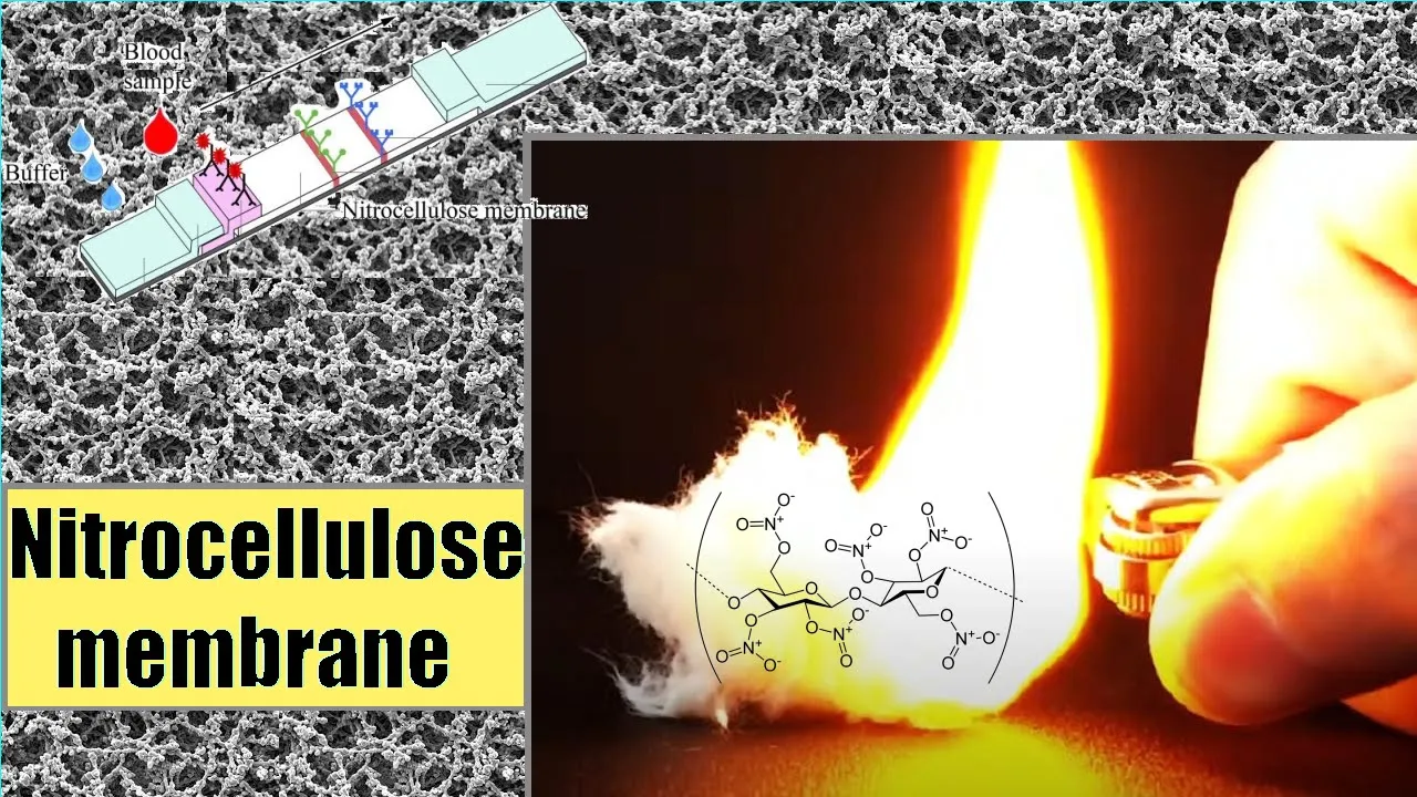
Nitrocellulose membrane is a widely used matrix in protein blotting due to its high protein-binding affinity, compatibility with multiple detection methods (chemiluminescence, chromogenic, and fluorescence), and ability to immobilize proteins, glycoproteins, or nucleic acids; it is a highly flammable compound formed by nitrating cellulose with nitric and sulfuric acids, where each glucose unit in the cellulose polymer is esterified with three nitrate groups that confer both the negative charge at neutral pH and the unusual flammability of dry nitrocellulose.
Click to watch →
Prerequisite Knowledge
- Concept 01Basic immunology, specifically the concept of antigen-antibody binding, epitopes, and binding specificity.
- Concept 02The physical principle of capillary action, which drives the passive flow of liquid through porous membranes.
- Concept 03An understanding of conjugate labels, such as colloidal gold nanoparticles or fluorescent dyes, and their role in signal detection.
- Concept 04The concept of membrane chromatography and how different biomolecules migrate through a solid matrix like nitrocellulose.
Subsequent Learning
- Step 01The competitive lateral flow immunoassay format, which is used for detecting small molecules (haptens) that cannot bind two antibodies simultaneously.
- Step 02Advanced multiplexing strategies, including how to design test strips that can detect multiple distinct pathogens or biomarkers at once.
- Step 03The 'Hook Effect' (prozone effect) and other chemical interferences that can cause false-negative or false-positive results in rapid diagnostic tests.
- Step 04Quantitative lateral flow assays, including the integration of digital optical readers and smartphone-based diagnostic applications.
Music
Music
0:00Playing Section
- 1
Video starts with music only.
- 2
No spoken content at this point.
- 3
Prelude before main discussion begins.
The Competitive Assay Format and the Hook Effect
While the sandwich format is highly effective for detecting large analytes with multiple binding sites, it is not universally applicable and suffers from a major vulnerability known as the 'high-dose hook effect.' For small molecules (haptens like drugs or hormones), a 'Competitive Format' must be used instead. In a competitive assay, the analyte blocks the binding of labeled antibodies to the test line, resulting in a signal that is inversely proportional to the analyte concentration. Furthermore, the sandwich format can produce dangerous false-negative results if the analyte concentration is extremely high (the hook effect). In such cases, excess unbound analyte saturates both the detection and capture antibodies individually, preventing the sandwich complex from forming at the test line. Understanding these architectural alternatives and physical limitations is crucial for robust diagnostic design.
Deep Dive
The competitive lateral flow immunoassay format, which is used for detecting small molecules (haptens) that cannot bind two antibodies simultaneously.

Food Analysis Lect 39 Dec 11 2020
136
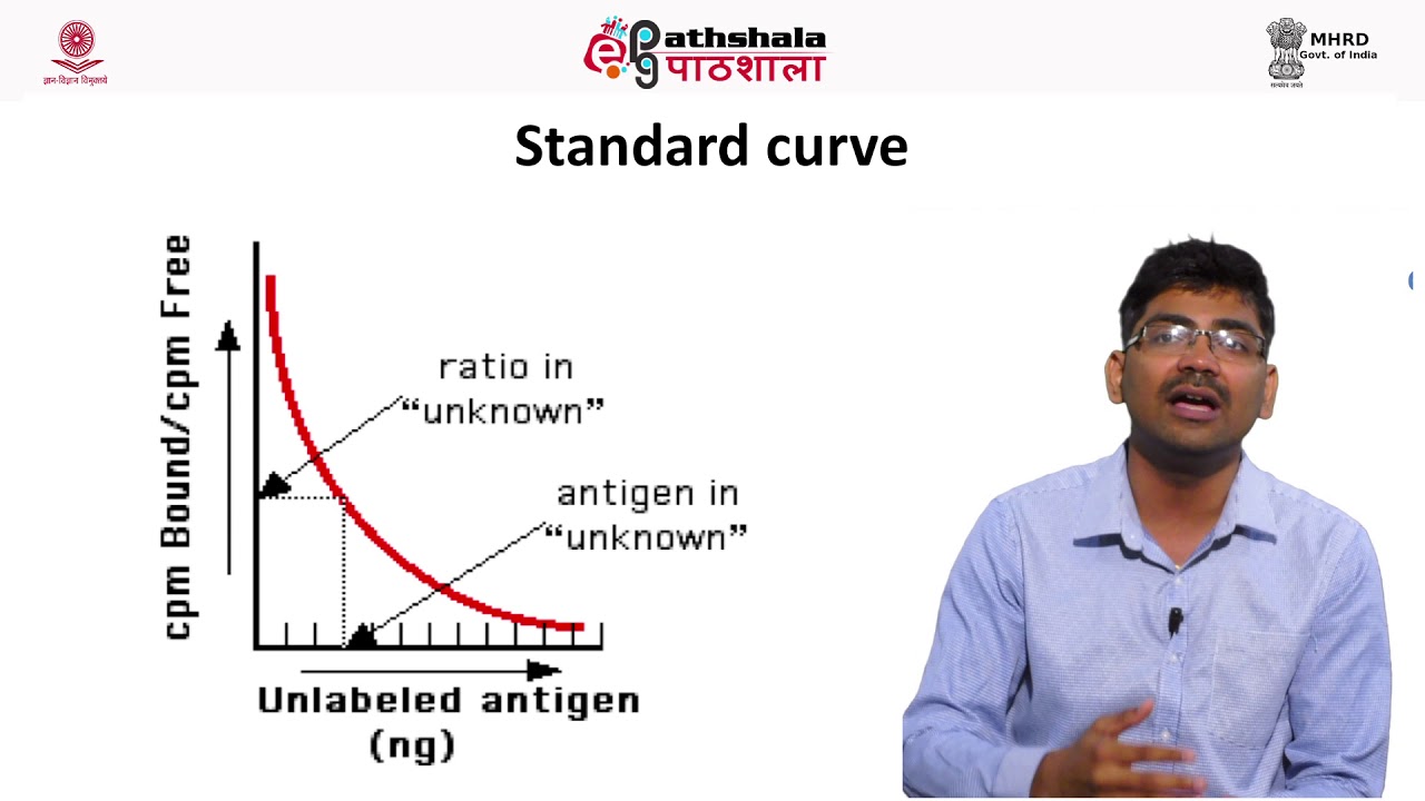
Competitive ELISA detects small molecules (<5000 g/mol) like pesticides and toxins that cannot be used in sandwich ELISA. The small molecule is linked to a carrier protein to form a hapten. Sample antigen and labeled standard antigen compete for antibody binding sites, producing inverse relationship between antigen concentration and absorbance. Bound hapten competitive ELISA binds hapten to the plate, uses labeled antibody, and free sample antigen competes for binding. The hapten used in the assay must differ from the hapten used to produce the antibody to prevent cross-reactivity. This format is essential for detecting small molecule contaminants in food samples, where higher antigen concentration results in lower absorbance.
Click to watch →
Immuno Assays and its Application in medicine
852
Sandwich ELISA detects large molecules by coating the plate with a capture antibody, adding sample, then adding a detection antibody that binds to a different epitope. Requires antigens with multiple epitopes; haptens cannot be detected. Competitive ELISA detects haptens (small molecules like thyroid hormones) by having unknown and known antigens compete for limited antibody binding sites. Color development is inversely proportional to analyte concentration. This technique is essential for small molecules that cannot be detected by sandwich ELISA.
Click to watch →

Lateral Flow Webinar: A Guide to Lateral Flow Immunoassay Development
53.4K
Lateral flow immunoassays are unidirectional diagnostic tests that detect analytes through antibody-antigen interactions, consisting of five key components: a sample application pad, conjugate release pad containing detection particles (gold nanoparticles or latex beads), reaction membrane with immobilized capture antibodies, wicking pad, and optionally a plastic cassette. The assay operates in two formats: direct assay for large analytes with multiple antigenic sites (producing two lines indicating positive results) and competitive assay for small analytes with single binding sites (producing only a control line for positive results). Successful assay development requires selecting antibodies with high binding affinity (low KD value) and fast on-rates, as well as ensuring uniform detection particle size and stable conjugation methods. Common troubleshooting issues include uneven lines, false positives/negatives, and inconsistent liquid fronts, which can be resolved through component optimization and quality control measures.
Click to watch →
Lateral Flow Assay Development Webinar: Introduction to the development of a lateral flow assay
32K
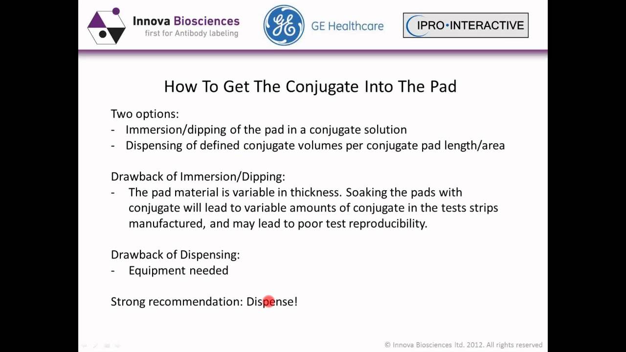
Lateral flow assays originated in the 1980s with pregnancy tests and have expanded into clinical, veterinary, environmental, and defense applications. These assays consist of membrane, sample pad, conjugate pad, absorbent pad, and reagents for test/control lines. Two main formats exist: sandwich assays for large proteins using double antibodies (signal proportional to analyte concentration), and competitive inhibition assays for small molecules like drugs (signal inversely proportional to analyte concentration). Antibody selection is critical: polyclonal antibodies offer high affinity but require pure antigens and immunoaffinity purification; monoclonal antibodies provide unlimited supply without purification but may have lower affinity. Commercial databases help identify existing antibodies. Immunization protocols for proteins above 5,000 MW differ from those for small molecule haptens, which require covalent coupling to carrier proteins with appropriate spacers. Antibodies should be evaluated on laboratory-based enzyme immunoassays before lateral flow development, as microtiter plate ELISAs don't correlate well with actual performance.
Click to watch →
Competitive ELISA Kit Operation procedure
157
Competitive ELISA is a detection method suitable for small molecule antigens or haptens, where the sample antigen competes with a pre-added biotinylated antibody for binding sites on the detection antibody, followed by streptavidin-HRP conjugate addition, TMB substrate incubation, stop solution addition, and absorbance reading at 450nm to quantify the target substance through standard curve analysis.
Click to watch →
Deep Dive
Advanced multiplexing strategies, including how to design test strips that can detect multiple distinct pathogens or biomarkers at once.

THE FOUNDATION DAY, ICMR-NIRTH
516
Multi-biomarker test strips can simultaneously measure six biomarkers using a single sample. To prevent interference between markers, flow direction is designed from bottom to top rather than left to right. Each marker line produces a test peak relative to a control peak, with the ratio indicating biomarker concentration. This multiplexed approach improves diagnostic discrimination between infected contacts and those who develop active disease.
Click to watch →

AMYC-BIOMED 2021 - Analytical Chemistry Session 2
163
Multiplex lateral flow strips can simultaneously detect general pathogen positivity and identify specific serotypes. Two types of sandwich immunoassays are used: homologous sandwiches (same antibody for capture and detection) for general detection, and heterologous sandwiches (different antibodies) for serotype-specific identification. Probe concentration optimization (0.5-2 OD) and test line positioning affect signal intensity, with closer lines showing higher signals due to increased interaction time between detection antibody and antigen.
Click to watch →

Havey Institute for Global Health – Ask Dr. Murphy: 03/01/23
158
Multiplex testing allows detection of multiple pathogens (such as flu A, flu B, and SARS-CoV-2) in a single test. This technology was accelerated through federal programs like RADx. Single-pathogen tests are becoming obsolete because respiratory infections often involve multiple potential causes, and appropriate treatment depends on accurate diagnosis. Future tests will likely detect four or more pathogens simultaneously.
Click to watch →

Recording: accelerating the development of point-of-care molecular diagnostic platforms (overview)
352
The RFP seeks multi-pathogen multiplex platforms but acknowledges that multiplexing may be part of an applicant's R&D plan if they currently singleplex. The optimal design specification requires detecting four or more pathogens simultaneously from a single sample, while the acceptable specification allows detection of one pathogen. Scoring guidelines award points based on the number of pathogens detectable in a single reaction. Multiplex capacity is weighted heavily (denoted by 3) and must be specifically addressed in applicant presentations and RD plans. Applicants with extensive test menus but no current multiplexing capability can still score points by describing their plans to multiplex key pathogens.
Click to watch →

Multiplexing with Unisart StructSure®️ Membranes Webinar
457
Multiplexing in lateral flow assays allows the simultaneous detection of multiple target analytes in a single test. This is achieved through three main formats: (1) Multi-line design where several test lines are printed behind each other with different capture molecules, (2) Parallel channel design with separate channels for each target molecule, and (3) Combined approaches using both multi-line and parallel channel designs. These formats enable applications like drug testing and disease diagnosis by detecting multiple biomarkers simultaneously.
Click to watch →
Deep Dive
The 'Hook Effect' (prozone effect) and other chemical interferences that can cause false-negative or false-positive results in rapid diagnostic tests.

Diagnosis of Syphilis - Types of tests, Principle, Uses, Short-comings
5.5K
Prozone phenomenon (also called hook effect or antibody excess phenomenon) is an immunological interference where high antibody concentrations prevent proper immune complex formation, causing false-negative results. In the zone of equivalence, adequate antibody-antigen ratios produce visible flocculation. In prozone, excess antibodies coat antigens incompletely, preventing precipitation. Post-zone phenomenon occurs with antigen excess. Both cause false negatives. This is particularly common in HIV/syphilis co-infection due to excessive B-cell stimulation, pregnancy, early syphilis stages, and neurosyphilis. Laboratories should dilute patient serum to reach the zone of equivalence for accurate results.
Click to watch →

Principle of an antigen detection rapid diagnostic test (including prozone and backflow effect)
20.6K
Antigen detection rapid diagnostic tests (RDTs) work through immunochromatography using a nitrocellulose membrane strip where a conjugate of anti-antigen antibody coupled to colloidal gold migrates with patient samples; if the antigen is present, it binds to the conjugate and gets captured by test line antibodies, producing a visible purple-red line, while the control line always appears purple regardless of test results. However, two critical artifacts can affect accuracy: the prozone effect occurs when high antigen concentrations cause free antigens to outcompete conjugate binding, leading to false negatives; the backflow effect happens when tests are read too late and dried strips allow reverse migration, potentially causing false positives.
Click to watch →

8th Webinar - Assay Interference by Dr. Gayan De Costa
186
This section covers antibody-related interferences and the hook effect. Heterophile antibodies can interfere with multiple assays including thyroid function tests, insulin assays, and rheumatoid factor assays, causing both positive and negative interference. Detection methods include precipitation, different analytical platforms, heterophile blocking anti-tubes, and serial dilutions. The hook effect affects single-step sandwich immunoassays where excess antigen causes falsely low results in prolactin, HCG, growth hormone, and tumor marker assays. A variant hook effect can occur with variant HCG forms in pregnancy, causing falsely negative pregnancy test results.
Click to watch →

CLINICAL CHEMISTRY 1 (INSTRUMENTATION) 3B
173
Five major sources cause immunoassay interference: (1) Human endogenous antibodies (heterophilic, anti-animal, autoantibodies, therapeutic antibodies) causing positive/negative interference; (2) Cross-reactivity with closely related proteins, varying with temperature; (3) Prozone/hook effect where high analyte concentrations reduce immune complex formation; (4) Elevated endogenous serum components (bilirubin, hemoglobin, lipids, proteins) interfering more in homogeneous assays; (5) Matrix effects from sample components interfering with antibody-protein binding. Resolution strategies include sample dilution (effective for prozone, matrix, and elevated components), recognizing interference patterns, and implementing automated analyzer flags for suspicious results.
Click to watch →

What is the Prozone Effect? (How to troubleshoot false negatives in immunoassays)
2.7K
The Prozone effect (also called the hook effect) is a phenomenon in immunoassays where an excess of target antigen saturates all available antibody binding sites on microspheres, preventing the formation of antibody-antigen bridges necessary for signal detection; this results in false negative readings despite high antigen concentrations, and can be mitigated through serial dilution protocols that allow accurate quantification across different concentration ranges.
Click to watch →
Deep Dive
Quantitative lateral flow assays, including the integration of digital optical readers and smartphone-based diagnostic applications.

TEDItalia - Un laboratorio grande quanto un francobollo: George Whitesides
106
Lateral flow immunoassays, exemplified by pregnancy tests, represent the most common paper-based diagnostic technology. These binary tests work well for conditions with clear yes/no outcomes but are limited for quantitative analysis. Wax printer technology enables mass production of paper-based diagnostic devices by printing wax patterns onto paper, which absorb when heated. These printers cost approximately $800 and can produce about 10 million tests per year when operating 24 hours daily. The printing process takes about two seconds per device. Smartphone camera phones can capture images of paper-based diagnostic devices, which are then sent to central laboratories for analysis. This approach eliminates the need for doctors to be present at the point of care, allowing healthcare workers to collect samples and transmit data while computers perform complex analysis.
Click to watch →

Webinar - Point of Care: Testing to Care, Monitor and Protect
187
Lateral flow tests offer significant advantages in portability, accessibility, and cost but face inherent trade-offs in sensitivity compared to molecular PCR testing. Negative results do not definitively rule out disease—they simply indicate viral loads below detection limits. When results reach or exceed detection thresholds, sensitivity and specificity achieve very high levels appropriate for their intended clinical applications. Future developments will include novel target diseases, integrated DNA/RNA amplification to enhance sensitivity, expanded reporter particle options beyond gold nanoparticles, and introduction of alternative capture mechanisms. Most significantly, digital readers utilizing smartphone image processing represent a transformative advancement, enabling automatic result interpretation, eliminating visual interpretation uncertainty, and supporting lay person use while maintaining clinical traceability and data integrity.
Click to watch →

Les Minutes de l'X : La détection ultra sensible de biomarqueurs et de pathogènes
151
Rapid diagnostic tests use lateral flow strips where samples migrate via capillary action through membranes containing capture antibodies and control lines. When target biomolecules bind to detection antibodies conjugated to nanoparticles, visible signals appear at test lines. This technology provides qualitative yes/no results similar to pregnancy tests but can be adapted for quantitative measurements. The integration of luminescent nanoparticle probes with smartphone readers enables portable, accessible diagnostic capabilities with laboratory-grade sensitivity.
Click to watch →

Geert Vanden Bossche, "Wave of the Variants", the surge in need for rapid antibody testing (FULL)
5.2K
Lateral flow assays can be used for both serology (antibody) testing and antigen testing, as well as detecting chemical and biological markers. The technology is scalable globally with manufacturers in Taiwan, India, Brazil, and the US. A universal lateral flow assay reader standardizes tests, improves sensitivity, eliminates human error, and collects metadata including vaccination status and disease history. The reading of each assay takes approximately 1-2 seconds, with machines capable of processing 500 samples per hour. Quantitative lateral flow assays can be developed by calibrating against PCR or ELISA results. Nucleocapsid antibodies are the best candidate for serology testing because they are completely discriminative between natural infection and vaccination. The nucleocapsid protein is more stable and does not mutate like spike, making it a reliable marker for natural immunity. Finger prick tests are not sensitive enough; venous blood draw is required.
Click to watch →

Considerations on the development of quantitative lateral flow assays
693
Quantitative lateral flow assays require comprehensive understanding of device architecture, test formats, and analytical principles. The device comprises four components: sample pad, conjugate release pad, nitrocellulose membrane with test/control lines, and absorption pad. Two primary test formats exist: sandwich tests for macromolecular analytes with multiple binding sites (producing increased signal intensity) and competitive tests for small molecules with single binding sites (producing decreased signal intensity). Quantitative tests differ from qualitative tests by determining exact analyte concentrations rather than mere presence/absence. Key parameters include Limit of Detection (LOD), Lower Limit of Quantification (LLOQ), and Upper Limit of Quantification. System uncertainty, representing standard variation around mean values, must be minimized. Sample variability significantly impacts performance, as demonstrated by human urine pH fluctuating between acidic and alkaline states. Material selection critically affects performance through capillary flow time: slow membranes cause false positives while fast membranes cause false negatives. Reagents including antibodies and hapten-carrier conjugates demand strict quality control with batch-specific standard curves.
Click to watch →
Music
Music
0:00- 1
Video starts with music only.
- 2
No spoken content at this point.
- 3
Prelude before main discussion begins.
The Competitive Assay Format and the Hook Effect
While the sandwich format is highly effective for detecting large analytes with multiple binding sites, it is not universally applicable and suffers from a major vulnerability known as the 'high-dose hook effect.' For small molecules (haptens like drugs or hormones), a 'Competitive Format' must be used instead. In a competitive assay, the analyte blocks the binding of labeled antibodies to the test line, resulting in a signal that is inversely proportional to the analyte concentration. Furthermore, the sandwich format can produce dangerous false-negative results if the analyte concentration is extremely high (the hook effect). In such cases, excess unbound analyte saturates both the detection and capture antibodies individually, preventing the sandwich complex from forming at the test line. Understanding these architectural alternatives and physical limitations is crucial for robust diagnostic design.
Basic immunology, specifically the concept of antigen-antibody binding, epitopes, and binding specificity.
The physical principle of capillary action, which drives the passive flow of liquid through porous membranes.
An understanding of conjugate labels, such as colloidal gold nanoparticles or fluorescent dyes, and their role in signal detection.
The concept of membrane chromatography and how different biomolecules migrate through a solid matrix like nitrocellulose.
The competitive lateral flow immunoassay format, which is used for detecting small molecules (haptens) that cannot bind two antibodies simultaneously.
Advanced multiplexing strategies, including how to design test strips that can detect multiple distinct pathogens or biomarkers at once.
The 'Hook Effect' (prozone effect) and other chemical interferences that can cause false-negative or false-positive results in rapid diagnostic tests.
Quantitative lateral flow assays, including the integration of digital optical readers and smartphone-based diagnostic applications.
393.4K views3.3Klikes1:53@AbingdonhealthdiagnosticsOriginal Release: 2019-04-08
[Music]
Up Next
4:42
Covalent Conjugation of Antibodies to Gold Nanoparticles Tutorial
@nanocomposix
16.8K views•2017-01-17

12:54
Triumph of Orthodoxy Icon: Byzantine Art & History Explained
@BenCallan
2.1K views•2024-08-06

9:49
FastAPI vs Flask vs Django: Choosing the Right Python Web Framework
@TechWithTim
302.5K views•2024-05-26

1:42
Game of Thrones Opening Credits: A Cinematic Analysis
@gameofthrones
46.3M views•2011-04-18
Related Study Plans & Knowledge Roadmaps
Structured learning paths in General & Interdisciplinary Studies
general-studies
Elevator Engineering: History, Physics & Cities
01From Hoists to Safety Brakes: The Dawn of Vertical Lift
02Hydraulic vs. Traction: The Engineering Physics of Elevators
03Modern Speed, Safety, and Algorithms
+ 2 more learning modules
12h5 modules
Explore Roadmap
general-studies
Rubber: Material Science, History & Geopolitics
01Origins of Natural Rubber & Mesoamerican History
02The Chemistry of Natural Rubber & Vulcanization
03The Geopolitics of the Rubber Boom & Colonization
+ 2 more learning modules
12h5 modules
Explore Roadmap
general-studies
Containerization: Evolution, Logistics & Trade
01Ancient Trade & Bulk Cargo: Amphorae to Barrels
02The Industrial Revolution & Cardboard Packaging
03The Containerization Revolution: Malcom McLean
+ 2 more learning modules
12h5 modules
Explore Roadmap
general-studies
Textile Tech: Evolution, Labor & Global Impact
01Pre-Industrial Textiles: Handloom & Cottage Industry
02Mechanization: The Spinning Jenny to the Power Loom
03The Factory System & Global Labor Transformation
+ 2 more learning modules
10h5 modules
Explore Roadmap