











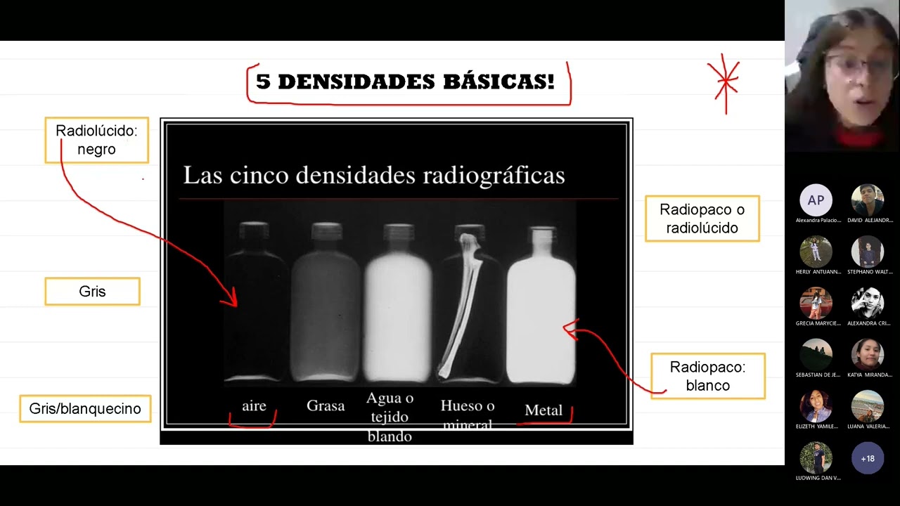









The five basic radiographic densities—metal, bone/calcium, soft tissue/fluid, fat, and air—are fundamental to interpreting conventional radiographs, as they determine how tissues appear on X-ray images based on their X-ray absorption properties, enabling radiologists to identify abnormalities by comparing their density characteristics against normal tissue patterns.
Radiographic Densities: The 5 Basics in Radiology Imaging
Added:The physical properties of X-rays as ionizing radiation within the electromagnetic spectrum.
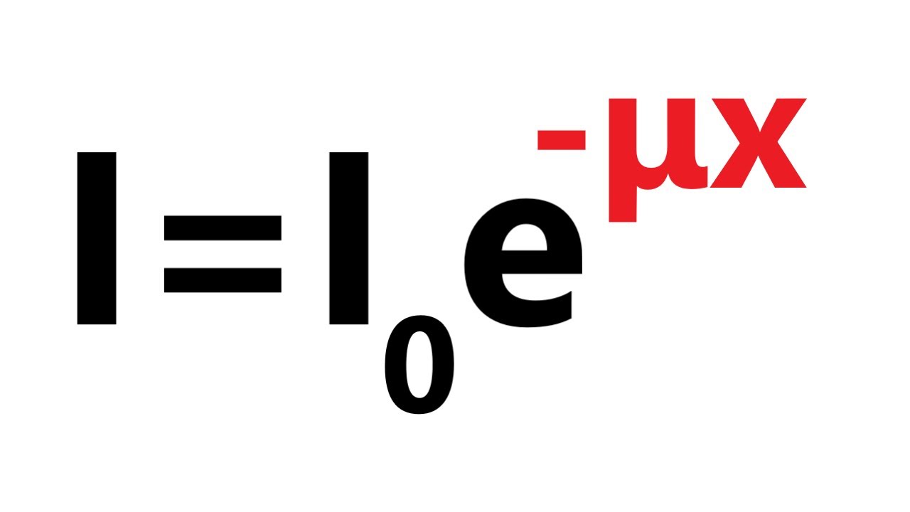
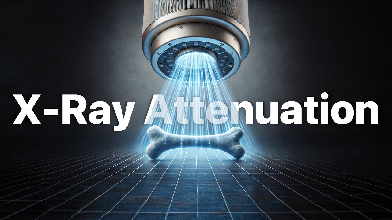
The concept of X-ray attenuation, which explains how different materials absorb or scatter photons to varying degrees.
The mechanics of radiographic image formation, including the difference between radiolucency (black/dark areas) and radiopacity (white/light areas) on a detector.
Basic human anatomy, specifically the physiological location of air, fat, soft tissue organs, and bones within the body.
Identifying pathological density alterations, such as fluid consolidation in the lungs (replacing air) or free air in the abdominal cavity.
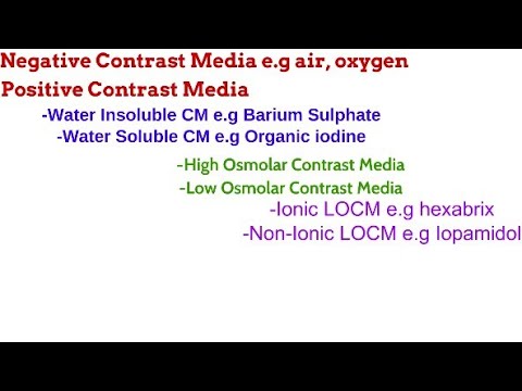
The clinical application of contrast media (e.g., barium, iodine) to artificially alter radiographic density and highlight specific anatomical structures.
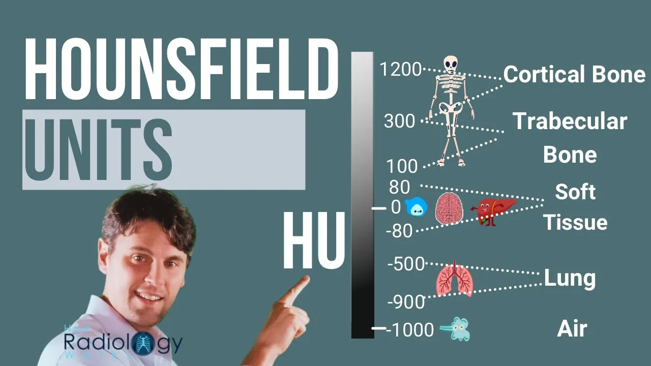
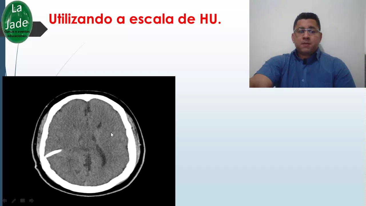
An introduction to Computed Tomography (CT) imaging and how these five basic densities translate into quantitative Hounsfield Units (HU).
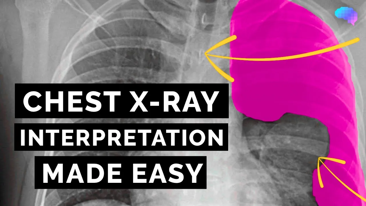
Systematic frameworks for clinical X-ray interpretation, such as analyzing chest radiographs using a structured approach (e.g., the ABCDE method).
81.9K views731likes5:40Original Release: 2010-08-23
[Music] welcome to video podcast one the five radiographic densities on learning radiology.com I'm William Herring from Albert Einstein Medical Center in Philadelphia so the five basic densities are important for our fundamental understanding of conventional radiographs they help us to tell the location of disease and they also will help us tell what those diseases may be the five densities are metal density bone or calcium density soft tissue or fluid density fat and air and the graphic on the left indicates approximately how they'll appear on a image metal absorbs the most X-rays and very few of them Reach the film cassette air on the other hand absorbs the least x-rays so most will reach the film cassette and be able to darken it calcium fluid and fat are somewhere in between so some are absorbed some are transmitted and they produce varying densities on the film cassette how this works in everyday practice is shown on this frontal chest radio graph we can see air density here in the left upper lobe of the lung can also see air density in the room surrounding the patient we can see fat density in the chest wall we can see fluid or soft tissue density in the right upper quadrant of the abdomen with the liver we see bone or calcium density in the clavicle scapula and ribs and there is metal density on this radioresepsjonen [Music] [Music] and they can also help to tell us where an abnormality is located and that we'll talk about in the next video podcast here's a chest x-ray that you can pause your computer or MP3 player to determine where the abnormality is and what you think it might be well you were correct if you identified an abnormal density superimposed on the right ailla if we examine the density of that abnormality more carefully we can tell that it is not a lymph node because it should be the same density as all the other lymph nodes in the ailla and it's actually much denser than soft tissue it's not a calcified lymph node because this is the density of calcium in the ribs and it's denser than that and yes if you correctly identify this as a far in body You' be correct and if you identified it as a bullet you would be even more correct so this is metal density here's another abnormality you can [Music] pause clearly there is abnormality in the right parat tral region and if we look at it carefully we can see that it is soft tissue density it's the same density as for example the heart it's denser than the air in the lungs it's adenopathy in the right parat tral region and did you also see that there was a mass in the lung which represents the bronchogenic carcinoma this patient had which metastasized to the regional lymph nodes so this is soft tissue density here's a mini quiz for you look at this radiograph pause your computer or MP3 player and decide what you think it is well there is a metallic density right in the middle of this little Kitty's chest and it's superimposed on the area where the tracha or the esophagus would be this metallic density is a coin in the esophagus and it actually represented a US Quarter so if if you correctly identified that it was metallic density you were using the five basic radiographic densities to recap they are air fat soft tissue calcium and metal and by knowing them we can recognize what an abnormality is frequently and determine where the abnormality [Music] is [Music] oh
Up Next

9:02
Kerley B Lines on Chest X-Ray: Heart Failure Signs Explained for Medical Students
@DrSharmaDO
23.7K views•2020-04-07

48:47
Graphic Medicine: Comics for Collaborative Healthcare Communication
@nationalpatientadvocate
189 views•2023-12-04

10:28
Neuroanatomy: Central and Peripheral Nervous System Divisions Explained
@AKLECTURES
136.2K views•2014-09-20

3:05
Stages of Labor and Vaginal Birth | Childbirth Animation
@nucleusmedicalmedia
52.1M views•2017-08-18
Related Study Plans & Knowledge Roadmaps
Structured learning paths in Medicine
medicine
Abnormal LFTs: Diagnostic Workup & Pathology
01Liver Anatomy and Basic Physiology
02Introduction to Liver Function Tests (LFTs)
03Hepatocellular vs. Cholestatic Patterns
+ 3 more learning modules
12h6 modules
Explore Roadmap
medicine
Analyzing Blood Smears: RBC, WBC & Platelets
01Introduction to Blood Components & Smear Preparation
02Normal Morphology of Blood Cells
03Erythrocyte Pathologies & Morphological Variants
+ 2 more learning modules
12h5 modules
Explore Roadmap
medicine
Mechanical Ventilation: Waveforms & Alarms
01Anatomy and Physiology of Breathing
02Introduction to Mechanical Ventilation & Core Modes
03Ventilator Settings and Waveform Interpretation
+ 2 more learning modules
12h5 modules
Explore Roadmap
medicine
Neuro Exam: Localization & Stroke Syndromes
01Foundations of Neuroanatomy and Vascular Supply
02The Neurological Examination: Step-by-Step
03Principles of Neurological Localization
+ 3 more learning modules
18h6 modules
Explore Roadmap