















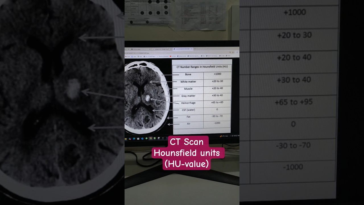






Hounsfield Units (HU), also known as CT numbers, are a normalized scale used to display CT images where values are calculated as HU = (μ_material - μ_water) / μ_water × 1000, with water serving as the reference point at 0 HU; this system allows radiologists to easily communicate tissue characteristics, with common values including air (-1000 HU), lung (-900 to -500 HU), soft tissue/fat (-80 to 80 HU), trabecular bone (100 to 300 HU), cortical bone (300 to 1200 HU), and iodinated contrast (150 to 300 HU).
Hounsfield Units Explained: CT Numbers & Attenuation Scale
Added:Basic physics of X-ray production and how electromagnetic radiation interacts with human tissue.
The concept of the linear attenuation coefficient, which mathematically describes how easily a material can be penetrated by an X-ray beam.
Fundamental principles of Computed Tomography (CT) hardware, including how a rotating gantry and detector array acquire projection data.
The algebraic concept of scaling and normalization relative to a standard baseline (specifically, the properties of water).
CT windowing and leveling techniques, which map specific ranges of Hounsfield Units to grayscale values on a monitor to optimize tissue visualization (e.g., bone, lung, and soft tissue windows).
Clinical interpretation of CT numbers, such as distinguishing between cysts, hemorrhages, and solid tumors based on their specific HU values.
Common imaging artifacts that distort HU accuracy, including beam hardening, metal streak artifacts, and the partial volume effect.
Advanced imaging technologies like Dual-Energy CT (DECT), which utilize multiple energy spectra to characterize tissue composition beyond standard HU values.
30.6K views517likes11:19Original Release: 2022-03-08
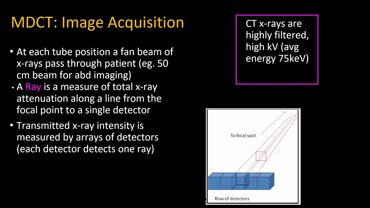
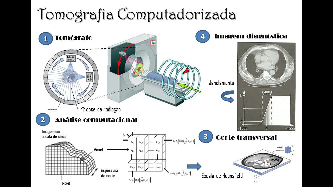
all right all right rad nation today we're talking about house field units ct number basically the way we display images coming out of a ct scanner and brian from how radiology works we have bite-sized content for those in the radiology field first off x-rays are going to pass through the body as they pass through the body we can talk about the attenuation of those x-rays that went through we say that's how many x-rays penetrated our body we do that for one view we can make an x-ray image right if we do it for lots of views we can make a ct image and what a ct image actually is is we want to quantify that process so x-rays are getting stopped throughout the body in general the process will look like this if it's one single given material and what we're looking for is the rate at which they're getting stopped here we call that the attenuation coefficient and the different materials inside your body are going to be absorbing the x-rays differently we have separate videos on photoelectric effect and on compton scattering that has to do with how the x-rays are going to interact in your body those two things add up the photoelectric and compton add up to this attenuation coefficient inside of your body and it's going to be different for each different type of tissue so the way that we can make an image inside your body is by making a map of those attenuation coefficients so if you think about breaking the image up i've shown here a relatively well sampled image which has five 12 by 512 pixels in it but if you think about each pixel i've drawn here at larger ones you would assign a pixel for that given material and you would assign one value inside of that and then you'd go the next pixel assign one value and you go next pixel assign one value and the values for these attenuation coefficients depending on the energy and depending on what type of material it is these values might be around uh 0.2 here video on penetration to see more about beer's law and the units for these attenuation coefficients so then after we make a map of the attenuation coefficients inside of our body we need to present that to the radiologist and the question is how should we do that should we just present it in kind of the standard units that you would expect to see on the nist website see we have a value of 0.2 and here we have a value of 0.227 these are in units of inverse centimeters so in general the different materials of the body can have pretty similar coefficients and these coefficients will obviously depend on the energy of the x-rays that are used not good for a radiologist to communicate in the third decimal right i know they're smart and all but we want to make it easier we want to make communication easier for people reading these cp images so what do we need a system that is a couple things number one it's normalized right so it's in reference to some material that's in our body what do you guys think we should use obviously when i was a kid i learned your body's mostly made up of water from the magic school bus of course and because your body's mostly made up of water and because most of the things that we're imaging in your body are actually soft tissue that means water is really a good candidate to use as the normalization because it's very similar to the soft tissues in your body so we got step one down we know we're going to use water to do the normalization step two is we need to make a scale that has small steps so that in our new unit system one step is relatively small in that way we can just use this unit system and we won't have to talk about fractions of it so we won't have to say this is 0.1 ct number this is point ct number we can just refer to it as one ct number five ct numbers and like we said the ct numbers are actually also referred to as house field units so 1 hu or 5 hu would be a way radiologists could communicate with one another our definition what it means is we take the attenuation coefficient of that material and we say how far is it away from water we're going to divide by the value of water that's what we call normalization so that things are all with reference to water so again this is linear so the images aren't going to look a lot different than if we displayed them with the native scale but the idea is that this is a really good way for the radiologist to communicate have normalization down then the other thing we said is we need to take small steps so that's where we multiply by a thousand so that we're going to have relatively small steps in this unit system and because we're talking about relative differences here you can think of these like point one percent steps so one house field unit is like a point one percent step the ability of ct is so good to measure quantitatively relatively low contrast structures we can have 0.1 steps and we can quantitatively have those really have meaning we're often looking at things that might only be 0.3 percent difference in terms of their attenuation values two really important materials that we want to talk about one is water and the other is air so first off with water if we plug in water for the material that we're concerned with you can see now we have mu water minus mu water so then we just have zero it doesn't matter what else we're multiplying by zero times anything is just going to be zero right so the idea now is that mu water is zero one is air mu air is not actually zero mu of a vacuum would be zero but mu air is so much smaller than mu water that we can relatively treat it like zero for the purpose of this calculation and discussion the idea is that if we take zero plug that in here you can see we have minus mu water divided by mu water so that would just be a minus one and then you multiply by a thousand so we just have minus one multiplied by a thousand is just going to be minus 1000 we'll fill in some of the other common materials in our body as we're going up the scale long is mostly filled with air right so it's relatively lower attenuation and on this scale you can see it's roughly minus 400 minus 900.
don't quote me on these precise numbers what we're trying to do here is establish the relative values of the different areas in the body and also establish that that's kind of a big range so i also haven't drawn everything perfectly to scale here because this is just a picture but you can think that when we get later on we're going to have another video about windowing and leveling as far as how to look at these images the lung has a relatively big range of tissue values you're going to need to look at it with a relatively wide image window with right around water is the soft tissue so some soft tissues are a little bit less and some soft tissues are a little bit more than the water attenuation value we're going to zoom in on the soft tissues in just a minute after we get through these values then we talk about the bones in the body right and we have different types of bone we have trabecular bone which is our softer spongier bone and we have cortical bone which is our harder bone and the softer spongier bone will have you know between 100 and 300 house filled units and then the cortical bone will go from there from 300 up to close to a thousand health food units on top of that if you have metal in the body that can have values that are off of the chart even off of what we record within our dicom images so these can be in the values of thousands for metal in the body and this is why metal can really cause artifacts in our ct images other things in the body such as calcifications which can be right in here and then also iodinated contrast within the vessels is also usually right in here within that range of 150 to 300 haunted units typically zoom in on the soft tissue remember water is our zero value that was our reference then we look at below water the soft tissue that's less attenuating than water is actually the fat right so if you remember you pour oil into a water that's gonna float the fat is actually less dense than the water so it is less attenuating than the water liberal spinal fluid is very close to water so it is just a little bit more dense than water and is a little bit more attenuating than water i'm going to talk about the gray matter and the white matter the white matter in the brain is actually a little bit less attenuating than the gray matter because the white matter has the tracks and those uh white matter tracks are myelinated which is basically including a fatty coating and since it has that fatty coating it's a little bit less attenuating the gray and the white matter is one of the more challenging tasks that we have in order to differentiate those within our ct images then we look at blood this is particularly important to know that blood is going to be more attenuating than typical soft tissue in the grain the white matter and this is very important to know that if you do have a bleed in your brain we will be able to detect that relatively easily on just a non-contrast ct scan because the blood will show up relatively bright the gut the values are also going to be similar for the soft tissue there you can see again there's a range of values in the kidney the pancreas in general is going to be a little bit more attenuating than the kidney and then the liver even a little bit more attenuating than the pancreas but in general you can see that the values are relatively close for all these soft tissue and that's why when we're looking at things that are soft tissue we're going to be looking at them in a more narrow display window with about that an upcoming video we'll have on the channel we'll post a link for you right here when it's ready about window width window level and viewing it's intricately related to these household units
Up Next

4:33
GCSE Physics: Mains Electricity, Live, Neutral & Earth Wires
@Freesciencelessons
523.5K views•2017-07-03

12:53
21cm Hyperfine Transition in Neutral Hydrogen: Radio Astronomy Basics
@AaronRobertParsons
12.4K views•2011-10-13

32:32
NMR Spin Physics I: Zeeman Effect, Resonance Condition & Larmor Frequency
@nptel-indianinstituteofsci8064
2.3K views•2024-01-17

27:14
Entropy and the Second Law of Thermodynamics Explained
@veritasium
27.5M views•2023-07-01
Related Study Plans & Knowledge Roadmaps
Structured learning paths in Physics
physics
Nuclear Physics (Decay, Fission & Reactors)
01Atomic Structure and the Binding Force
02Radioactive Decay and Half-Life
03Nuclear Fission Mechanics and Chain Reactions
+ 2 more learning modules
12h5 modules
Explore Roadmap
physics
Soft Matter (Polymers, Colloids & Rheology)
01Foundations of Soft Matter & Thermal Forces
02Polymer Physics and Macromolecules
03Colloids, Surfactants, and Self-Assembly
+ 2 more learning modules
38h5 modules
Explore Roadmap
physics
Analytical Mechanics: Lagrangian & Hamiltonian
01Mathematical Foundations: Calculus of Variations
02From Newtonian Mechanics to Constraint Dynamics
03The Principle of Least Action & Lagrangian Mechanics
+ 3 more learning modules
28h6 modules
Explore Roadmap
physics
General Relativity (Spacetime & Cosmology)
01Foundations of Relativity: Special Relativity
02The Geometry of Spacetime: Equivalence Principle & Curvature
03Einstein's Field Equations: The Mathematics of Gravity
+ 2 more learning modules
55h5 modules
Explore Roadmap