













![Cette pilule pourrait remplacer le sport ?! [Avec des Sci]](https://i.ytimg.com/vi_webp/xoWKAFn0JmM/maxresdefault.webp)
![КАКИЕ КНИГИ ЧИТАТЬ, ЧТОБЫ ПОНЯТЬ МИР? [ч.1, Эволюция, нейробиология, антропология]](https://i.ytimg.com/vi/MC1CxPMkcvM/maxresdefault.jpg)
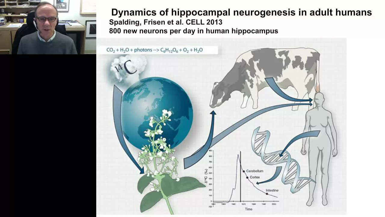
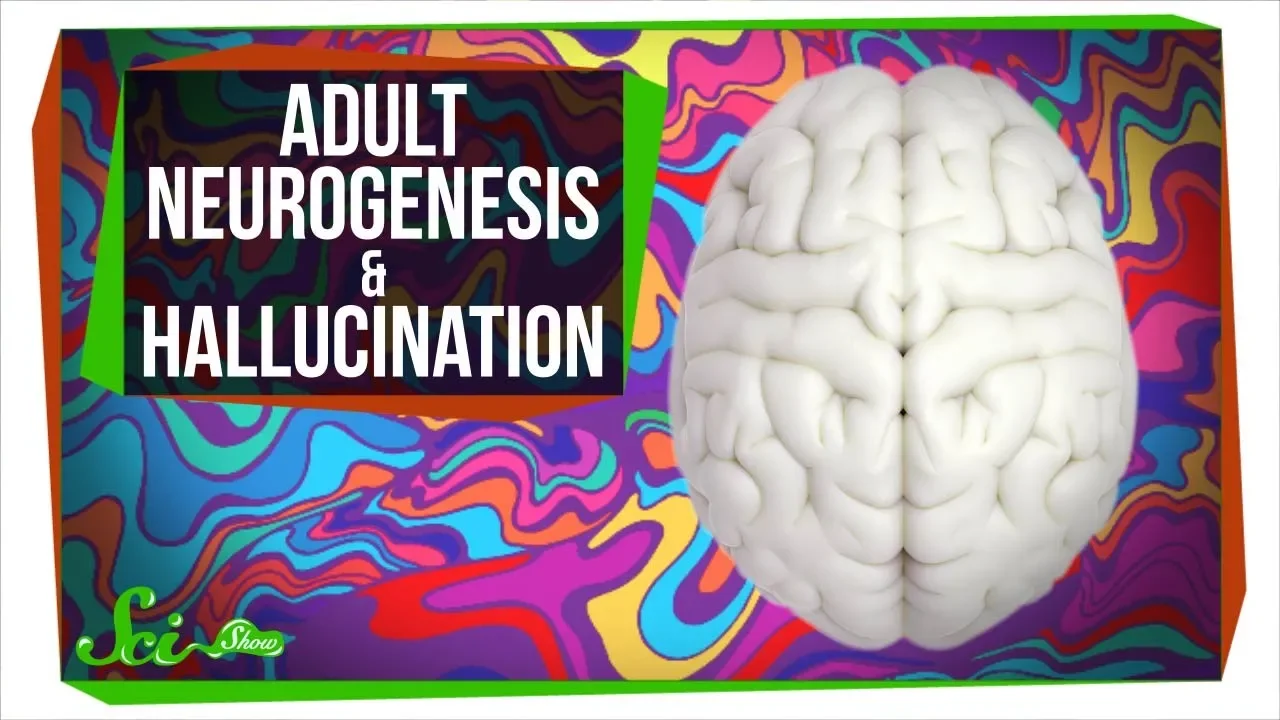
Adult hippocampal neurogenesis—the continuous generation of new neurons in the hippocampus—is a robust phenomenon in the human brain that persists until at least the ninth decade of life, but this process is disrupted in neurodegenerative diseases including Alzheimer's, ALS, Huntington's, Parkinson's, dementia with Lewy bodies, and frontotemporal dementia. These diseases impair the neurogenic niche homeostasis by affecting neural stem cells, proliferation rates, immature neuron maturation, and microglial phagocytic capacity, suggesting that dysfunction of adult neurogenesis may represent a therapeutic target for treating these conditions.
Neurogénesis Hipocampal Adulta y Enfermedades Neurodegenerativas
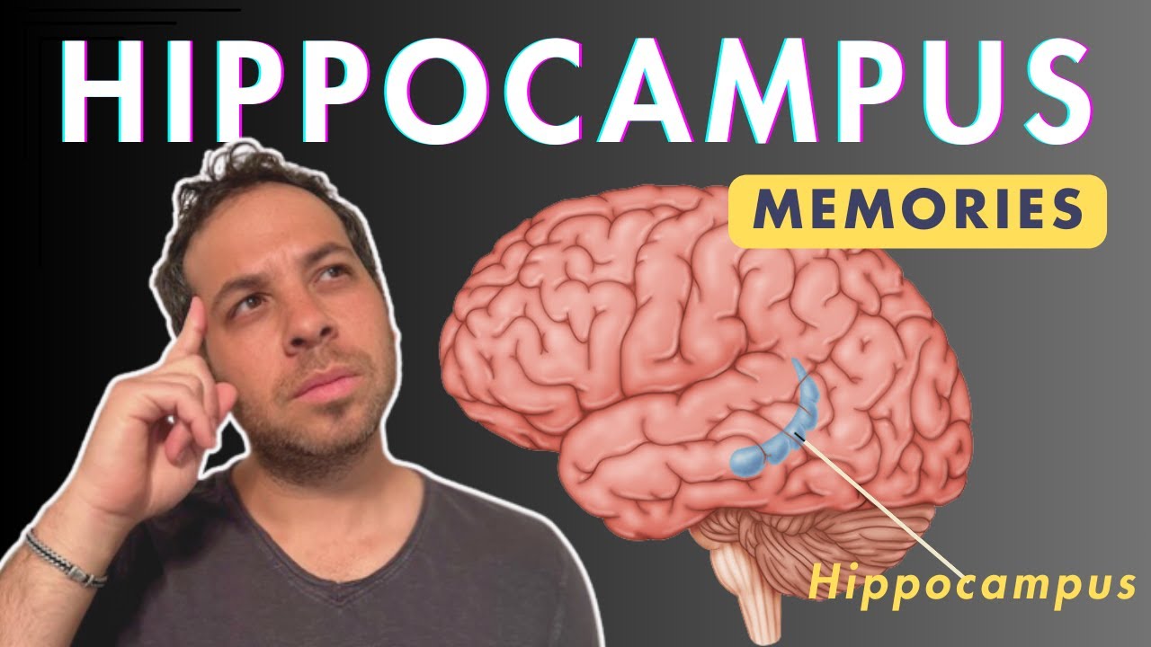
Added:Basic neuroanatomy of the limbic system, specifically the structure and primary functions of the hippocampus in memory formation and spatial navigation.
The fundamental concepts of cellular neurobiology, including neural stem cells, progenitor cells, and the general process of neurogenesis.
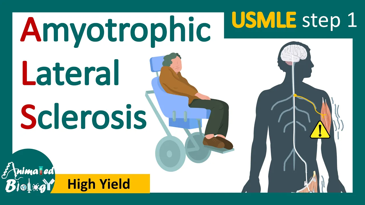
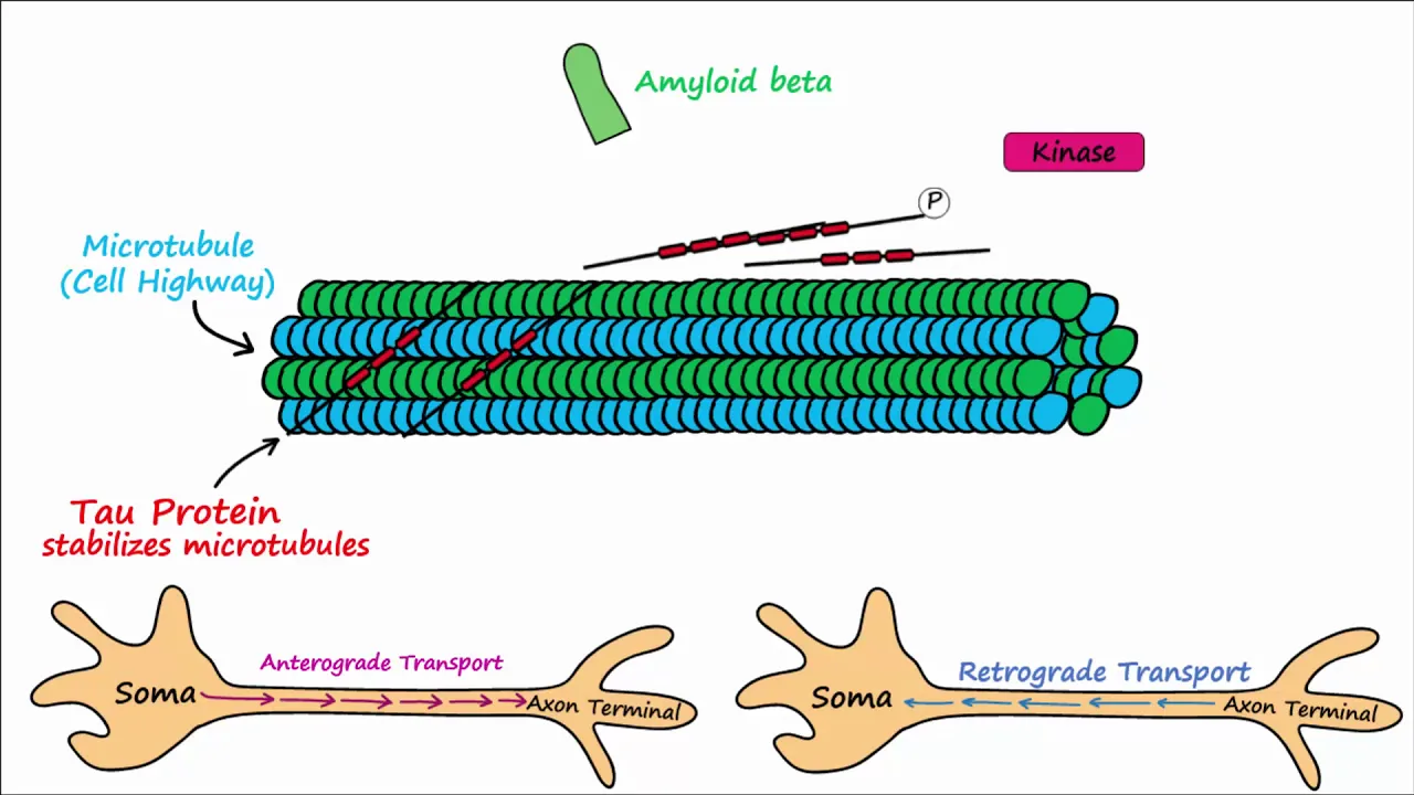
General pathophysiology of major neurodegenerative disorders, particularly the classic hallmarks of Alzheimer's Disease and Amyotrophic Lateral Sclerosis (ALS).
Translational therapies and pharmacological interventions aimed at stimulating adult neurogenesis to halt or reverse cognitive decline.
The influence of epigenetic and environmental factors (such as exercise, diet, chronic stress, and aging) on the regulation of hippocampal neurogenesis.
Advanced neuroimaging technologies and biomarker identification methods used to measure and track neurogenesis in living human patients.
The comparative neurobiology debate regarding the rate, functional significance, and existence of adult neurogenesis in humans versus rodent models.
2K views34likes1:00:54Original Release: 2022-01-31
welcome everyone to this new session of the cycle biology in the media that we organize at the school of biology at computancy university our goal as most of you know with this seminar series is to get a deeper understanding on those news related to biology that have a big impact on mass media on non-specialized media we want to provide more information on those news with rigor and a little more detail than the one that is given in newspapers or in the news today's talk is about a piece of news that made it to mass media at the end of last year regarding a discovery related to an old dogma in neurobiology that stated that no neurons are produced in the adult brain we have known for years that that idea was not accurate and that there are in fact specific areas in our adult brains capable of generating new neurons but we didn't know until now was the origin of those cells and their involvement in neurological diseases or the other way around the effect of those diseases on this cell population the answers to these questions were provided by our speaker in a research paper published last october in the journal science it is a great pleasure for me to introduce maria durens martin the main responsible for that work maria studied biology in our school and did her phd at the cajali institute where she started working on adult hype compiled neurogenesis she continued her research in this field in jesus abuela's lab at the center of molecular biology surveillance as a postdoctoral fellow and then she moved to japan for a few months to the university of of sukuba with a very prestigious japanese postdoctoral fellowship for foreign researchers in 2016 she returned to spain to the cbm where she currently holds a 10-year position as scientifico titular it was then and i repeat 2016 just five years ago when she set up her independent lab which focuses on the study of the basic biology and neuroprotective potential of adult and hypocampal neurogenesis for the treatment of various diseases since the establishment of our own group she has been the principal investigator of several national international research grants an erc consolidator grant just mentioned one and have published 27 papers in high impact journals such as science nature medicine nature protocols and others she has received three national research awards the young investigation investigator award in 2014 miguel catalan young investigator award in 2019 and young female italian award of the spanish royal academy of sciences in 2019 and she's a full member of the spanish young academy as you can see it's pretty obvious that we have an exceptional guest with us today and before passing the torch to her let me tell you all that this talk is very special for us not only because of maria's high profile and because she studied here but also because this is not the first time that we invite her roughly two years ago maria published another paper confirming the existence of adult hypocal neurogenesis that also had huge impact in the media we invited her then to give a talk that was actually scheduled for may 2020 but we had to cancel because we were immersed in the worst peak of the pandemic so having maria here today is not only an honor for us but also a sign that we are somehow and slowly going back to to normal so now and without any further ado welcome to your home maria and and thank you for accepting our invitation not once but twice so whenever you want thank you christina it's a big pleasure and a big honor for me to be to be here to be as you said at my home i feel the university of congress like my home where i started my scientific journey in some way so as i said it's it's a pleasure it was a pleasure to accept your invitation two years ago and and it is now so as christina said today i would like to talk about an extraordinary phenomenon that occurs in the mammalian brain which is adult neurogenesis and i would like to start specifically by defining what adult neurogenesis means and what does this phenomenon involve the incorporation of new neurons is an extraordinary phenomenon of plasticity given that the generation of new functional neurons implicates that the existing circuitry needs to be remodeled each time that a new neuron is incorporated so as you can imagine at all neurogenesis does not occur in widespread in our brain but in very specific regions of the brain named neurogenic niches some of these regions have been studied for the last decades in romans and i want to show you here the two ones that now everybody have knowledge to be neurogenic to hold this neurogenic potential in the rodent brain one of them is the the walls of the lateral ventricles where neural progenitors divide and give rise to to transiently to to migrate the neuroblast which which goes through the roster and migratory stream and migrate towards the old factory ball but they integrate into into this circuit the other neurogenic niche is the subgranular sun of the hippocampus and which is the region on most of the work from my lab is photospon here you can see a representation of the general anatomy of the hippocampus both in humans and in mice as you can see there are similarities but also the structure the whole structure of this part of the brain is slightly different anyway we can perfectly identify this layer this dentitus which is formed by the presence of dental granule cells branding neurons and this other region named cornu amonis which is supervised in two different fields named ca1 ca2 ca3 and ca4 the main difference or one of the main differences between these two areas is that the dental gyrus holds the capacity to generate new neurons whereas the ca fields are not neurogenic the hippocampus is involved in learning and memory and is also a key node of emotional regulation moreover the hippocampus is the target of several neurodegenerative diseases and is affected in psychiatric disorders as you can imagine the addition of new neurons in this structure plays a key role since it participates in each of the functions in which the hippocampus piecing mode in this general scheme i want to show you a representation of what the process of adult neurogenesis looks like here we have made a composition of several images showing the presence of a very special cellular type which is the neural stem cell type which is the origin of the process of adult neurogenesis these cells hold the capacity to generate new neurons through asymmetric cell division the daughter cells which are the transit amplifying progenitors go through different to various differentiation stages that transform these cells both from the morphological and the functional points of view and finally after the cells have completed their maturation process newly generated neurons are integrated into the hippocampal circuitry the way in which this process this process of adult hippocampal neurogenesis has been studied is based on the use of specific cell markers which are molecules that are expressed during during a limited periods of time and allow us to identify specific stages of this process here this is a just a very simple scheme of uh the better known molecules that are expressed during each of these stages as you can see the process is really complex i don't want to to get too much into detail but as you can see some of these markers are expressed during long periods of time whereas other cells are expressed during shorter periods of time so by combining these markers we can know at which at which stage is one individual cell another feature that is very important in this process is the presence of these accompanying cells which form the so-called neurogenic niche this structure is formed by astrocytes microglia blood vessels interneurons and other variety of cells which give the trophic support to the newly generated neurons for them to mature and integrate moreover another key strategy that has been used to study adult neurogenesis is the birth dating of new cells it was necessary for adult neurogenesis to be demonstrated to actually occur to be able to label the cells at the moment in which they were generated so by using these methods no one questioned the fact that adult neurogenesis was a robust phenomenon in the roman brain however due to technical and ethical difficulties the study of this process was a bit more complicated in the human brain so the question of whether at all neurogenesis takes place and to what extent this process is robust in the adult human brain was an important focus of controversy in the last years here i represented in green color all the papers that supported the occurrence of adult neurogenesis in the human brain but also in red color three key papers that showed the absence of markers of adult neurogenesis in the adult human dentals the problem here was that by using very similar methodologies different labs came to opposite conclusives so we wondered whether methodological differences even though they might be very subtle could underlay the discrepancies between the results obtained from different labs in order to answer this question which looks very trivial but it's not at all one has to to have a clear idea of the so many stages that a human sample has to go through before being observed in the microscope so as you can see here this process as i said is complex and there are many choices that that make the different pathways non-compatible so we couldn't study the effect of all the slight modifications in these steps but we focused our attention on the fixation process which is the inversion of the brain samples in a fixative solution which generally is made of distinct aldehydes paraformaldehydes vormaline etc and which in our hand was critical to study several components of the adult neurogenesis process in romans so we wondered whether fixation could influence our capacity to detect markers of adult neurogenesis in the human brain to answer this question in an initial study what we did was to obtain the whole hippocampus from several subjects and divide this hippocampi into small fragments and each of this fragment was fixed for a different period of time in a freshly prepared solution of para formaldehyde so what we did was something very simple we wanted to compare the number of double quotes in positive cells the number of immature neurons that we were able to to detect in samples obtained from the same subject but that have been fixed for different periods of time as you can see here when we fix the samples for short periods of time only 12 hours and and why do i say these are short times of fixation well the standard protocol that is followed at the brain banks worldwide is to fix the sample for several months or even years informally so these are extremely short fixation times that were that matched what we did with the the mouse symbols in the lab so here you can see in red color the presence of an abandoned population of immature neurons in the dentatums but surprisingly when we fix the samples obtained from the same subjects for 12 more hours so 24 hours in total the beautiful signal obtained with this anti-double protein antibody completely disappeared and was replaced by this and a specific background signal that embedded identification of positive cells so we started to work very hard to figure out what was happening there and finally identified a combination of histological treatments that we have to to subject the tissue to that removed this excessive fixation of of the produced by the aldehydes present in the in the fixative solution so by using this simple protocol we reverted these negative consequences of the fixative and were able to visualize not only these immature neurons but also some morphological characteristics of these cells so the conclusion of these two works by our lab is the fact that depending on how we treat the human tissue we can conclude that adoleogenesis is present or not but the true can only be one so the fact was that if we fixed for longer periods of time the samples that allowed visualization of markers of immature neurons that signal completely disappeared so this was our first conclusion that fixation is a critical factor that should be taken into account when working with the adult human brain to study adult neurogenesis but another important factor was the specificity of the signal that we obtained so it was very important to demonstrate that in those places of the of the hippocampus that are not known or that are known not to generate new murals such as the ca1 ca2 ca3 feels there was no presence of double quartet positive neurons as you can see here but in contrast when we analyzed specifically the dendrites the neurogenic zone of the hippocampus we observe this robust presence of immature neurons although the numbers of these neurons showed some variations between the subjects and this is something that we were very curious about also it was very important to demonstrate that the morphology of the cells that we were observing with these antibodies was that one that is classical for the granule neurons so we were not detecting with our staining protocol other cells or we were not detecting double cortine expression in other cell types which would make no sense at all so double curtain is not expressed in astrocytes in microglial cells or in blood vessels so as i mentioned we found this variability in the number of immature neurons and which were the factors that could account for this variability the first one was the postmortem delay which is the time elapsed between the person dies and the sample is extracted and inversed in fixed during this period of time as you can imagine most enzymes are working and are degrading proteins so most of our samples have short postmortem delays in green color but we also have some samples with longer postmortem delays in this case we did not find a reduction in the number of immature neurons that we detected with the with longer postmortem delays we also did not found any uh change in the number of double quotes in positive cells depending on the gender of the subjects but in contrast when we represented the number of cells and the eights of the subjects we found this phenomenon which is the reduction of the rate of adult neurogenesis during aiding which also served as a validation of our results given that this reduction have been described in numerous mammalian species and something that caused very powerfully our attention was the fact that not all the double protein positive neurons appear to be the same they show different location in the granules and layer different presence of a different number of neurites an orientation of the same and also the soma size of these cells appear to to be different so the only way we could check if this seemed to seem to seem to mean something was to co-localize uh the expression of double chordine which identifies these cells as inventory neurons with other markers that are specific for shorter periods of time during the maturation of the neurons in this way we aimed at comparing whether cells very very immature were different from other cells that were at more advanced stages of maturation as you can see here most of the recording positive cells were positive for the specific marker for granule neurons named pros1 but importantly a small population of well not that small population of double quote in positive cells was possible for markers of proliferation which indicated that this cell's whole proliferative capacity as they did in romans and these percentages of cells which hold the capacity to proliferate is maintained through aging other cells in this case are small percentage of cells expressed markers of indifferentiated neurons or named neuroblasts whereas other cells express markers of intermediate stages of maturation and how can we affirm that these cells are at intermediate stages of maturation well we repeated something that had been done in mice which was to compare the intensity of the expression of other markers such as nuan in cells that were positive for double quotient and in cells that were negative which were the mature the fully mature neurons as you can see here double chordine positive cells did express nuance but a lower rate compared to fully mature neurons which indicated that they were somehow an at an intermediate stage of maturation and finally other double quote in positive cells expressed markers characteristic of more differentiated neurons so what we did was to compare the two cells that were at the extremes of this maturation process and to see if the morphology of these cells was also represented of their maturation states as you can see here the most immature cells those that express carotene were horizontal small and have the presence of several apical neurons but in contrast the cells that are positive for calvinism look much more mature bigger and with the presence of one single primary apical neurone which is characteristic of mature planning neurons however something that was somehow criticized about our previous work published in 2019 was that we were not able to detect um neural stem cells in our tc and it was true that at that time when we attempted staining with markers of neural stem cells such as nesting we did not observe any convincing staining but what happened at that time we were using a standard immunostochemistry protocol with the modifications i mentioned before but the key point was that we were using some uh this molecule this triton x100 which is a strong detergent we have very recently been aware that we have to substitute triton x100 by saponin which is a milder detergent and which allows visualization of several markers of neural stem cells in the adult human tissue by using this modified or improved protocol protocol version 2.0 we can observe cells that are positive for nesting subscribe or dfap which are all of them markers of astrocyte um like uh radial glia like cells so these stem cells are known to share several features with astrocytes in the adult brain and some of these features include the expression of some markers of astrocytes so this raised the question of whether these cells were actually neural stem cells which of course can only be uh completely checked when these cells are played in vitro to see whether they are actually pluripotent pluripotent but speaking in immunochemical terms all these markers could be expressed also by astrocytes so in fact we also detected some nesting positive cells which were positive for 800 beta which is an universal marker of astrocytes but what was most important was that we were able to identify a population of nesting positive s hundred beta negative cells which indicates that these cells are not astrocytes and share phenotypic and morphological properties with the neural stem cells that we know from rodents and other mammalian species for example these cells have these long processes which transfers the granum cell layer and moreover 99 of these cells are located at the subgranular superannuation with neural stem cells in normals are known to to be located so these characteristics spoke about the population of nesting positive s hundred beta negative radial clear lex cells which shared phenotypic characteristics with mammalian neural stem cells moreover it was important also to detect that these cells were able to proliferate so we studied markers of proliferation in order to reconstruct the whole process of adult neurogenesis in the human brain we detected markers of proliferation mitosis and proliferative neuroblasts in the human dental tiers but to what extent were these cells reflecting proliferation of neuroblasts or cells committed to the neuronal limits well we quantified different parameters and determined that 90 or 80 percent of um atuc hud positive neuroblasts were double quartering positive and were also located at the superannuation which indicated that phenotypically these proliferative cells were committed to the neuronal dynamics in the human dentals so this reconstruction uh was talking about the dynamic process represented by adult neurogenesis also in humans as in many other mammalian species we detected immature neurons neural stem cells or cells with characteristics of neural stem cells and also proliferative cells in the dental gels so together with all the previous evidences represented in green color in my original table supported the notion that adult neurogenesis is a robust phenomenon in the human dental gerus until the ninth decade of life but all these so far is about uh physiological conditions let's say that these subjects that we studied until this point were all neurologically healthy so what um well what happened uh specifically with the other components of the of the of the adult neurogenesis process um if you remember we talked about the presence of astrocytes microglia and vascular elements and the functioning of all these elements is crucial for adult neurogenesis not only to exist but also to be adapted to the external conditions of the of the subject in particular we know from rodent's work that the interaction between adult neurogenesis and microglial cells is crucial for these cells to survive here you can see in green color microglial cells which are approaching to this pygnotic nucleus from an apoptotic cell and are attempting engulfing or phagocyting this nucleus in fact we know that 35 percent of uh apoptotic cells in the dental chairs are being phagocytosed by microglia and this interaction between these two types of cells seem to be important given that the morphology of microglial cells changes depending on the presence of these phallocytic bones which are the structures that these cells use to pharmacy those dysfunctional cells we know that the lower the number of microglial phagocytic causes the higher the number of cells of the apoptotic cells that remain to be falsities so the better the microglial cells work the better the adult neurogenesis process also seemed to work and it was important given that even though the number of microglial cells was not modified during aging the capacity of these cells to pharmacitos apoptotic neurons was decreased so we wondered whether this decrease in the phalocetic capacity of microglia that we observe in neurologically healthy subjects could also be related to to sound dysfunction of the whole hippocampal dentals in patients with neurodegenerative diseases so at first time in 2019 we started studying maybe the most obvious neurodegenerative disease that attacks or that targets the hippocampus which is alzheimer's disease we started characterizing these 45 this cohort of 45 patients with alzheimer's disease that were at distinct stages of severity of the disorder i forgot to mention that all this work both this in in neurologically healthy subjects that i mentioned before and all the work with patients with neurodegenerative diseases has been done in collaboration with dr alberto at the foundation field here in martin we are very proud of this collaboration and the the high quality samples we obtain in collaboration with the brain bank so by using this collection of samples which were processed in the same way that the those from neurologically healthy subjects we could determine that the number of immature neurals as you can see here was progressively reduced during the advance of the disease but what is most remarkable is the fact that even at fractal stage one which is the first severity states of the disease in which most of the patients do not present clinical symptoms the rate of adonirogenesis or at least the presence of immature neurons was reduced by a 33 as compared to controls moreover the number of immature neurons was further reduced when when the disease continues its progress this reduction is specific of immature neurons given that the number of total mature neurons remain unchanged however the cells that were reputatively being generated in the vendajarus expressed with less frequency markers that indicated that they were correctly advancing in their maturation process it means that aldo neurogenesis can be targeted by although by alzheimer's disease at multiple levels and one of these levels could be the final maturation of these cells so as a conclusion about neurogenesis was altered in patients with alzheimer's disease but somehow this was not surprising as i said given that the hippocampus is one of the main targets of this disease but we were also interested in studying whether other diseases that do not target at least in a direct form the hippocampus could also affect adult neurogenesis in humans and some of the diseases that we studied recently in our 2021 paper where his patients suffered als for example which is a motor neural disease that targets a very distant part of the brain or the spinal cord even huntington's disease in which the basal ganglia are degenerated as you can see here the enlargement of the lateral ventricles parkinson's disease in which the substantia and the basal ganglia are degenerated with these laws of teroxin hydroxylase positive neurons and then we studied further two diseases that were more [Music] sparse if you want so they are not focused on a single region of the brain but are characterized by dispersed neural degeneration in different brain areas these two diseases are dementia with levy bodies with which also shares some similarities with parkinson's disease from the um both the clinical and the neuropathological points of view although there are relevant differences between these diseases and from the temporal dimension which is characterized by a general alteration in movement and and also and also behavior and cognitive capacities so this were the initial number of diseases that we studied we started to study several years ago but now we are increasing our scope and studying also psychiatric diseases in our lab so i would like um to to to summary in a simple way there's so complex data that we obtained we when we studied these diseases and to do that i think that following this scheme will be somehow useful so we can start by defining what happens with the population of neural stem cells in these diseases as a general markers of neural stem cells we use nesting and cells too but we also determine the person the the number of distance that did not express s hundred beta so we quantified specifically the neural stem cells uh properly speaking so in cases with als we found an increase in both the number of nesting positive s and repeat and negative and solves to positive cells which reflect the fact that the population of neural stem cells was increased in patients with these diseases so one could think that this is something beneficial but increasing the number of neural stem cells is something very dangerous in adult mammals because this expansion of the population is usually related to an exhaustion of the of the neurogenic capacity of these neural stem cells in patients with huntington's disease we also found an increase in the number of nesting positive s and negative cells uh which also reflected this possible increase in the number of neural stem cells which was also replicated in patients with parkinson's disease so look these three diseases which do not target the hippocampus directly are repetitively altering the neurogenic capacity of the neural stem cells that are in this structure in contrast patients with dementia with levy body disease and frontotemporal dementia did not experience changes in the number of neural stem cells so what happens with proliferation one would expect that the levels of proliferation were also changed in parallel to those of the of the neural stem cells is this what is really happening in this case we use the two classical markers of proliferation and you can see how surprised we we were when detected that the number of proliferative cells was not modified in patients with als huntington's disease but in this case they were increased in patients with parkinson's so similarly we did not detect changes in patients with dementia with lewy body diseases with levy bodies but a reduction in the number of proliferative cells in patients with frontotemporal dementia so this scenario is starting to draw this balance between the number of radial clear-like cells that are present in these diseases and the amount of proliferation so how can this be explained we don't have an answer for that question and it's a very complex problem to solve but it could perfectly be that the number of gravial glia like cells is increased because these cells are somehow after being increased remaining in acquiescent states or because they cannot complete their proliferation cycle so the fact is that adult neurogenesis is not always regulated in a parallel way you know the multiple stages and this is something that we know from rodents and also some reports point to these same phenomenon in humans so accompanying this this balance uh [Music] initial stages of the process what happens with the what happened with the immature neurons labeled with double quoting well in this case in several of these diseases we again find an increase in the number of immature neurons but other diseases do not show this tendency this mean that the number of neurons that are being generated is increased so keep please this question in mind and i will try to to give an answer later because if this number of cells that are being generated were constant the size of the dental jars of these patients should also be progressively increased is this the case do you think obviously not so the point is that most of these newly generated neurons or immature neurons also exhibited altered morphology as you can see here we found alterations in the number of neurites or in the orientation of these neurons which somehow speaks about problems in the maturation process of these cells which are similar to those that we observe in patients with alzheimer's disease so in most of these diseases we found a remarkable alteration of the morphology or positioning of these cells so the process of adonirogenesis could seem to be increased but the fact is that when we quantify the number of apoptotic cells the cells that are actually dying in the dentatus it is the this number is increased in all the diseases so despite we may have increased number of radial glia like cells or immature neurons probably we are never going to observe an increase in the number of um total a mature neurons and that's in fact what we observe so the cells are being generated but because they cannot mature probably they die what happens in addition the neurogenic niche which is composed by microglia astrocytes and blood vessels could not be functioning correctly and that is the case in fact the phagocytic capacity of the microglial cells the capacity of these cells to remove those cells that are being that are dying uh through apoptosis is decreased so we have more immature neurons more apoptotic cells like the microglia cannot read of them the number of astrocytes probably as a consequence of these impairments in the pharmaceutical capacity of the microglia are more present in the dentatus of these basins and also we observe changes in the thickness of the capillaries which could be also indicating inflammatory processes that are taking place in in their dentals and this is something perfectly compatible with the neural degeneration that is occurring in digital brain areas of these spaces so in general we can say that the homeostasis of the of the dental tiaras niche is impaired in patients with neurodegenerative diseases so as an attempt to somehow order in our mind or in the reader's mind the alterations in the distinct stages of the disease of the of the process that is observed in patients with different neurogenerative diseases we made this model in which the color the of the arrows represent the disease in which these populations are altered either increased or decreased so the conclusion is not whether in als this step in particular is increased or not but in contrast the fact that in all these diseases apoptosis is increased which reflects a malfunctioning of the dental diaries neurogenic niche components given astrocytes microglia for blood vessels and this affects the behavior the proliferative behavior of neural precursors and neural stem cells which in turn which indeed leads to impairments in the differentiation of then that grant himself so in the future um well in the future we will do many things but by now we can conclude the presence of cells with phenotypic and morphological characteristics of neural stem cells proliferative cells and immature neurons in the human dentals we can also conclude the existence of a similar structure that than that described in province which is the dental neurogenic niche that neurodegenerative disease is impaired at all neurogenesis and the homeostasis of this niche and as i said in the future with the support of the crc consolidator grant in our lab we want to to unveil which are the molecular and cellular mechanisms that control adult neurogenesis not only during physiological aging but also in different neurodegenerative conditions and just to conclude i would like to to introduce you the most important part of this talk which is the group who made this work as christina said we are at the cbm in madrid and we are always willing to accept new students and postdocs whenever it's possible due to space limitations in our lab i would like to thank all our collaborators who made this work possible our sponsors and of course thank you very much for your attention and now i will be happy to to answer your questions thank you so much maria it was a fascinating talk what a what an interesting topic and what a fascinating line of research and congratulations not only on this last paper but on the whole career that you have established for yourself and your research group congratulations i'm gonna ask you to stop sharing so that any everybody can see your face like a big screen thank you so much and i'm gonna start asking you the questions that the audience is asking and i'm gonna remind the audience first that they can ask the questions through the tools that it's in the bottom part of the screens you can type your questions there and i will read them to maria and we have already two questions from one of our dear professors here at the school of biology agustin zapata i'm sure you know him and the first one is why there are two adult neural niches and how are the mutual relationships between them if they exist yeah thank you i was being happy to say hello to you i think no one knows why there are only two neurogenic nieces in fact i have to say that some researchers affirm that there are a vicious neurogenic niches such as the hypothalamus for example but i would say that these ones are the most studied ones why are they the most studied ones i would say that the hippocampus because it's involved in many behaviors and in many diseases that affect humans and probably the the the ventricular sun niche because it's very prominent when you use brvu or proliferation markers and observe the the whole mouse brain the proliferation is much more abundant i would say in the subgram in the subventricular sun than in the subliminal so i think that's the the maybe the reason why these are the two most studied niches and regarding their relationships they respond similar to several stimuli but they are regulated independently by many other external factors for example it's known that physical exercise specifically specifically increase at all neurogenesis in the dental gyrus but not in the ventricular symmetrical zone but other factors such as inflammation can affect the behavior of neural stem cells in both neurogenic nations um it has also been demonstrated that abolishing the neurogenesis in one of these niches does not abolish neurogenesis neurogenesis in the other one so it seems that of course they are mutually related but also that they are regulated in some other ways in a totally independent manner thank you maria and i'm gonna go back to his first question and you mentioned that these are the most studies uh niches but do you think that the improvement in histological techniques which was one of the grounds of your papers do you think that that improvement could lead to the discovery of new areas for example or yeah what i would say is that the lack of evidence for adoleogenesis in other regions is not approved for the absence of neurogenesis i think we should apply the same document that has been being discussed for the dental jars for several decades now to other regions so honestly i don't see much neurogenesis in rodents in other brain areas but i would never say that that means there is no neurogenesis in those areas because i consider that there are many groups uh doing very sort of studies of those regions and and probably there are some indicators of neurogenesis in in other regions such as the hypothalamus and why not i think it's important to improve the protocols but in parallel it's also important to improve the controls and austin has another question for you and it has to do with the relationship between microbiota and central nervous system and he is asking how the central nervous system feels the changes in microbiota well there are very very nice studies on very funny [Music] conclusions about the the alterations in microbiota and how do they influence adult neurogenesis i think it's now clear for the field that microbiota influences adult neurogenesis and that factors external factors such as adult adult physical exercise influences microbiota and through that pathway it influences other immunogenesis but i think that we still don't have a map of the most beneficial bacteria and the most detrimental ones specifically for adult neurogenesis we have some clues for new inflammation and neurodegeneration and even even for emotional aspects of the behavior but it's not been yet so well characterized how the how it influences uh above neurogenesis and related to that and i'm gonna skip the line of the questions i'm sorry i apologize and which are the main stimuli that uh trigger adult neurogenesis do you know and related to this and your answer your potential answer how how do you envision modulating that in order to potentially treat neurodegenerative diseases in the future well the the best studied ones or the most powerful ones in rodents are physical exercise cognitive stimulation and social interaction so it was demonstrated in 1997 that the combination of these three stimuli applied in in what we named we call an enriched environment uh was one of the most powerful stimuli that potentiates adult neurogenesis it has been demonstrated later uh in the in the history that individually each of these factors can trigger neurogenesis our group for example demonstrated that increasing social interaction alone in the absence of higher physical activity or cognitive stimulation increases the maturation of new neurons so i would say that those are very powerful stimulators learning is also a good way to to increase adult neurogenesis spatial navigation in particular in rodents and also there are many well-known negative regulators of adult neurogenesis for example stress sleep deprivation diet can influence both in a positive and in a negative way the rate of adult neurogenesis hormones estrogen cycle maternity so there are many many many factors and regarding your second question i think that one of them we have two problems two maybe two big problems we related to neurodegenerative diseases one is that we don't know the etiology of these diseases so we don't know what is malfunctioning several decades before the appearance of these of these diseases the clinical symptoms symptoms of these diseases so that makes things a bit complicated and the second problem we have is that we apply generally all the interventions after the clinical symptoms have appeared so i wouldn't dare to say that increasing adult neurogenesis is going to to cure any neurodegenerative diseases especially these diseases that do not target the hippocampus we are not going to cure parkinson alone with stimulating other neurogenesis because there are other neurons that are lying in other parts of the brain however what we know from rodents is that maybe if we could prevent the loss of new neurons in the hippocampus we could ameliorate or or delay some of the symptoms of these diseases some secondary symptoms of these diseases but i think neurodegenerative diseases are so terrible for the whole brain there are so many things that are not working well that probably reversing only along neurogenesis wouldn't be sufficient even though we started very early we would need to treat the disease in a more direct way thank you so much for your answer we have another question from maria gomez our colleague from the school of medicine i think you know her too and she's asking you if you think that an increase in the central nervous system or an increase in adult neurogenesis rate in general would be beneficial or detrimental and if that would depend for example on the disease itself well that's a very interesting question and in fact we we know some conditions in which increasing adult neurogenesis worsens the course of the disease for example in the case of epilepsy we know that having more immature neurons is amplifying the consequences of scissors so i think it of course depends on what are the neuropathological mechanisms that are taking place in in those diseases um i think well it because it's a it's a very interesting question and it's a very difficult to answer because there are some papers and some groups who have been studied the participation of adult neurogenesis in forgetting in the opposite process that all of us have been studying which is the the incorporation or the generation of new memories it's known or it has been suggested that new neurons are also important for forgetting so regarding for example ptsd what would we want more neurons would it be beneficial to forget the traumatic memories or would it be somehow beneficial to keep those memories so i think it's complicated to answer whether it would be good or bad and i have another question and i apologize because it's gonna sound very silly and i'm aware of that but um i mean your studies are very clear and very solid in describing uh with markers with the use of markers the existence the structure and the positioning of the cells but you obviously cannot do functional styles so everything is pointing to a good proper function of the of the cells in the brain but you're still lacking that other piece of information and i know right now it's virtually impossible to approach these studies but how do you see the future in that aspect yeah absolutely this this is the main limitation of all postmortem studies that you can you have to limit yourself to observe what is happening to observe the final picture that is printed in the in the brain of those patients there are some attempts or there there have been some attempts to visualize markers or biomarkers of adult neurogenesis in vivo some strategies are being developed by different groups including functional magnetic spectroscopy or um a positive emission tomography which by now haven't developed a widely accepted indicator of adult neurogenesis but i do ambition that that has to be developed if possible because i think it would seem impossible for us to visualize a very small tumor several years ago and now we can even count the number of cells that this tumor has in vivo so i trust on those strategies on the potential of those strategies to to really visualize what is happening and to be able to correlate the functional aspects of neurogenesis which as far as we know are related to the acquisition of immune memories and to see those cells function and functioning in vivo of course that would be uh the dream for every researcher in this field okay i don't see my questions in the chat and i think i'm gonna ask you the last one and is i mean when i when i define researchers i tend to do like two categories like in the soccer leagues it's the first teams and then it's the rest of us so i i i see you as a player in the main league and and one of the difference between you and us is the kind of questions that you ask to yourselves and i think you ask yourself like big questions what i define as big questions in biology so what is this next big question that you are asking yourself well first let me say that i love small questions i think everyone should ask small and big questions and i think that the league in which all of us play i would say it's the same circumstances are different for all of us during different periods of our career but i consider all of us doing all the time big and small questions one of our biggest question is to what extent the process of adult neurogenesis in humans is actually unique so to what extent this process shares features between species and which are the things that are different and the biggest question is why those things are are different but i don't consider i mean it's as big or as small as any other question that anyone can have that's the one that interests us now uh the most well thank you so much it was well thank you for your answers specifically this one and and thank you again for accepting the invitation it has been a fantastic lecture on neurobiology and it has been a real pleasure to have you back at home not physically and we hope we can have you here soon probably to talk about your next science paper on nature medicine paper so good luck with that and i'm sure it will happen and we will invite you again and well thank you and thank you all at home for participating in these events and for keeping this seminar series alive and we will be back soon with more stuff we will talk about farms we will talk about all the things that are in the media and are related to biology and we hope to see you there thank you maria and thank you and please take care thank you very much bye
Up Next

58:51
Epilepsy Types and Treatment: Clinical Features Explained
@DoctorNajeeb
104.2K views•2022-04-09

48:47
Graphic Medicine: Comics for Collaborative Healthcare Communication
@nationalpatientadvocate
189 views•2023-12-04

10:28
Neuroanatomy: Central and Peripheral Nervous System Divisions Explained
@AKLECTURES
136.2K views•2014-09-20

3:05
Stages of Labor and Vaginal Birth | Childbirth Animation
@nucleusmedicalmedia
52.1M views•2017-08-18
Related Study Plans & Knowledge Roadmaps
Structured learning paths in Medicine
medicine
Abnormal LFTs: Diagnostic Workup & Pathology
01Liver Anatomy and Basic Physiology
02Introduction to Liver Function Tests (LFTs)
03Hepatocellular vs. Cholestatic Patterns
+ 3 more learning modules
12h6 modules
Explore Roadmap
medicine
Analyzing Blood Smears: RBC, WBC & Platelets
01Introduction to Blood Components & Smear Preparation
02Normal Morphology of Blood Cells
03Erythrocyte Pathologies & Morphological Variants
+ 2 more learning modules
12h5 modules
Explore Roadmap
medicine
Mechanical Ventilation: Waveforms & Alarms
01Anatomy and Physiology of Breathing
02Introduction to Mechanical Ventilation & Core Modes
03Ventilator Settings and Waveform Interpretation
+ 2 more learning modules
12h5 modules
Explore Roadmap
medicine
Neuro Exam: Localization & Stroke Syndromes
01Foundations of Neuroanatomy and Vascular Supply
02The Neurological Examination: Step-by-Step
03Principles of Neurological Localization
+ 3 more learning modules
18h6 modules
Explore Roadmap