











![Tomographic Image Reconstruction: Introduction (Part 1) [L28]](https://i.ytimg.com/vi/KjYnjlFLzvA/maxresdefault.jpg)


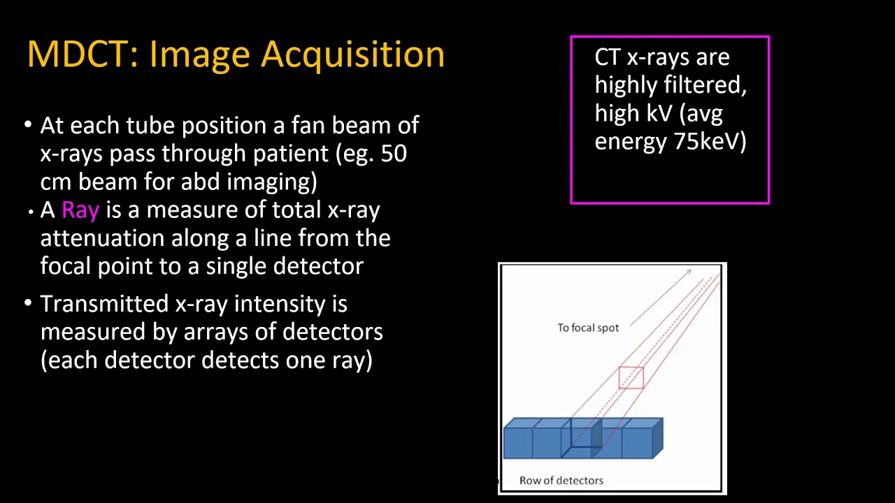
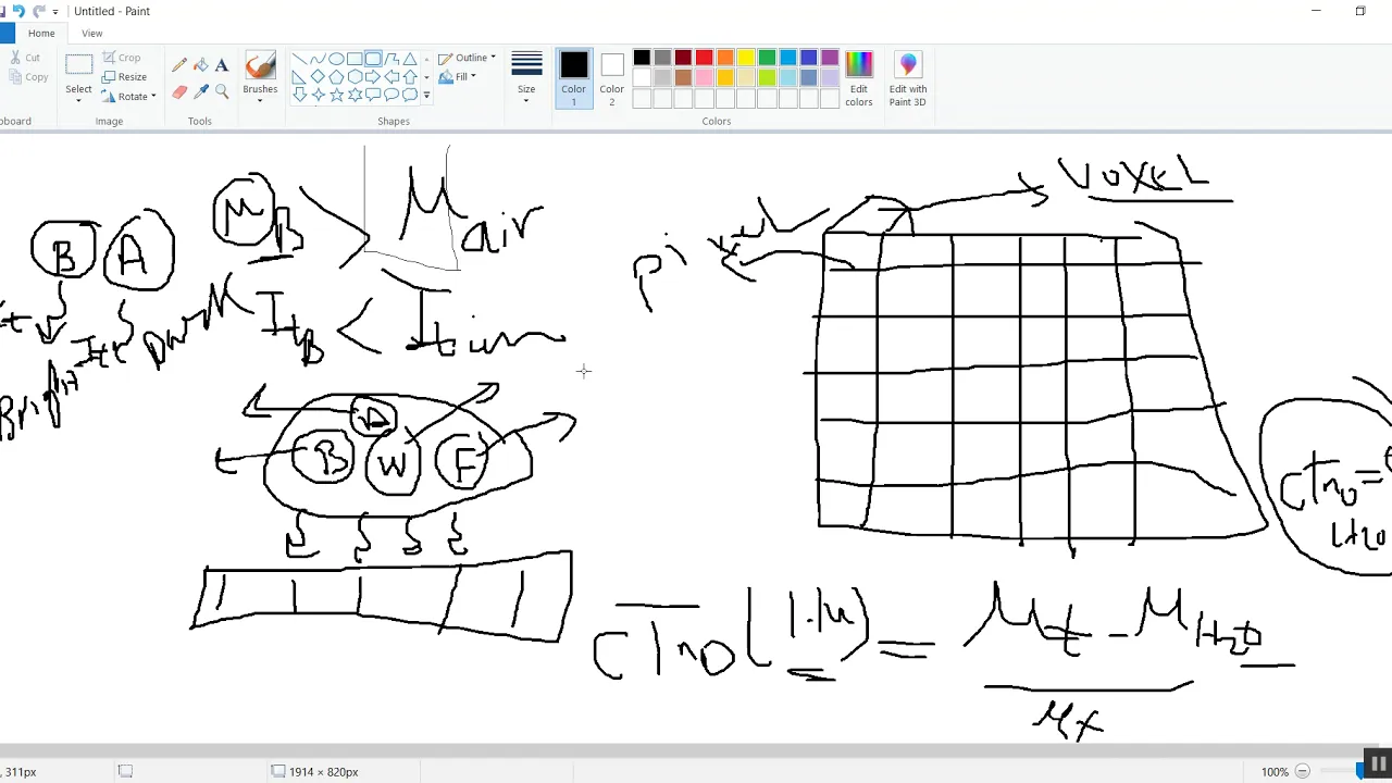
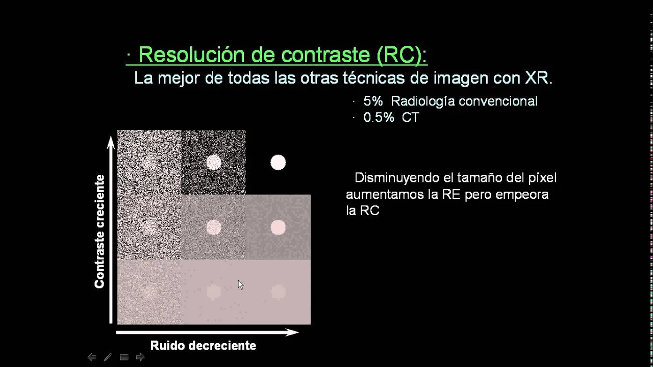


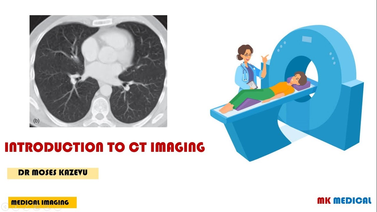

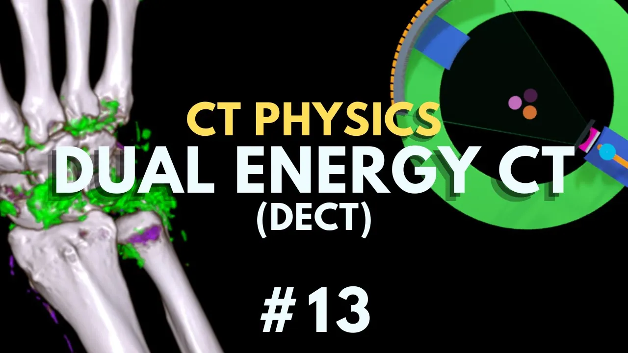

CT scanners differ from conventional radiography by acquiring volumetric data in axial slices using voxels (3D volume elements) rather than pixels, enabling superior low-contrast resolution; modern third-generation scanners use rotate-rotate motion with multi-row detector arrays, incorporating slip ring technology for continuous helical scanning, along with beam optimization components including metal filters, bowtie filters, collimators, and anti-scatter grids to enhance image quality and reduce patient radiation dose.
CT Scanner Components and Imaging Principles | Radiologic Technology Tutorial
Added:Basic physics of electromagnetic radiation and X-ray production, including how X-ray tubes generate radiation.
The concept of tissue attenuation and how different biological structures (bone, soft tissue, air) absorb X-rays.
Fundamentals of human anatomy in cross-section, particularly the axial (transverse) anatomical plane.
Basic electrical and mechanical engineering principles, such as rotational mechanics and continuous electrical contact.
Mathematical methods of image reconstruction, including Filtered Back Projection (FBP) and Iterative Reconstruction.
CT image quality parameters and the physics behind common image artifacts (e.g., beam hardening, motion, and metal artifacts).
Radiation dosimetry and patient safety protocols, specifically understanding ALARA, CTDIvol, and Dose Linear Product (DLP).
Advanced CT applications and modalities, such as Dual-Energy CT (DECT), CT Angiography (CTA), and multi-detector row CT (MDCT) technology.
29.7K views699likes7:59Original Release: 2021-01-23
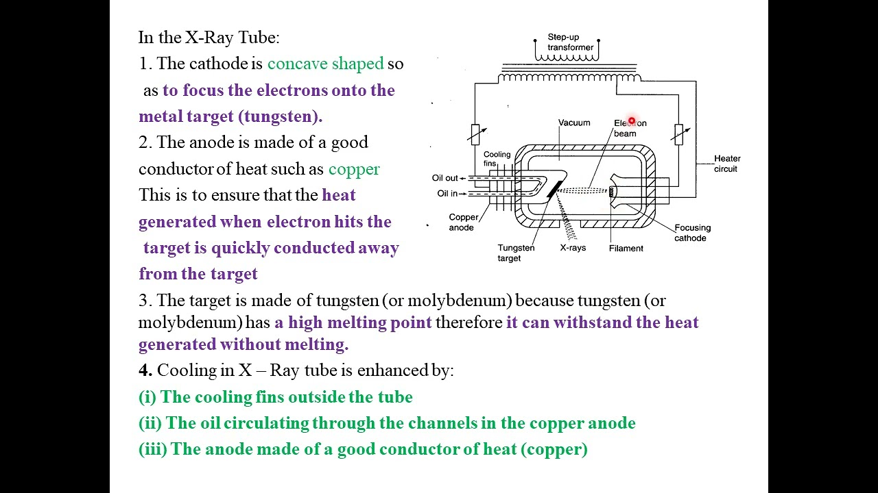
in this video we're going to discuss the major components and a brief history of ct scanners when compared to conventional radiography ct has the advantage of eliminating superimposition of structures by acquiring data in what we describe as slices in the axial plane which can then be reconstructed into a volumetric data set with three-dimensional information so where conventional radiography has us thinking in pixels or picture elements ct has us thinking in voxels or volume elements a voxel is like a pixel but with coordinates in 3d space ct also provides us with a much greater ability to differentiate between very small differences in density between anatomical structures in imaging terms this means ct has very good low contrast resolution the first ct scan was performed by sir godfrey hounsfield in 1971 and the first generation of scanners used a translate rotate motion where a single detector element would be exposed sequentially by a pencil beam while translating width waves across the patient then the whole system would rotate one degree at a time and repeat the process needless to say this took a very long time the pencil beam was soon replaced with a fan beam which maintained the translate rotate movement but covered a wider area the fan beam endured into modern day scanners but in the late 80s we lost the translate rotate nonsense and replaced it with the rotate rotate system with which we're familiar today where the tube and detector assembly spin in unison on opposing sides of the patient we've upgraded the single slit shaped fan to a thicker fan beam exposing a multi-row detector where the detector instead of being a single row of detector elements is now composed of multiple rows of detector elements so it looks more like a curved version of a conventional digital radiography detector it's just much wider than it is long this type of system is referred to as a third generation scanner and is the most common type of ct scanner we use today from the outside a ct scanner is composed of the generator which provides high voltage constant potential to the x-ray tube in the same manner as conventional x-ray system and also governs the speed of anode rotation the gantry which is the ring-shaped body of the scanner housing the tube detector array a control panel and laser lights to align the patient to the table or couch which the patient is positioned upon and which slides in and out of the scanner along the z-axis so the x-axis being left to right the y-axis being up and down and the z-axis being in and out of the scanner and the computer and console from which the technologist acquires the scan which is in the control booth outside of the scan room on the inside of the scanner is the x-ray tube which generates x-rays the detector array which detects the x-rays transmitted through the patient and the data acquisition system which receives data from the detectors and communicates that data to the scan computer also housed throughout the inside of the gantry are a technology called slip rings slip rings are used in many modern electronics requiring rotating parts through which an electrical current or signal needs to pass they function like a bearing where two separate components can turn relative to one another or one can turn while one stays stationary but while turning there is a contact surface either a smooth ring or metal brushes which maintains conduction of electrical current or signal throughout the rotation without the need for wires which would twist up under that type of motion this is significant because it allowed for the continuous acquisition of data in what's called helical scanning where the scanner acquires data in a spiral pattern wrapping around the patient acquiring continuously while the patient moves through the scanner along the z axis this is in contrast to axial scanning or step and shoot scanning where the scanner would acquire one rotation move the table in acquire the next rotation move the table and again and so on now the terminology here can be confusing because when conducting a helical scan we are still considering that to be acquiring data in the axial plane in ct we're always scanning in the axial plane but the difference between helical scan mode and an axial scan mode just refers to this difference of continuous scanning in a spiral versus step and shoot scanning in discrete slices there are various ways in which the x-ray beam is optimized within the gantry between the tube and the patient are metal filters which absorb low energy x-ray photons increasing the average photon energy or hardening the x-ray beam and one type of filter unique to ct is the bow tie filter which is shaped like a bow tie so it's narrow in the middle and thicker at the edges this serves to remove a higher number of photons at the edge of the patient where less photons are needed while allowing a higher number of photons to pass through the centre of the patient where the additional beam penetration is required this leads to a more homogeneous beam exiting the patient while reducing unnecessary radiation dose to the patient's periphery as in conventional x-ray there is a collimator between the tube and the patient which shapes and defines the edges of the x-ray beam leaving the tube and unlike conventional x-ray there's also a post-patient or pre-detector collimator which aligns to the edges of the detector area being used again shaping the beam before it hits the detector and absorbing scatter radiation to improve image quality also absorbing scatter we have an anti-scattered grid again much like in conventional radiography made up of lead strips which absorb any x-rays which aren't traveling straight towards the detector since any x-rays incident at an odd angle to the detector are scattered x-rays unlike in conventional x-ray in some ct systems the grid lines are oriented in both horizontal and vertical directions much like what nuclear medicine technologists might call a collimator different professions different language and the reason we can do this is because we know that the tube and detector will never be angled relative to one another in ct where in x-ray we're used to angling in one direction relative to the detector so we have grid lines only pointing in that direction the detector itself is composed of multiple rows of detector elements and the number of rows in the z-axis is the number of slices that the scanner can acquire at once most modern scanners have at least 64 rows and many vendors are pushing the limit of 256 rows or even higher the detector is an indirect type digital detector meaning that x-rays incident on the detector are absorbed by a scintillation layer and converted into visible light this visible light hits a photodiode which converts that light into an analog electrical signal that analog signal is then converted into a digital signal by an analog to digital converter or adc and at this point we have raw image data which is ready to be communicated to our workstation and processed into an image we'll cover the course concepts of producing an image in ct in the next video including field of view pitch window width and level linear attenuation coefficients ct numbers and house field units if you have any questions on the content of this video you're welcome to comment or send me an email and thanks for watching you
Up Next
114:06
A2 Medical Physics Review: MRI, Ultrasound, X-Ray & CT | 9702
@ETphysics
13.6K views•2021-04-30

12:53
21cm Hyperfine Transition in Neutral Hydrogen: Radio Astronomy Basics
@AaronRobertParsons
12.4K views•2011-10-13

32:32
NMR Spin Physics I: Zeeman Effect, Resonance Condition & Larmor Frequency
@nptel-indianinstituteofsci8064
2.3K views•2024-01-17

27:14
Entropy and the Second Law of Thermodynamics Explained
@veritasium
27.5M views•2023-07-01
Related Study Plans & Knowledge Roadmaps
Structured learning paths in Physics
physics
Nuclear Physics (Decay, Fission & Reactors)
01Atomic Structure and the Binding Force
02Radioactive Decay and Half-Life
03Nuclear Fission Mechanics and Chain Reactions
+ 2 more learning modules
12h5 modules
Explore Roadmap
physics
Soft Matter (Polymers, Colloids & Rheology)
01Foundations of Soft Matter & Thermal Forces
02Polymer Physics and Macromolecules
03Colloids, Surfactants, and Self-Assembly
+ 2 more learning modules
38h5 modules
Explore Roadmap
physics
Analytical Mechanics: Lagrangian & Hamiltonian
01Mathematical Foundations: Calculus of Variations
02From Newtonian Mechanics to Constraint Dynamics
03The Principle of Least Action & Lagrangian Mechanics
+ 3 more learning modules
28h6 modules
Explore Roadmap
physics
General Relativity (Spacetime & Cosmology)
01Foundations of Relativity: Special Relativity
02The Geometry of Spacetime: Equivalence Principle & Curvature
03Einstein's Field Equations: The Mathematics of Gravity
+ 2 more learning modules
55h5 modules
Explore Roadmap