













![[19/05/2021] Severo Ochoa Seminar by C. Lee; "Shape prior metal artifact reduction algorithm..."](https://i.ytimg.com/vi/OhKPSg2r7ws/maxresdefault.jpg)



![[TCTAP 2021 Virtual] Hot Topics - III. CTO](https://i.ytimg.com/vi/BJ4y8LMJqk8/maxresdefault.jpg)

CT image reconstruction converts raw detector data into diagnostic images through mathematical processes including back projection (mapping attenuation pathways from multiple angles), filtered back projection (applying sharpening or smoothing filters to enhance spatial resolution or reduce noise), iterative reconstruction (repeatedly refining estimates to reduce artifacts and noise), multiplanar reformatting (viewing data from different orientations), and rendering modes (volume averaging, MIP, MinIP, and 3D surface rendering) to visualize anatomical structures with varying properties.
CT Image Reconstruction: Back Projection, Filters, MPR & 3D Rendering
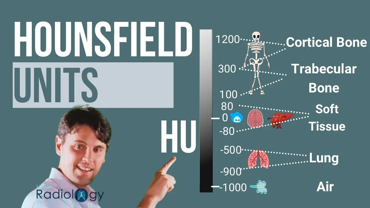
Added:Basic physics of X-ray attenuation, tissue density, and the concept of Hounsfield Units (HU).
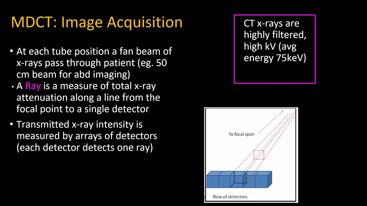
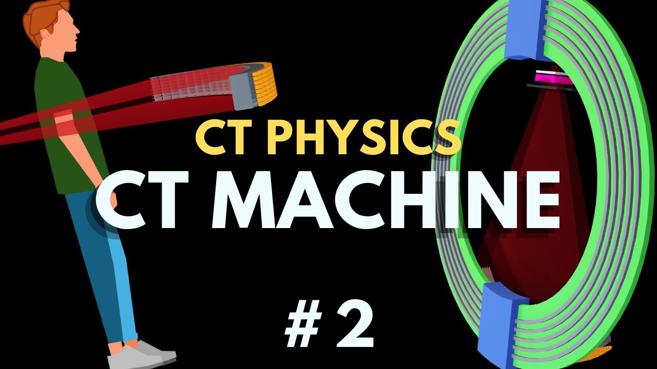
The mechanics of CT data acquisition, including how a rotating gantry and detector array generate raw projection data (sinograms).
Fundamental mathematical concepts in signal and image processing, particularly Fourier transforms and spatial convolution.
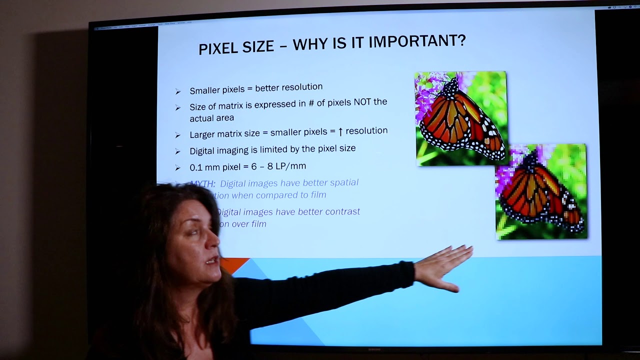
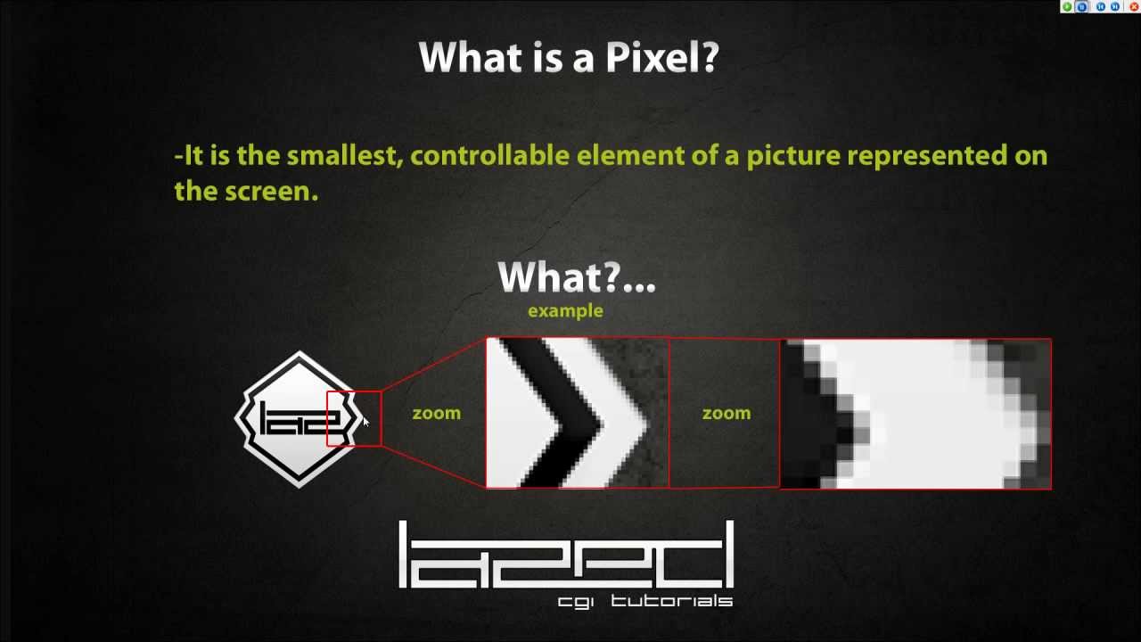
An understanding of digital imaging fundamentals, such as pixels, voxels, and spatial resolution.
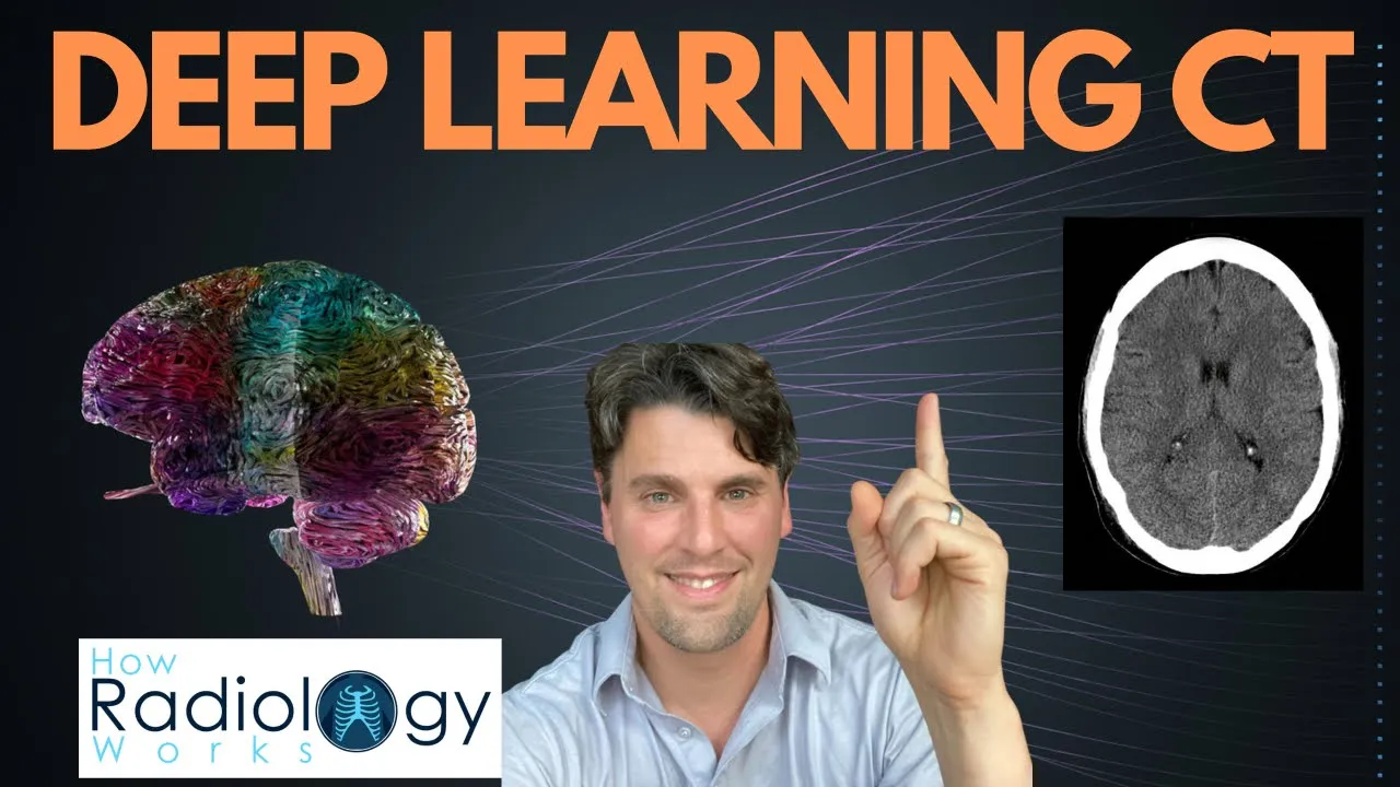
Advanced deep learning and AI-driven CT image reconstruction algorithms that reduce noise and radiation dose.
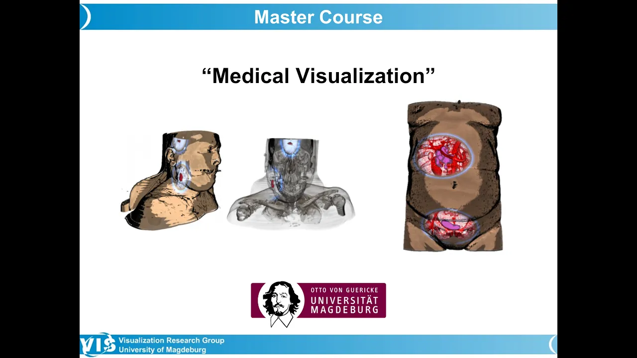
Clinical applications and techniques of advanced visualization, such as Maximum Intensity Projection (MIP) and Virtual Endoscopy.
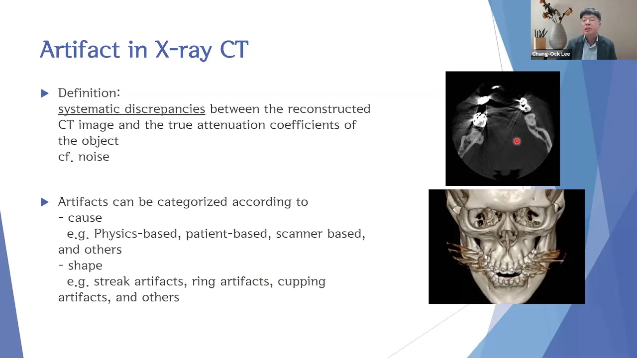
CT image artifact identification and correction methods, such as beam hardening, motion correction, and metal artifact reduction.
The trade-offs between image reconstruction quality, reconstruction speed, and patient radiation dose optimization (ALARA principle).
72.8K views1.4Klikes9:24Original Release: 2021-08-23
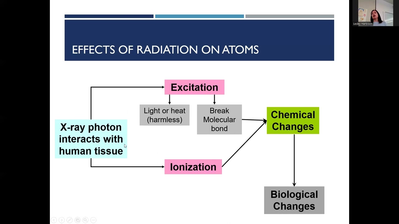
image reconstruction is the process of turning braw data collected by the detectors into image data which can be viewed on a screen both raw data and image data can be manipulated in different ways to create images with differing properties the most rudimentary process of turning raw data into image data is called back projection this is the process of mathematically mapping the attenuation pathway at every angle measured through a scan to locate where in a patient attenuation is occurring if there is a dense object at this location in a patient then a projection at this angle will show us attenuation in this area because less photons will be arriving at the detector through this attenuation pathway when we apply that same principle from various angles through the patient we can map where that dense object is within the patient and generate a cross-sectional image that shows the object the star pattern you see here is a natural drawback of the back projection method but it's reduced to an extent by the fact that we have many other angles showing where there is or isn't attenuation the beam in this direction for example tells us that there is no dense object here this means that the more projection angles we have in our data set the greater our image quality will be the relatively simple process known as back projection is now applied within another process called filtered back projection all that this means is that a filter is applied to all of the data that's put through the back projection process a filter also referred to as a kernel or sometimes just as an algorithm is a form of data pre-processing and it's an algorithmic manipulation which Alters the value of a pixel and its neighboring pixels relative to one another so for example if we want to see very sharp edges and details we would apply a sharpening filter also known as an edge enhancement or high pass filter and what that does is it drives the value of each pixel away from the value of its neighboring pixels so in this example we have an interface between a dark gray and a light gray pixel and we're going to apply a sharpening filter and when that filter is applied to this middle pixel it's going to take its neighbors and drive their values away from the average value of these three so the dark pixel becomes darker and the light pixel becomes lighter conversely if we use a smooth filter also known as a blurring or a low pass filter we're going to do the opposite and make this sharp black white interface into a much smoother gray to gray transition so we're averaging this pixel out with the value of its neighbors with filtered back projection we're applying these mathematical transformations to our whole data set when reconstructing it so every pixel is individually going through some kind of algorithmic n ation relative to its neighbors to either give us a sharper or a smoother set of images this has a direct impact on the properties of our image because a sharpening algorithm is going to give us higher spatial resolution and crisp sharp edges but it's also going to make the noise more prominent whereas a smoother algorithm is going to blur and smooth out that noise but at the cost of also blurring out our nice sharp edges so as with everything in CT there is this trade-off between whether we want to see sharp or smooth images and in some scans you're going to reconstruct both sharp and smooth sets of data which can each be used to view different structures within one scan for example with a chest CT you're going to reconstruct both lung and soft tissue data sets from your raw data the lung images will be reconstructed with a sharp filter so we can see small nodules and fine details in the lung tissue with high spatial resolution and the soft tissue images will be reconstructed with a smooth filter so we can look at the structures of the har and the media Signum with reduced noise and therefore with better contrast resolution in a previous video we discussed windowing and each of these different data sets are also going to have different Windows applied as well because we're looking at different structures so two different filters are used and two different windows are used to view these two different reconstructions from one raw data [Music] set Beyond filtered back project ction another method of reconstructing raw data into image data is called iterative reconstruction iterative reconstruction is a repetitive process which starts with a filtered back projection which is treated as an estimate of the true data set and then each data point in this estimate is compared to the scan data to see how close to the True Value it is this process happens in a cycle repeatedly until a certain predetermined number of iterations has happened and at that point the assumed image has then repeatedly refined and is going to be very close to a true representation of the data the advantage of iterative reconstruction is that both noise and certain artifacts in the image can be reduced because both of these things are non-true representations of what should be in the data set so with every iteration we're reducing these inconsistencies in our data the increasing proliferation and effectiveness of iterative reconstruction algorithms has meant that we are able to reduce the radiation doses used in CT scanning because any form of noise reducing technology means we can settle for more noise in our data and then reduce that noise with the power of computing so a lot of the dose reduction strategies which various vendors have developed recently in CT have been driven by improvements in iterative reconstruction software alongside a growth in computing power that has enabled these complex algorithms to run much more quickly after reconstructing our image data from the raw data we can apply several other manipulations to those images multi plary formats or mprs are reconstructed in almost every CT scan perform today and what this means is simply putting our data together to view it from a different orientation because a set of axial images is basically a lined up row of voxal displayed as pixels we can effectively stackle those slices on top of each other and view the voxal from any angle so we can manipulate those axial images to be viewed in the coronal plane or the sagittal plane or even an oblique plane through the body in most scan protocols the scanner will have pre-loaded instructions to generate an axial image series from the raw data and then to generate mprs in the coronal and sagittal planes from that axial data if thick axial slices are used to reconstruct the nprs the coronal and sagittal images will have a jagged appearance known as a stairstep artifact at the interfaces of each of those slices so routine practice is to reconstruct an axial data set in the thinnest possible slices from the raw data with overlap and then to use that thin data set solely for the purpose of generating mprs when generating reconstructions in any plane from a thin set of image data there are a few ways to render these slices if for example we're using a set of 0.5 mm slices to generate a 3mm slice the simplest and most common way to render that 3mm slice would be to take all those stacked 0.5 mm voxal and to average them to to give us a value for the voxal represented by that 3mm slice this is how normal images are made and it's referred to as volume averaging because you're averaging all the values within that volume this isn't the only way to render that data however we could also take the maximum value represented within that 3mm volume and take that maximum value to represent the whole voxal this is called a maximum intensity projection or a MIP similarly we could take the minimum value represented within this volume and take that to represent the whole voxal this is called a minimum intensity projection or a minip while volume averaging is the most common mode of rendering image reconstructions mips and to a lesser extent minips do have certain applications in which they can be useful for example if you're looking for tiny lung nodules a MIP may be used to bring out and highlight nodules which are smaller than your two mm slice for example on the other hand if you want to only look at the air space in the lung a minimum intensity projection will give you a very black image to see air spaces without obstruction from lung tissue which might otherwise occupy the same voxel the last manipulation to discuss is 3D reconstructions because a set of thinly stacked axial images is comprised of volumetric information we can use that volumetric information to generate a 3D image that represents the volume surface rendering techniques can be used to detect a continuous surface within a volume of data volume rendering techniques can be used to render the full scope of 3D data and see various tissues in conjunction with one another as always thanks for watching and keep up the hard work
Up Next
16:01
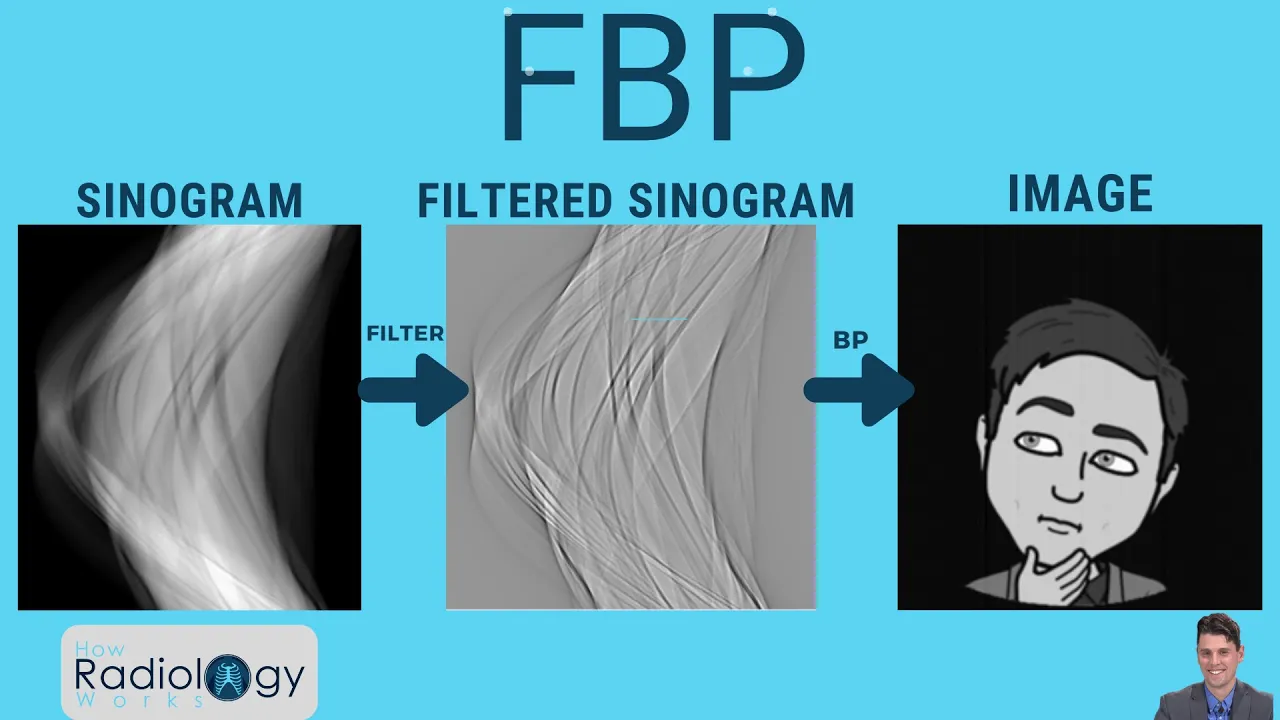
Filtered Backprojection in CT: A Guide for Technologists
@HowRadiologyWorks
39.6K views•2020-11-27
30:32
Ultrasonic Transducers: Resonant Frequency Measurement and Horn Design
@imajeenyus42
229.9K views•2017-02-22
54:16
Polymer Environmental Degradation: Mechanisms & Stabilization
@iit
1.8K views•2012-07-10
33:39
How a Student's Question Saved a NYC Skyscraper from Collapse
@veritasium
22.8M views•2025-04-26
Related Study Plans & Knowledge Roadmaps
Structured learning paths in Engineering
engineering
Building Acoustic Levitators: Physics & Arduino
01The Physics of Sound and Standing Waves
02Piezoelectric Transducers and H-Bridge Drivers
03Arduino Programming: Generating 40 kHz Signals
+ 2 more learning modules
14h5 modules
Explore Roadmap
engineering
Building Peltier Cloud Chambers: Particle Tracks
01Cosmic Rays & Cloud Chamber Physics
02Thermoelectric Cooling (TEC) Fundamentals
03Custom Liquid-Cooling Loop Design
+ 2 more learning modules
18h5 modules
Explore Roadmap
engineering
Yagi-Uda Antennas: Modeling, Build & VNA Test
01Introduction to RF and Antenna Fundamentals
02Yagi-Uda Antenna Design Principles
03Antenna Modeling and Simulation in 4NEC2
+ 2 more learning modules
18h5 modules
Explore Roadmap
engineering
ECG Monitor Design: Op-Amps, Filters & Noise
01Electronics & Breadboard Basics
02Op-Amps and Instrumentation Amplifiers
03Bioelectricity and ECG Fundamentals
+ 2 more learning modules
18h5 modules
Explore Roadmap